


Article Figures & Data
Figures
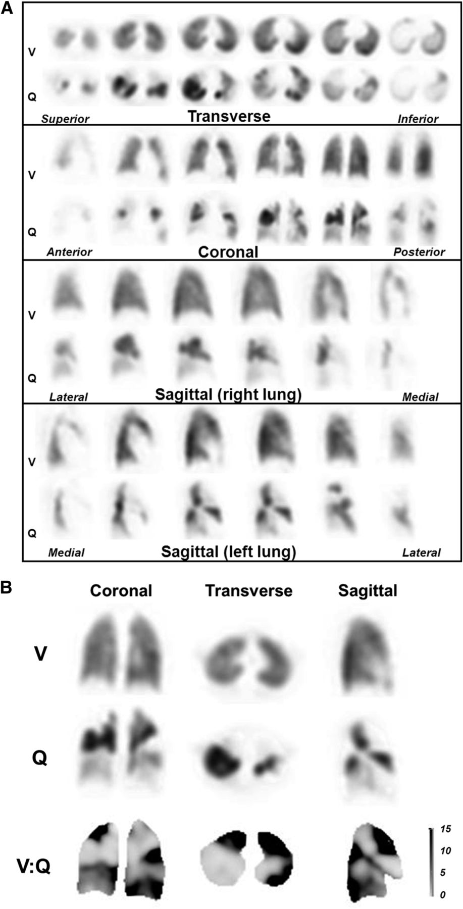
- FIGURE 1.
(A). Example of patient with multiple bilateral PE. Ventilation and perfusion images show multiple mismatched perfusion defects. (B) Representative ventilation, perfusion, and V/Q quotient images. Dark areas on V/Q quotient images, denoting high V/Q ratio, are indicative of V/Q mismatch. (Reprinted with permission from (19).)
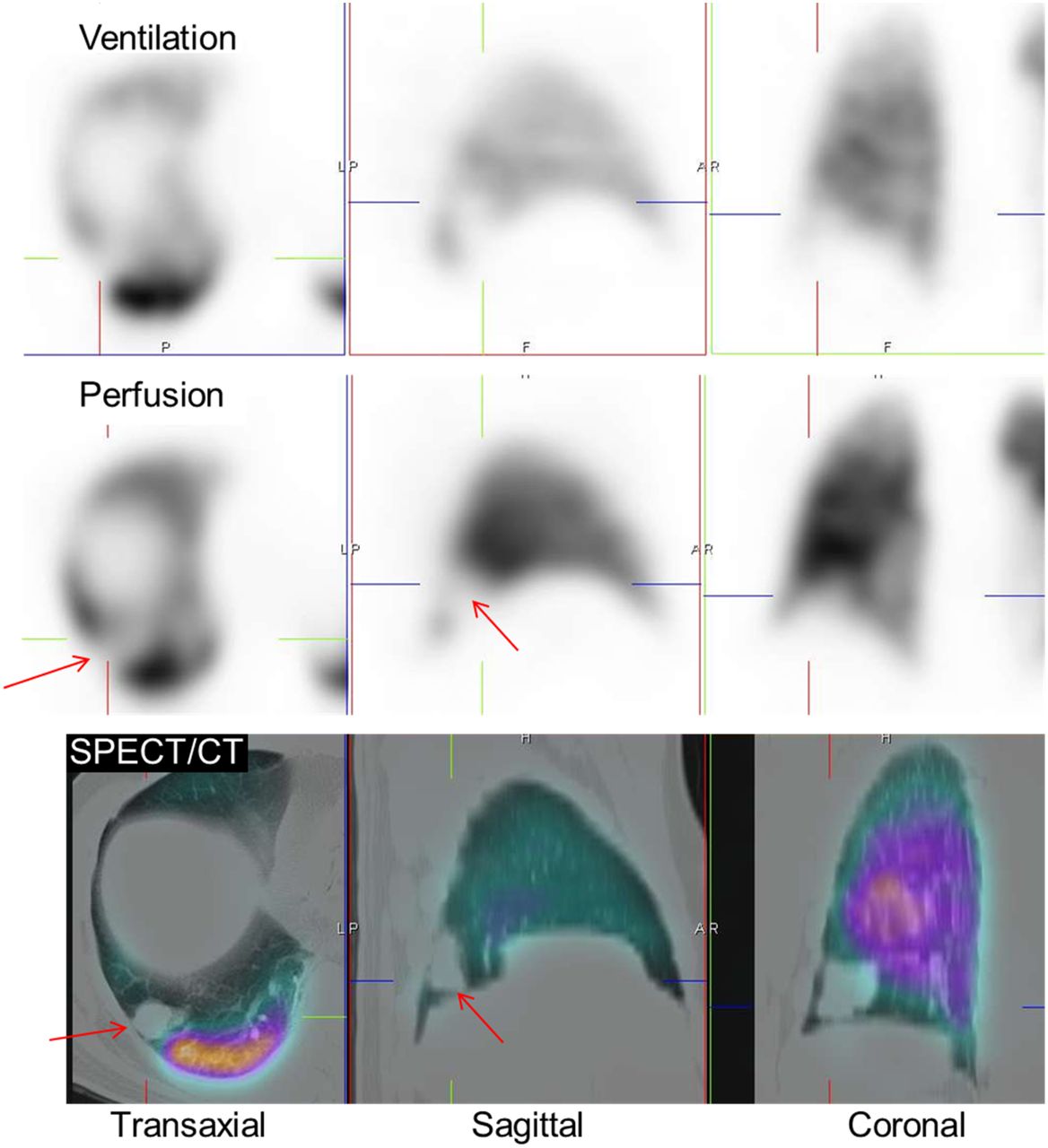
- FIGURE 2.
(A–D) False-positive V/Q scan due to emphysema. Mismatch is evident in right upper lobe (arrows), but CT (E) shows cause to be emphysematous bulla. R = right; A = anterior; L = left. (Reprinted with permission from (14).)
- FIGURE 3.
Representative SPECT/CT images in patient with colon cancer and dyspnea. SPECT shows matched defect in right lower lobe (arrows). CT shows this finding to correspond to previously undiagnosed metastasis.
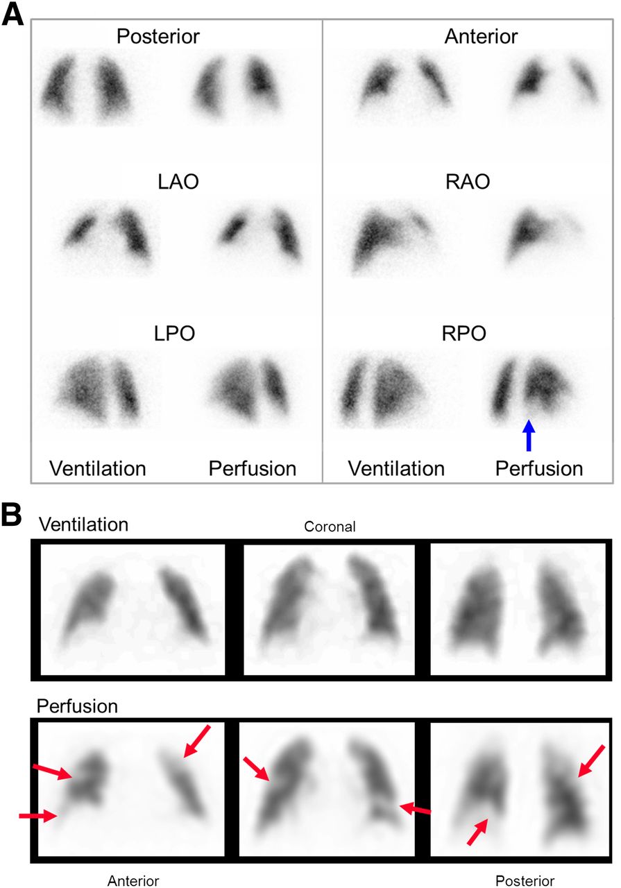
- FIGURE 4.
Representative SPECT/CT images in patient with multiple PE. Several mismatched defects are evident (arrows). CT shows no underlying structural abnormalities.
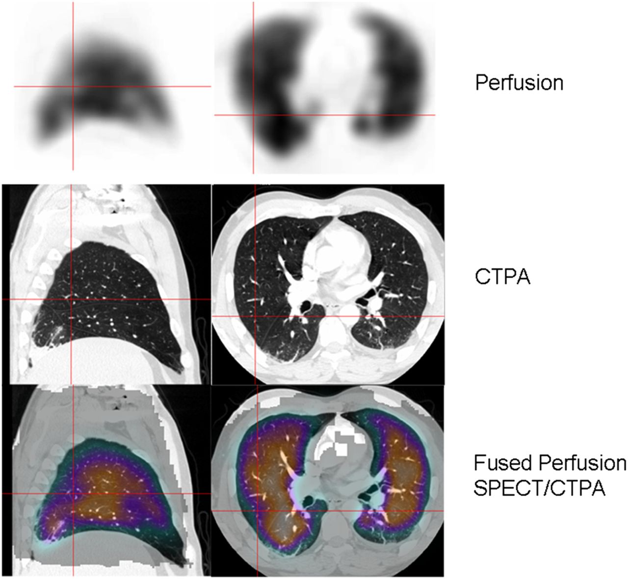
- FIGURE 5.
Sagittal (left) and transaxial (right) perfusion, CTPA, and fused slices in patient with PE and lower lobe volume loss due to atelectasis. Although defect (red crosshairs) was initially localized to superior segment of right lower lobe, fusion accurately localizes defect to posterior segment of right upper lobe. (Reprinted with permission from (52).)
- FIGURE 6.
Coregistered CTPA and perfusion SPECT scans (transverse slice) demonstrating extensive perfusion defects on SPECT. Findings correspond to proximal bilateral PE shown on CTPA (arrows) (Reprinted with permission from (52).)
- FIGURE 7.
(A) Planar V/Q scan in patient with dyspnea. Single mismatched defect is seen at right base (arrow), classifying study as intermediate probability of PE. (B) Representative coronal SPECT slices show multiple mismatched defects (arrows) indicative of widespread PE. Patient had extensive deep venous thrombosis.
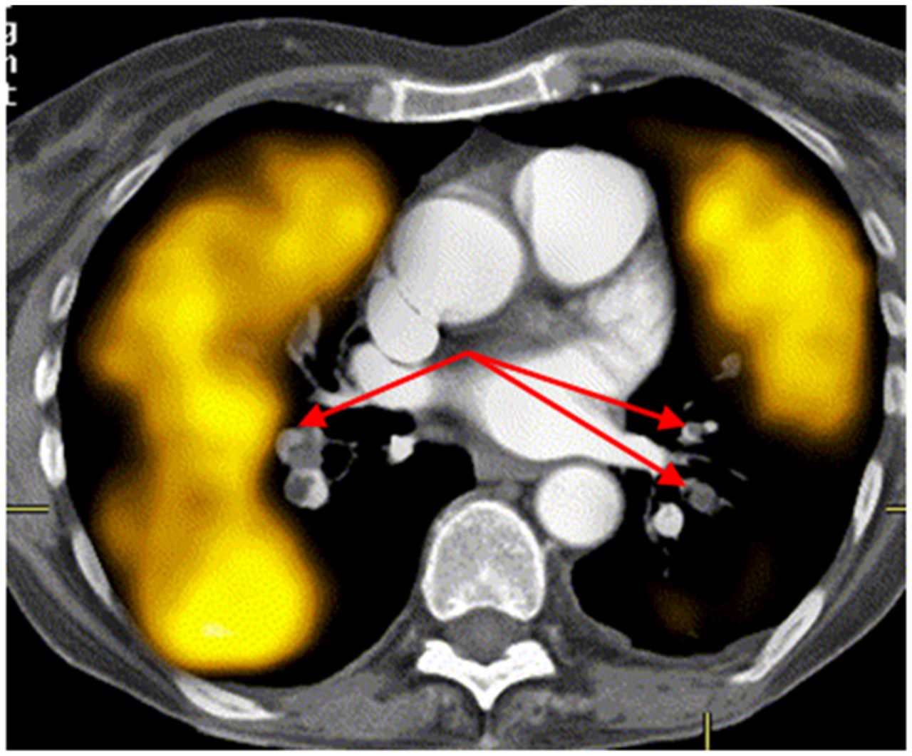
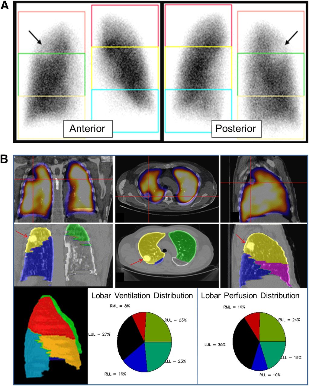
- FIGURE 8.
(A) Anterior (left) and posterior (right) planar images in patient with right lung carcinoma (arrows). Boxes over upper, middle, and lower thirds of each lung approximate relative contribution of each region. Because of overlap of segments and differences in individual anatomy, accuracy is lacking. (B) Fused perfusion/CT images (top row) in coronal (left), transverse (middle), and sagittal (right) planes show perfusion defect (due to tumor, denoted with red crosshairs) in right upper lobe. Patient’s individual CT scan can be used to generate patient-specific lobar slices (middle row) in corresponding orthogonal slices and rotating maximum-intensity-projection images (left image, bottom row). SPECT/CT allowed accurate determination of each lobe’s relative contribution to overall ventilation (middle image, bottom row) and perfusion (right image, bottom row). LLL = left lower lobe; LUL = left upper lobe; RLL = right lower lobe; RML = right middle lobe; RUL = right upper lobe.


Tables
Parameter Description SPECT acquisition 3° steps over 360° Acquisition time per projection 12 s (ventilation); 8 s (perfusion) Collimator Low-energy, high resolution Matrix size 128 × 128 (64 × 64 can also be used) Reconstruction Ordered-subset expectation maximization (8 iterations, 4 subsets) Postreconstruction filter 3-dimensional Butterworth; cutoff, 0.8 cycles/cm; order, 9 ↵* Protocol from Royal North Shore Hospital, Sydney.
Parameter CTPA V/Q SPECT V/Q SPECT/CT Sensitivity Moderate-high High High Specificity Very high High Very high Accuracy with abnormal radiograph finding Unaffected Sometimes affected Sometimes affected Ability to provide other diagnoses Frequent Rare Frequent Incidental findings requiring follow-up Frequent Rare Less frequent Radiation dose High Low Low-moderate Possible allergic reaction Yes No No Risk of contrast nephropathy Yes No No Technical failure rate Higher Rare Rare Availability (especially outside routine hours) High Usually lower Usually lower Accuracy in pregnancy Lower High High Accuracy in chronic PE Lower High High Performance in obstructive lung disease Unaffected May be affected May be affected Role and accuracy in follow-up Limited Very good Very good


Jump to section
Related Articles
Cited By...
- Comparability of Quantifying Relative Lung Ventilation with Inhaled 99mTc-Technegas and 133Xe in Patients Undergoing Evaluation for Lung Transplantation
- Pulmonary embolism
- V/Q SPECT and SPECT/CT in Pulmonary Embolism
- A Technical Overview of Technegas as a Lung Ventilation Agent
- Imaging of pulmonary hypertension in adults: a position paper from the Fleischner Society
- The effects of lung volume reduction treatment on diffusing capacity and gas exchange
- Nuclear Medicine Clinical Practice in the United States During the COVID-19 Era and Beyond
- Pulmonary Arterial Hypertension With Abnormal V/Q Single-Photon Emission Computed Tomography
- The Importance of Quality in Ventilation-Perfusion Imaging
- SPECT V/Q for the diagnosis of pulmonary embolism: protocol for a systematic review and meta-analysis of diagnostic accuracy and clinical outcome
- Correlation of 68Ga Ventilation-Perfusion PET/CT with Pulmonary Function Test Indices for Assessing Lung Function
- Pulmonary Scintigraphy for the Diagnosis of Acute Pulmonary Embolism: A Survey of Current Practices in Australia, Canada, and France