
Main menu

  • Home
  • Content
    • Current
    • Ahead of print
    • Past Issues
    • JNM Supplement
    • SNMMI Annual Meeting Abstracts
    • Continuing Education
    • JNM Podcasts
  • Subscriptions
    • Subscribers
    • Institutional and Non-member
    • Rates
    • Journal Claims
    • Corporate & Special Sales
  • Authors
    • Submit to JNM
    • Information for Authors
    • Assignment of Copyright
    • AQARA requirements
  • Info
    • Reviewers
    • Permissions
    • Advertisers
  • About
    • About Us
    • Editorial Board
    • Contact Information
  • More
    • Alerts
    • Feedback
    • Help
    • SNMMI Journals
  • SNMMI
    • JNM
    • JNMT
    • SNMMI Journals
    • SNMMI

User menu

  • Subscribe
  • My alerts
  • Log in
  • My Cart

Search

  • Advanced search
Journal of Nuclear Medicine
  • SNMMI
    • JNM
    • JNMT
    • SNMMI Journals
    • SNMMI
  • Subscribe
  • My alerts
  • Log in
  • My Cart
Journal of Nuclear Medicine

Advanced Search

  • Home
  • Content
    • Current
    • Ahead of print
    • Past Issues
    • JNM Supplement
    • SNMMI Annual Meeting Abstracts
    • Continuing Education
    • JNM Podcasts
  • Subscriptions
    • Subscribers
    • Institutional and Non-member
    • Rates
    • Journal Claims
    • Corporate & Special Sales
  • Authors
    • Submit to JNM
    • Information for Authors
    • Assignment of Copyright
    • AQARA requirements
  • Info
    • Reviewers
    • Permissions
    • Advertisers
  • About
    • About Us
    • Editorial Board
    • Contact Information
  • More
    • Alerts
    • Feedback
    • Help
    • SNMMI Journals
  • View or Listen to JNM Podcast
  • Visit JNM on Facebook
  • Join JNM on LinkedIn
  • Follow JNM on Twitter
  • Subscribe to our RSS feeds
Review ArticleContinuing Education

PET Imaging of Proliferation with Pyrimidines

Omid S. Tehrani and Anthony F. Shields
Journal of Nuclear Medicine June 2013, 54 (6) 903-912; DOI: https://doi.org/10.2967/jnumed.112.112201
Omid S. Tehrani
Department of Oncology, Karmanos Cancer Institute, Wayne State University, Detroit, Michigan
  • Find this author on Google Scholar
  • Find this author on PubMed
  • Search for this author on this site
Anthony F. Shields
Department of Oncology, Karmanos Cancer Institute, Wayne State University, Detroit, Michigan
  • Find this author on Google Scholar
  • Find this author on PubMed
  • Search for this author on this site
  • Article
  • Figures & Data
  • Info & Metrics
  • PDF
Loading

Article Figures & Data

Figures

  • FIGURE 1.
    • Download figure
    • Open in new tab
    • Download powerpoint
    FIGURE 1.

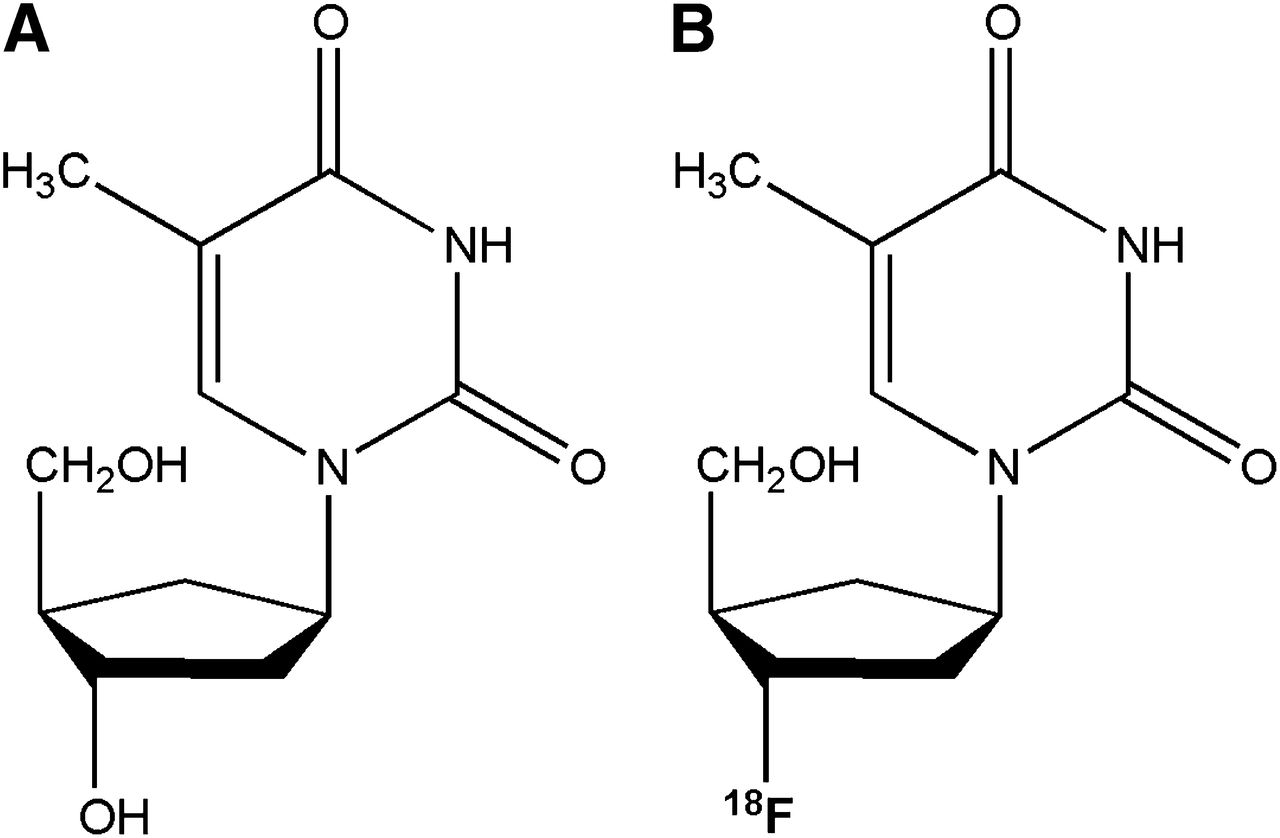
    Structures of thymidine (A) and 18F-FLT (B) with fluoride in position of hydroxide. Fluorine substitution allows labeling with 18F.

  • FIGURE 2.
    • Download figure
    • Open in new tab
    • Download powerpoint
    FIGURE 2.

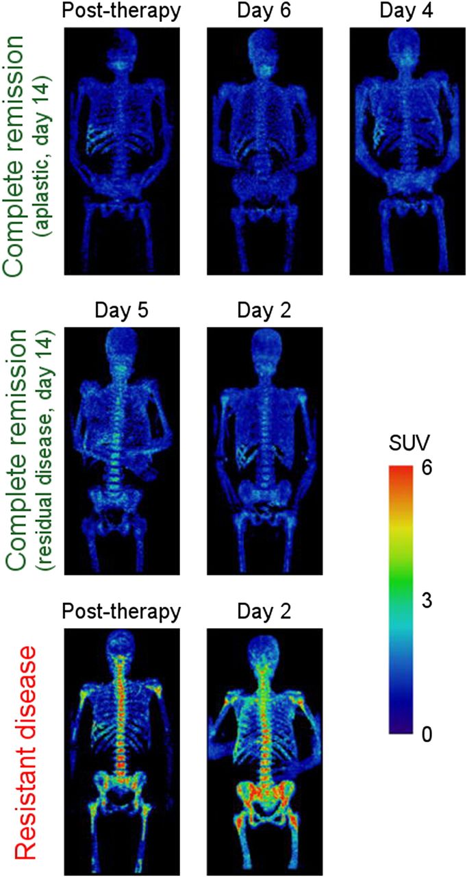
    18F-FLT PET images of bone marrow of 7 AML patients grouped by clinical response. PET scans were acquired at different time points of therapy, but results were consistent within each clinical response group (complete remission and resistant disease), independent of time of assessment. Resistant disease exhibited elevated uptake, whereas complete remission displayed low uptake. (Reprinted with permission of (20).)

  • FIGURE 3.
    • Download figure
    • Open in new tab
    • Download powerpoint
    FIGURE 3.

    Transaxial views of 18F-FLT PET (top), helical CT (middle), and 18F-FDG PET (bottom) for 2 patients with DLBCL. (A) A 41-y-old man with retromandibular lymphoma showing intense 18F-FDG and 18F-FLT uptake in projection of retromandibular lymph node. This stage 1A patient revealed disease progression under therapy. (B) A 40-y-old woman (stage IVA) with histologically proven lymphoma in right iliac bone and sacrum. 18F-FDG PET shows intensely increased uptake in right ilium and sacrum. Corresponding 18F-FLT PET images allow detection of increased asymmetric uptake in right ilium and sacrum despite high physiologic 18F-FLT uptake in proliferating bone marrow. Transaxial views of helical CT display osteodestruction of right ilium. Restaging after end of therapy revealed complete response. (Reprinted from (22).)

  • FIGURE 4.
    • Download figure
    • Open in new tab
    • Download powerpoint
    FIGURE 4.

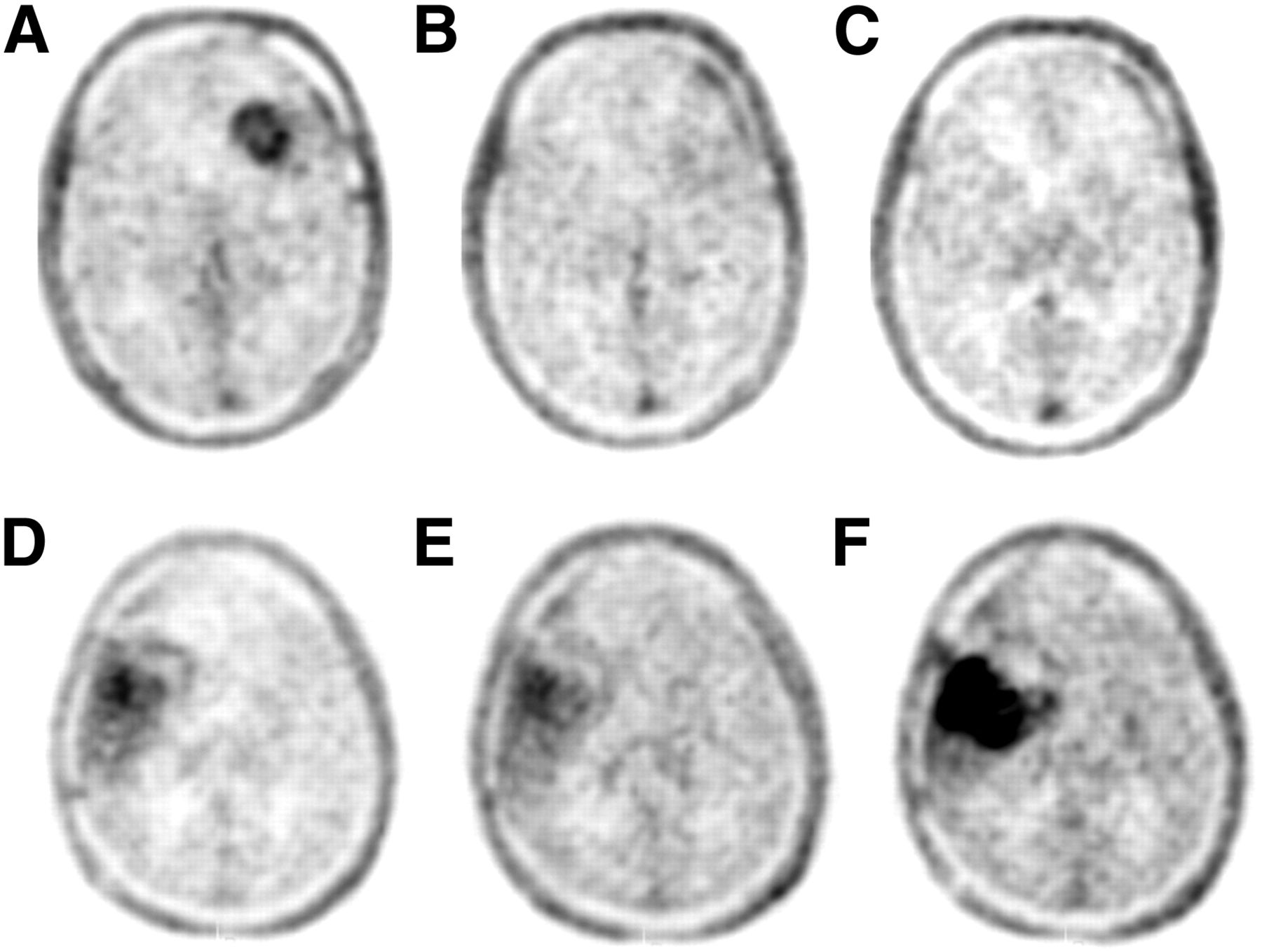
    18F-FLT PET at baseline, 2 wk, and 6 wk for responding patient (A–C, patient 25) and nonresponding patient (D–F, patient 9). (Reprinted from (34)).

  • FIGURE 5.
    • Download figure
    • Open in new tab
    • Download powerpoint
    FIGURE 5.

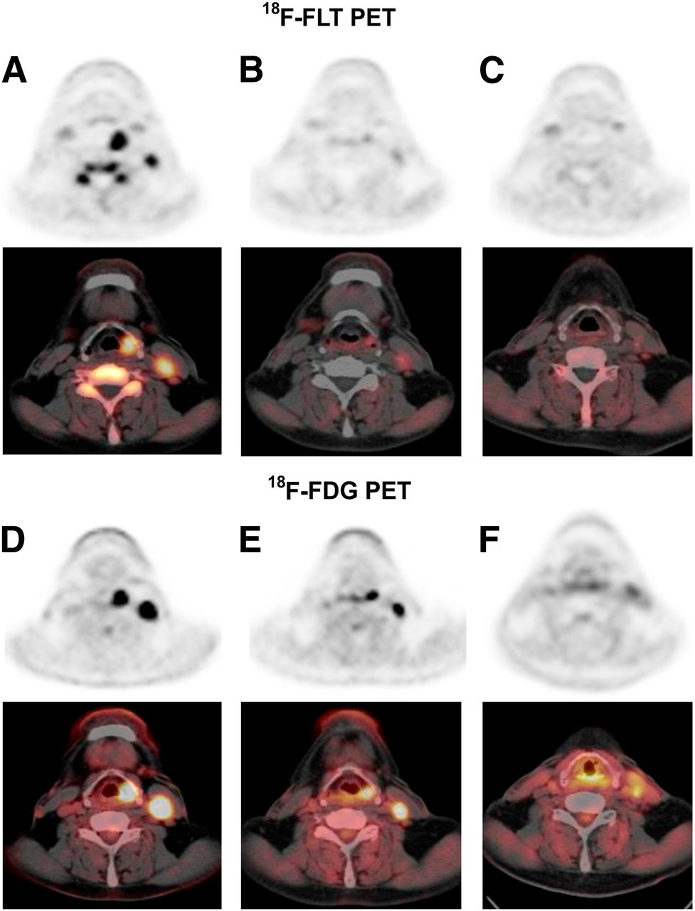
    PET images of patient with hypopharyngeal cancer (patient 14) before radiation therapy (A and D), 3 wk after initiation of radiation therapy (B and E), and 4 wk after end of radiation therapy (C and F). Pretreatment 18F-FLT and 18F-FDG axial PET images showed increased metabolism in primary tumor and metastatic lymph node (18F-FLT SUVs, 9.16 and 6.06, respectively; 18F-FDG SUVs, 21.81 and 13.37, respectively). 18F-FLT and 18F-FDG SUVs decreased after 30 Gy of irradiation (18F-FLT SUVs, 2.86 and 2.14, respectively; 18F-FDG SUVs, 11.44 and 6.39, respectively). 18F-FLT uptake in primary site and lymph nodes was same as in surrounding muscle (SUVs of 0.93, 0.9, and 0.9, respectively) at 4 wk after completion of treatment, whereas increased uptake of 18F-FDG persisted (SUV of 4.66 in primary lesion and 3.75 in lymph node). Patient was alive and without evidence of recurrent disease 19 mo after therapy. (Reprinted from (66).)

  • FIGURE 6.
    • Download figure
    • Open in new tab
    • Download powerpoint
    FIGURE 6.

    Patient 5, with non–small cell lung cancer in left upper lobe. (A) Transaxial 18F-FLT PET scan demonstrates high 18F-FLT uptake (arrow) in tumor margin. 18F-FLT uptake in vertebral column, scapula, and ribs represents proliferating bone marrow. (B and C) Corresponding CT and 18F-FDG PET scans show high 18F-FDG uptake in tumor margin and primary lung tumor. (D) On Ki-67 immunohistochemistry, Ki-67–positive nuclei (brown) demonstrate high proliferation rate of 54%, and hematoxylin background staining reveals Ki-67–negative nuclei (blue). (Reprinted from (78).)

PreviousNext
Back to top

In this issue

Journal of Nuclear Medicine: 54 (6)
Journal of Nuclear Medicine
Vol. 54, Issue 6
June 1, 2013
  • Table of Contents
  • Table of Contents (PDF)
  • About the Cover
  • Index by author
Print
Download PDF
Article Alerts
Sign In to Email Alerts with your Email Address
Email Article

Thank you for your interest in spreading the word on Journal of Nuclear Medicine.

NOTE: We only request your email address so that the person you are recommending the page to knows that you wanted them to see it, and that it is not junk mail. We do not capture any email address.

Enter multiple addresses on separate lines or separate them with commas.
PET Imaging of Proliferation with Pyrimidines
(Your Name) has sent you a message from Journal of Nuclear Medicine
(Your Name) thought you would like to see the Journal of Nuclear Medicine web site.
Citation Tools
PET Imaging of Proliferation with Pyrimidines
Omid S. Tehrani, Anthony F. Shields
Journal of Nuclear Medicine Jun 2013, 54 (6) 903-912; DOI: 10.2967/jnumed.112.112201

Citation Manager Formats

  • BibTeX
  • Bookends
  • EasyBib
  • EndNote (tagged)
  • EndNote 8 (xml)
  • Medlars
  • Mendeley
  • Papers
  • RefWorks Tagged
  • Ref Manager
  • RIS
  • Zotero
Share
PET Imaging of Proliferation with Pyrimidines
Omid S. Tehrani, Anthony F. Shields
Journal of Nuclear Medicine Jun 2013, 54 (6) 903-912; DOI: 10.2967/jnumed.112.112201
Twitter logo Facebook logo LinkedIn logo Mendeley logo
  • Tweet Widget
  • Facebook Like
  • Google Plus One
Bookmark this article

Jump to section

  • Article
    • Abstract
    • PROLIFERATION IMAGING AND ITS ROLE IN PRACTICE
    • BONE MARROW AND HEMATOLOGIC MALIGNANCIES
    • SARCOMAS
    • BREAST CANCER
    • CENTRAL NERVOUS SYSTEM CANCERS
    • GASTROINTESTINAL CANCERS
    • GENITOURINARY CANCERS
    • HEAD AND NECK CANCERS
    • LUNG CANCER
    • SKIN CANCERS
    • DISCUSSION
    • CONCLUSION
    • Footnotes
    • REFERENCES
  • Figures & Data
  • Info & Metrics
  • PDF

Related Articles

  • This Month in JNM
  • Erratum
  • PubMed
  • Google Scholar

Cited By...

  • 18F-FLT PET/CT Adds Value to 18F-FDG PET/CT for Diagnosing Relapse After Definitive Radiotherapy in Patients with Lung Cancer: Results of a Prospective Clinical Trial
  • Using Radiolabeled 3'-Deoxy-3'-18F-Fluorothymidine with PET to Monitor the Effect of Dexamethasone on Non-Small Cell Lung Cancer
  • Sex as a Biologic Variable in Preclinical Imaging Research: Initial Observations with 18F-FLT
  • Differential diagnosis of granulomatous lung disease: clues and pitfalls: Number 4 in the Series "Pathology for the clinician" Edited by Peter Dorfmüller and Alberto Cavazza
  • Preclinical Evidence That 3'-Deoxy-3'-[18F]Fluorothymidine PET Can Visualize Recovery of Hematopoiesis after Gemcitabine Chemotherapy
  • Prognostic Value of 18F-FLT PET in Patients with Neuroendocrine Neoplasms: A Prospective Head-to-Head Comparison with 18F-FDG PET and Ki-67 in 100 Patients
  • PET of Glucose Metabolism and Cellular Proliferation in Prostate Cancer
  • 18F-FLT PET/CT in the Evaluation of Pheochromocytomas and Paragangliomas: A Pilot Study
  • 18F-FLT PET Evaluation of Radiation Response
  • VEGF-Targeted Therapy Stably Modulates the Glycolytic Phenotype of Tumor Cells
  • Google Scholar

More in this TOC Section

  • Treatment Response Evaluation in Prostate Cancer Using PSMA PET/CT
  • Approaches to Imaging Immune Activation Using PET
  • Large Language Models and Large Multimodal Models in Medical Imaging: A Primer for Physicians
Show more Continuing Education

Similar Articles

Keywords

  • PET
  • proliferation
  • thymidine
  • FLT
SNMMI

© 2025 SNMMI

Powered by HighWire