


Article Figures & Data
Figures
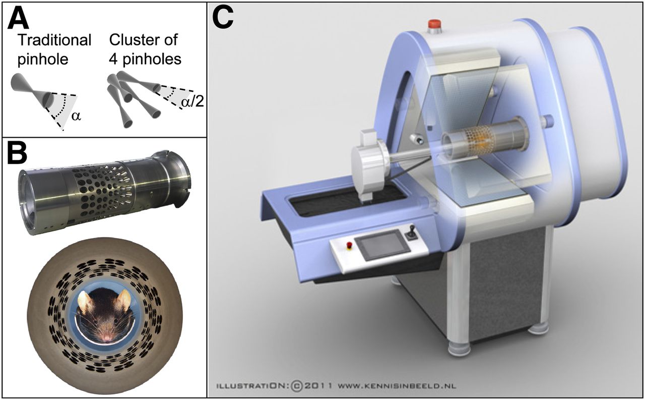
- FIGURE 1.
Integration of clustered-pinhole collimator into existing SPECT/CT platform. (A) Traditional pinhole with opening angle α and cluster of 4 pinholes with approximately same field of view and opening angle α/2. (B) Clustered-pinhole collimator optimized for imaging SPECT and PET tracers, into which mouse is placed. (C) Collimator mounted in SPECT/CT system.
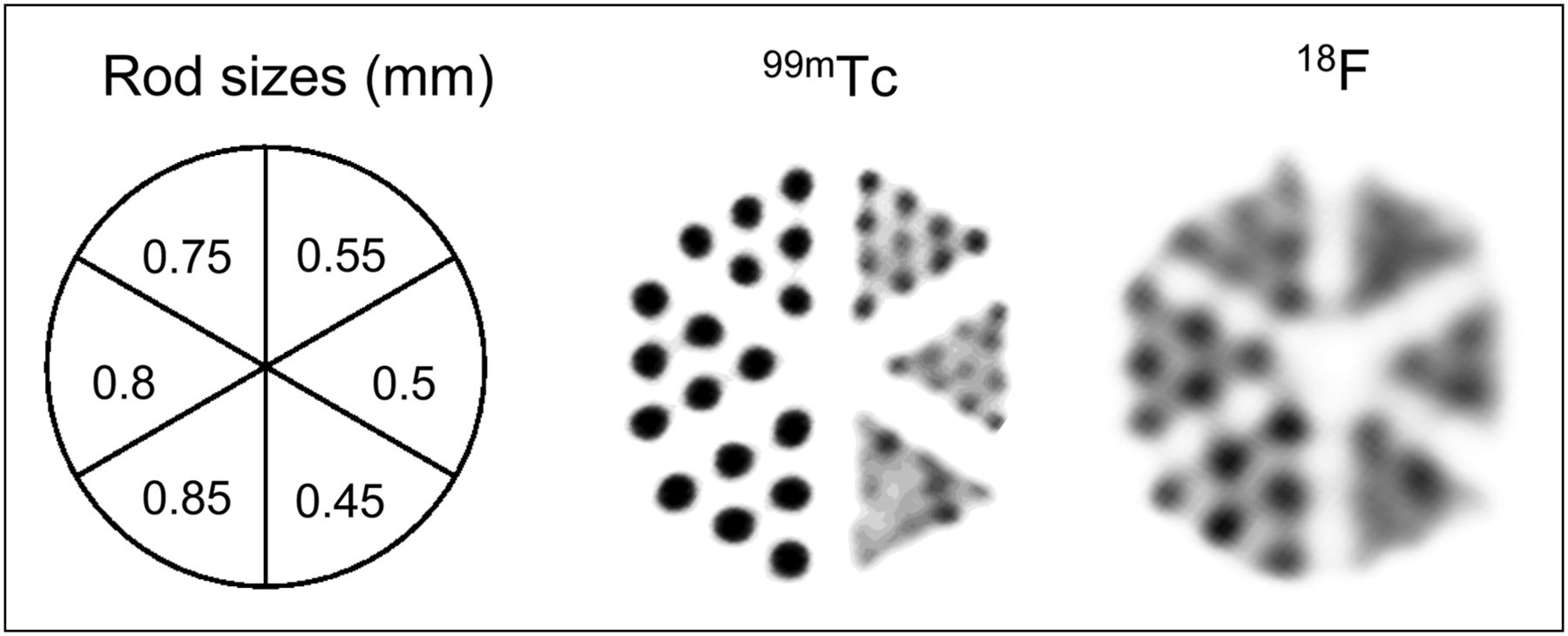
- FIGURE 2.
Simultaneously acquired images of SPECT and PET tracers in phantom with 6 segments with capillary diameters of 0.85, 0.80, 0.75, 0.55, 0.50, and 0.45 mm. At start of this scan, capillaries contained 14 MBq of 18F solution and 8.3 MBq of 99mTc solution. Phantom was imaged for 60 min. All images shown had slice thickness of 3.2 mm.
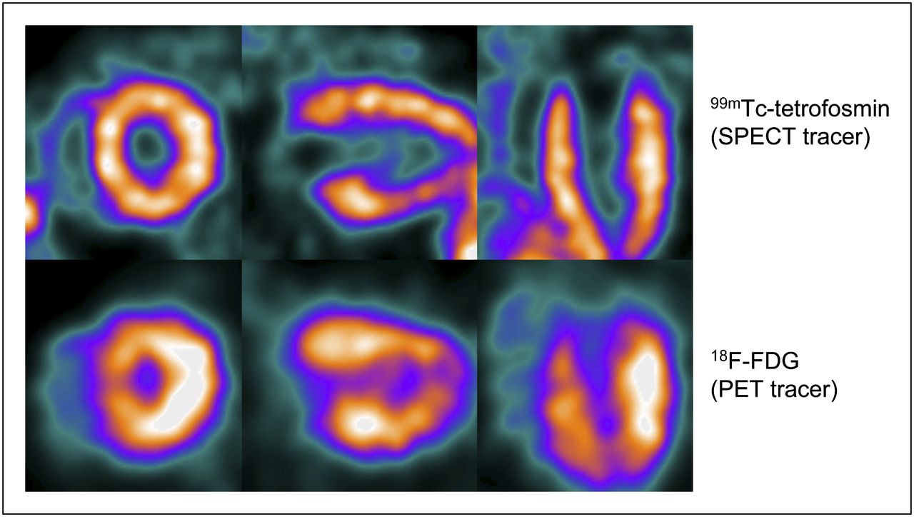
- FIGURE 3.
Simultaneous SPECT/PET cardiac scan. We imaged mouse, injected with 130 MBq of 99mTc-tetrofosmin and 24 MBq of 18F-FDG, for 60 min. Here, we show 3 mutually perpendicular slices through mouse’s heart. Images represent average over cardiac cycle. Supplemental Video 1, of beating heart, was reconstructed from same scan.
- FIGURE 4.
Simultaneously acquired SPECT/PET mouse brain images (color) overlayed on CT (gray). Coronal (A), sagittal (B), and horizontal (C) slices at identical levels are shown. Mouse was injected with 30 MBq of 123I-FP-CIT and 40 MBq of 18F-FDG and imaged for 60 min starting 105 min after injection. Uptake of 123I-FP-CIT (SPECT, top) in small brain structures such as striatum (1), olfactory tubercle (2), and Harderian glands (3) can be resolved. 18F-FDG (PET scan, bottom) is seen in multiple structures, including olfactory bulbs (4), cerebral cortex (5), and thalamic and midbrain areas (6).
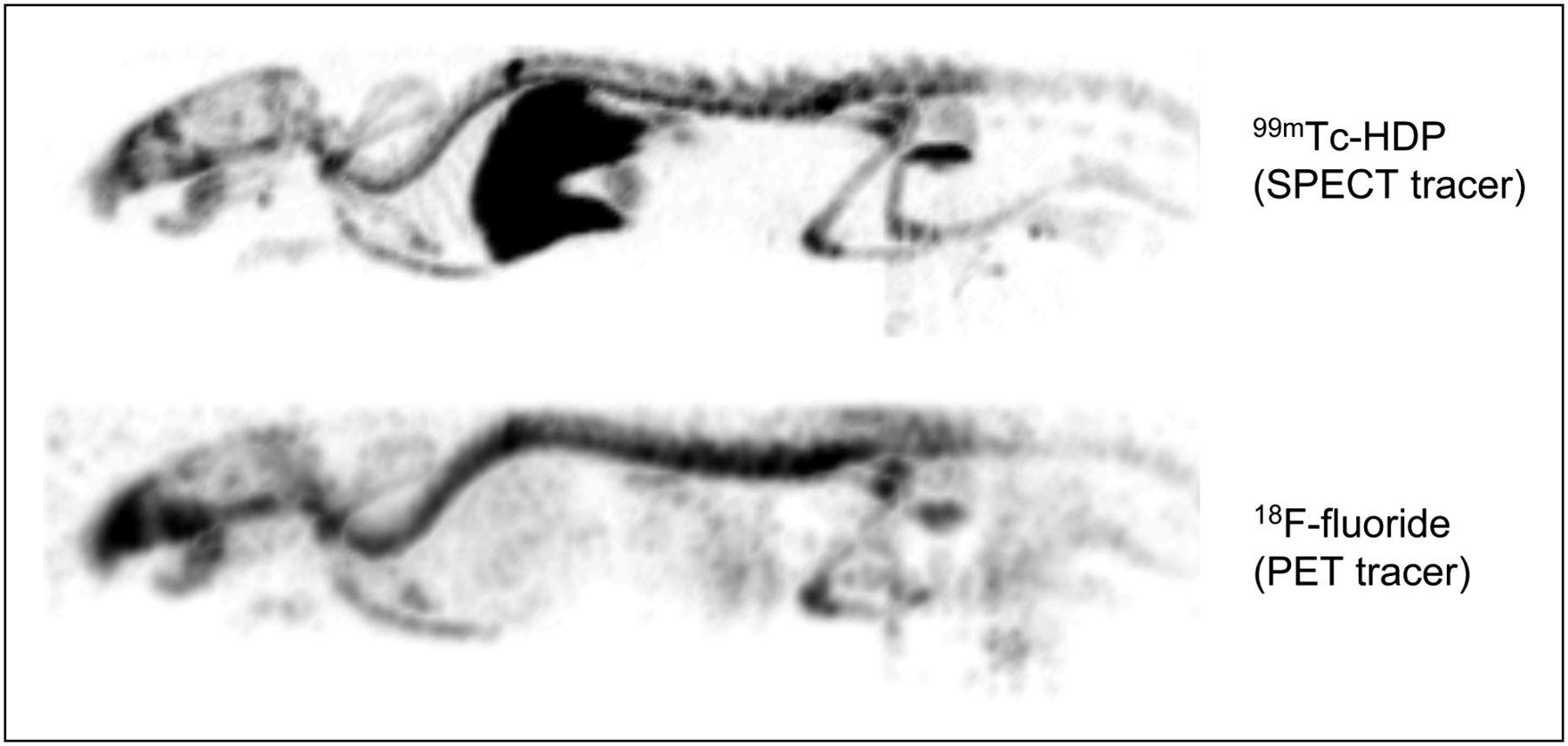
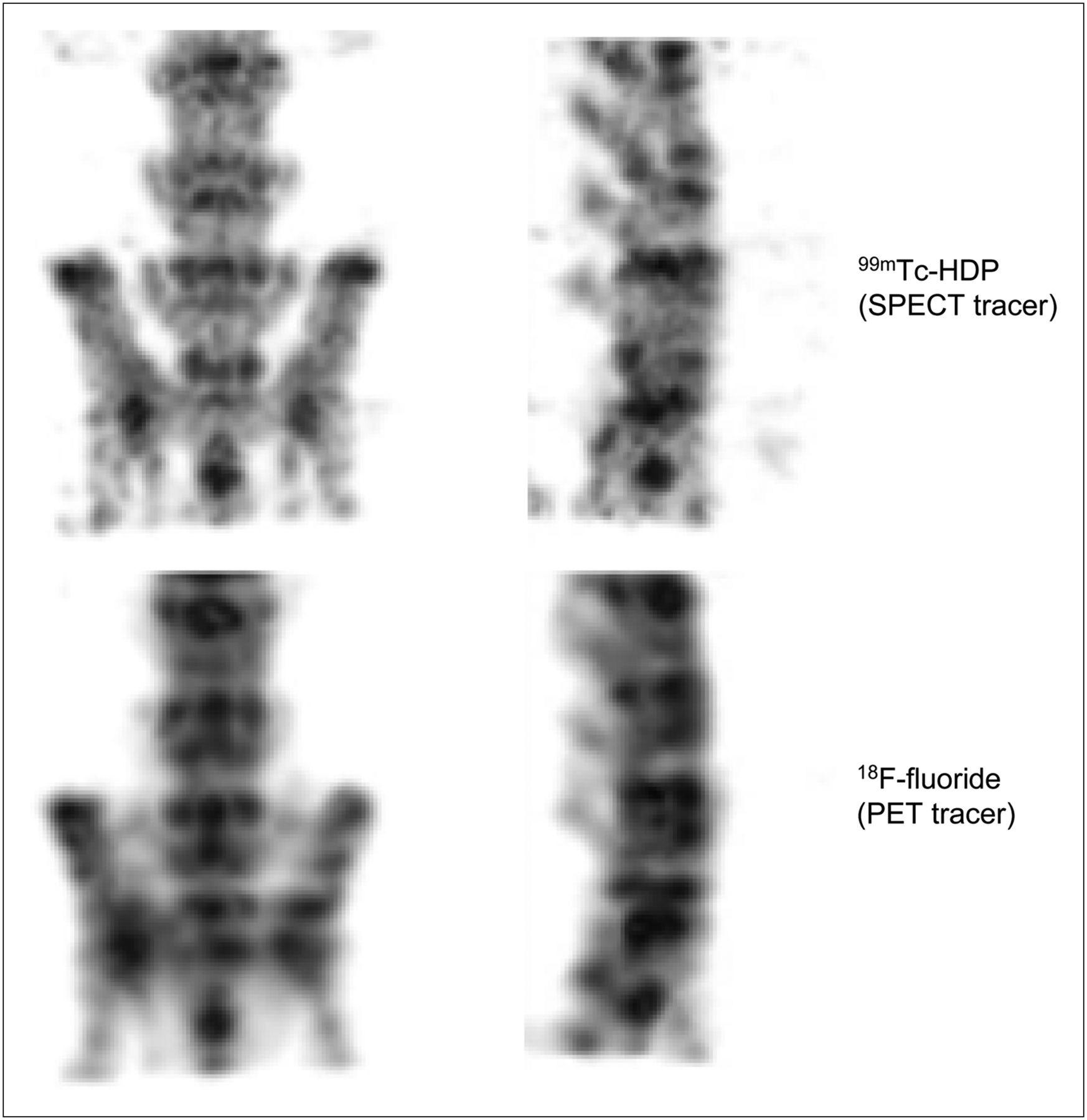
- FIGURE 5.
Example of simultaneous SPECT/PET scan obtained with 2 tracers that target same biologic function. Images represent MIPs of mouse injected with 2 bone tracers, 220 MBq of 99mTc-HDP (top) and 60 MBq of 18F-fluoride (bottom), and scanned for 60 min. Supplemental Video 2 shows rotating MIPs.
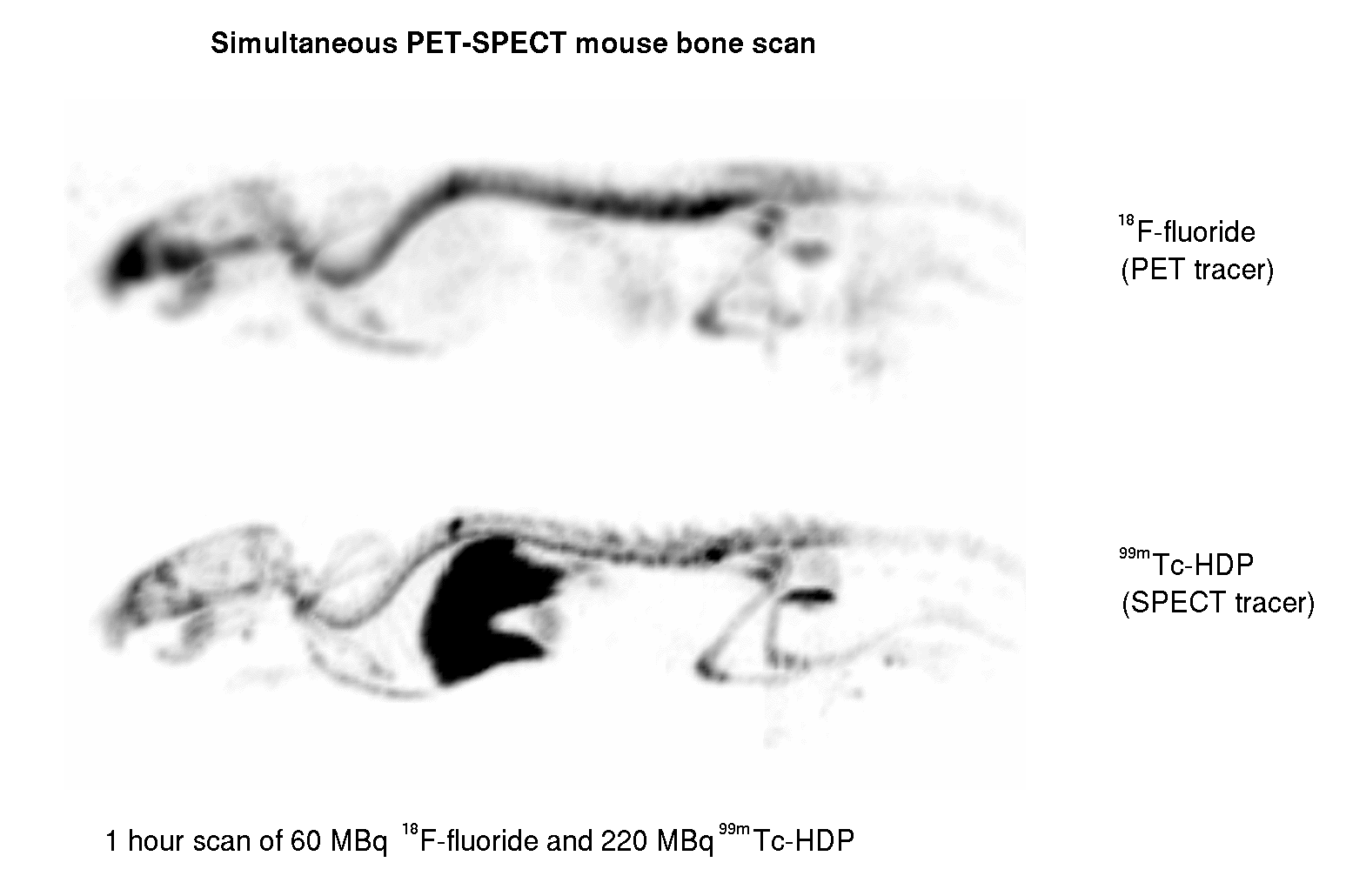
- FIGURE 6.
Example of simultaneous SPECT/PET bone scan of lumbar spine and pelvis. MIPs of mouse injected with 130 MBq of 99mTc-HDP (top) and 85 MBq of 18F-fluoride (bottom) and scanned for 30 min are shown. Supplemental Video 3 shows rotating MIPs.


Tables
Sensitivity Photopeak window width 99mTc 18F 30% 0.25% 0.31% 25% 0.25% 0.30% 20% 0.24% 0.29% 15% 0.22% 0.28%
Supplemental Data
Files in this Data Supplement:
Supplemental Videos
Files in this Data Supplement:


Jump to section
Related Articles
Cited By...
- Human monoclonal antibodies against Staphylococcus aureus surface antigens recognize in vitro biofilm and in vivo implant infections
- Utilizing High-Energy {gamma}-Photons for High-Resolution 213Bi SPECT in Mice
- An Integrated Quad-Modality Molecular Imaging System for Small Animals
- Performance Assessment of a Preclinical PET Scanner with Pinhole Collimation by Comparison to a Coincidence-Based Small-Animal PET Scanner
- Three-Dimensional Histologic Validation of High-Resolution SPECT of Antibody Distributions Within Xenografts
- Imaging Capabilities of the Inveon SPECT System Using Single-and Multipinhole Collimators