
Main menu

  • Home
  • Content
    • Current
    • Ahead of print
    • Past Issues
    • JNM Supplement
    • SNMMI Annual Meeting Abstracts
    • Continuing Education
    • JNM Podcasts
  • Subscriptions
    • Subscribers
    • Institutional and Non-member
    • Rates
    • Journal Claims
    • Corporate & Special Sales
  • Authors
    • Submit to JNM
    • Information for Authors
    • Assignment of Copyright
    • AQARA requirements
  • Info
    • Reviewers
    • Permissions
    • Advertisers
  • About
    • About Us
    • Editorial Board
    • Contact Information
  • More
    • Alerts
    • Feedback
    • Help
    • SNMMI Journals
  • SNMMI
    • JNM
    • JNMT
    • SNMMI Journals
    • SNMMI

User menu

  • Subscribe
  • My alerts
  • Log in
  • Log out
  • My Cart

Search

  • Advanced search
Journal of Nuclear Medicine
  • SNMMI
    • JNM
    • JNMT
    • SNMMI Journals
    • SNMMI
  • Subscribe
  • My alerts
  • Log in
  • Log out
  • My Cart
Journal of Nuclear Medicine

Advanced Search

  • Home
  • Content
    • Current
    • Ahead of print
    • Past Issues
    • JNM Supplement
    • SNMMI Annual Meeting Abstracts
    • Continuing Education
    • JNM Podcasts
  • Subscriptions
    • Subscribers
    • Institutional and Non-member
    • Rates
    • Journal Claims
    • Corporate & Special Sales
  • Authors
    • Submit to JNM
    • Information for Authors
    • Assignment of Copyright
    • AQARA requirements
  • Info
    • Reviewers
    • Permissions
    • Advertisers
  • About
    • About Us
    • Editorial Board
    • Contact Information
  • More
    • Alerts
    • Feedback
    • Help
    • SNMMI Journals
  • View or Listen to JNM Podcast
  • Visit JNM on Facebook
  • Join JNM on LinkedIn
  • Follow JNM on Twitter
  • Subscribe to our RSS feeds
Research ArticleClinical Investigations

Addition of 18F-FDG PET/CT to Clinical Assessment Predicts Overall Survival in HNSCC: A Retrospective Analysis with Follow-up for 12 Years

Vasavi Paidpally, Abdel K. Tahari, Stella Lam, Krishna Alluri, Shanthi Marur, Wayne Koch, Richard L. Wahl and Rathan M. Subramaniam
Journal of Nuclear Medicine December 2013, 54 (12) 2039-2045; DOI: https://doi.org/10.2967/jnumed.113.121285
Vasavi Paidpally
1Russell H. Morgan Department of Radiology and Radiological Sciences, Johns Hopkins University, Baltimore, Maryland
  • Find this author on Google Scholar
  • Find this author on PubMed
  • Search for this author on this site
Abdel K. Tahari
1Russell H. Morgan Department of Radiology and Radiological Sciences, Johns Hopkins University, Baltimore, Maryland
  • Find this author on Google Scholar
  • Find this author on PubMed
  • Search for this author on this site
Stella Lam
1Russell H. Morgan Department of Radiology and Radiological Sciences, Johns Hopkins University, Baltimore, Maryland
  • Find this author on Google Scholar
  • Find this author on PubMed
  • Search for this author on this site
Krishna Alluri
1Russell H. Morgan Department of Radiology and Radiological Sciences, Johns Hopkins University, Baltimore, Maryland
  • Find this author on Google Scholar
  • Find this author on PubMed
  • Search for this author on this site
Shanthi Marur
2Department of Medical Oncology, Johns Hopkins University, Baltimore, Maryland; and
  • Find this author on Google Scholar
  • Find this author on PubMed
  • Search for this author on this site
Wayne Koch
2Department of Medical Oncology, Johns Hopkins University, Baltimore, Maryland; and
3Department of Otolaryngology and Head and Neck Surgery, Johns Hopkins University, Baltimore, Maryland
  • Find this author on Google Scholar
  • Find this author on PubMed
  • Search for this author on this site
Richard L. Wahl
1Russell H. Morgan Department of Radiology and Radiological Sciences, Johns Hopkins University, Baltimore, Maryland
2Department of Medical Oncology, Johns Hopkins University, Baltimore, Maryland; and
  • Find this author on Google Scholar
  • Find this author on PubMed
  • Search for this author on this site
Rathan M. Subramaniam
1Russell H. Morgan Department of Radiology and Radiological Sciences, Johns Hopkins University, Baltimore, Maryland
2Department of Medical Oncology, Johns Hopkins University, Baltimore, Maryland; and
  • Find this author on Google Scholar
  • Find this author on PubMed
  • Search for this author on this site
  • Article
  • Figures & Data
  • Info & Metrics
  • PDF
Loading

Article Figures & Data

Figures

  • Tables
  • FIGURE 1.
    • Download figure
    • Open in new tab
    • Download powerpoint
    FIGURE 1.

    Reader study. Number of indeterminate study results was reduced from 12.3% to 9.7%, with increase in negative study results from 70.1% to 72.2%.
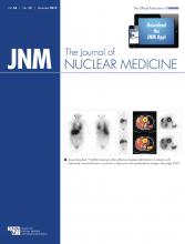
  • FIGURE 2.
    • Download figure
    • Open in new tab
    • Download powerpoint
    FIGURE 2.

    Kaplan–Meier survival plots. PET 1 scans were performed 4–6 mo after treatment; PET 2 scans, 7–12 mo after treatment; and PET 3 scans, 13–24 mo after treatment. OS differed significantly between patients with PET/CT positive for tumor and patients with PET/CT negative for tumor.
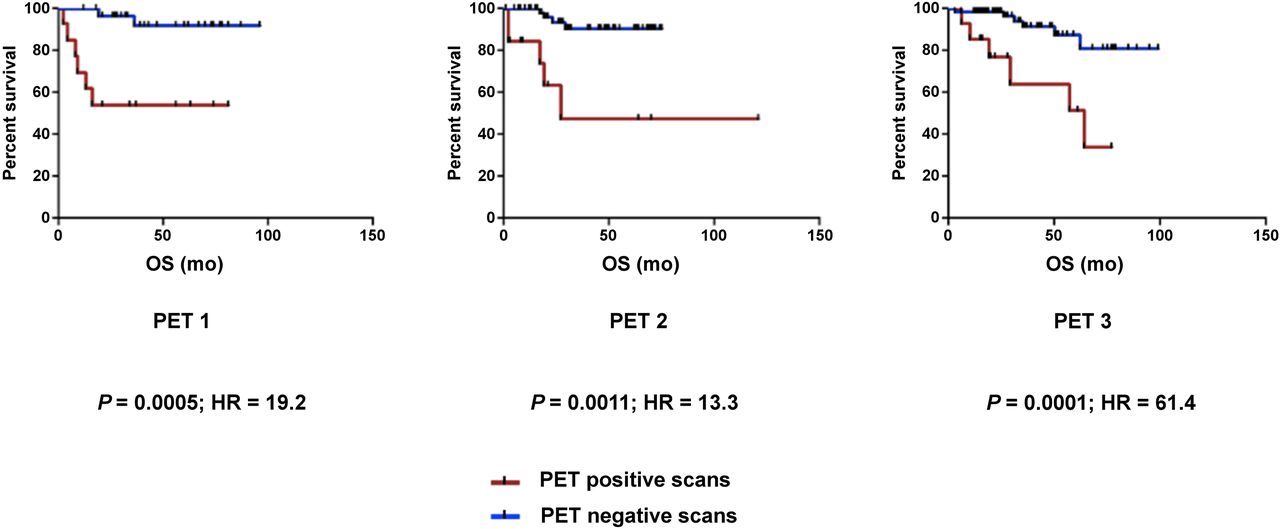
  • FIGURE 3.
    • Download figure
    • Open in new tab
    • Download powerpoint
    FIGURE 3.

    Added value of PET/CT for clinical assessment. PET/CT was helpful for excluding tumor in 51.5% of patients who had clinical suspicion of recurrence or uncertainty and for identifying recurrence in 4.6% of patients with no prior clinical suspicion.

  • FIGURE 4.
    • Download figure
    • Open in new tab
    • Download powerpoint
    FIGURE 4.

    Negative PET/CT scan at 6–12 mo of follow-up. A 43-y-old man had TXN3M0 squamous cell carcinoma on right side of neck (arrows) that was strongly positive for p16 but HPV-16 negative. (A) Staging PET/CT did not identify primary site. Patient completed concurrent chemoradiation. He initially received cisplatin, docetaxel, and 5-fluorouracil with induction chemotherapy but had significant nausea, vomiting, and dehydration. He was then switched to carboplatin and 5-fluorouracil, which was dose-reduced to 650 mg/m2. (B) Follow-up PET/CT scan obtained using same views at 7 mo after therapy showed no evidence of recurrence.
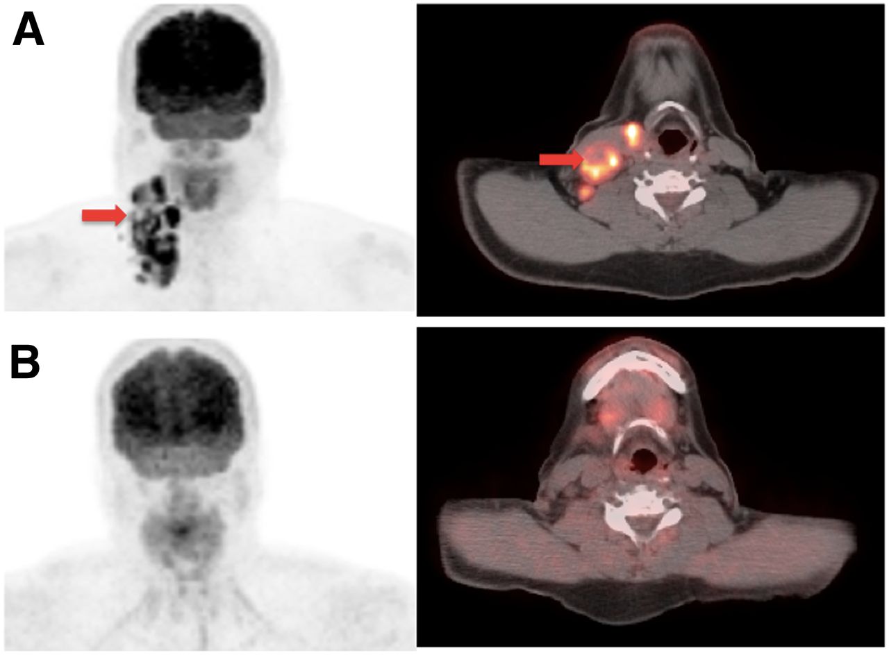
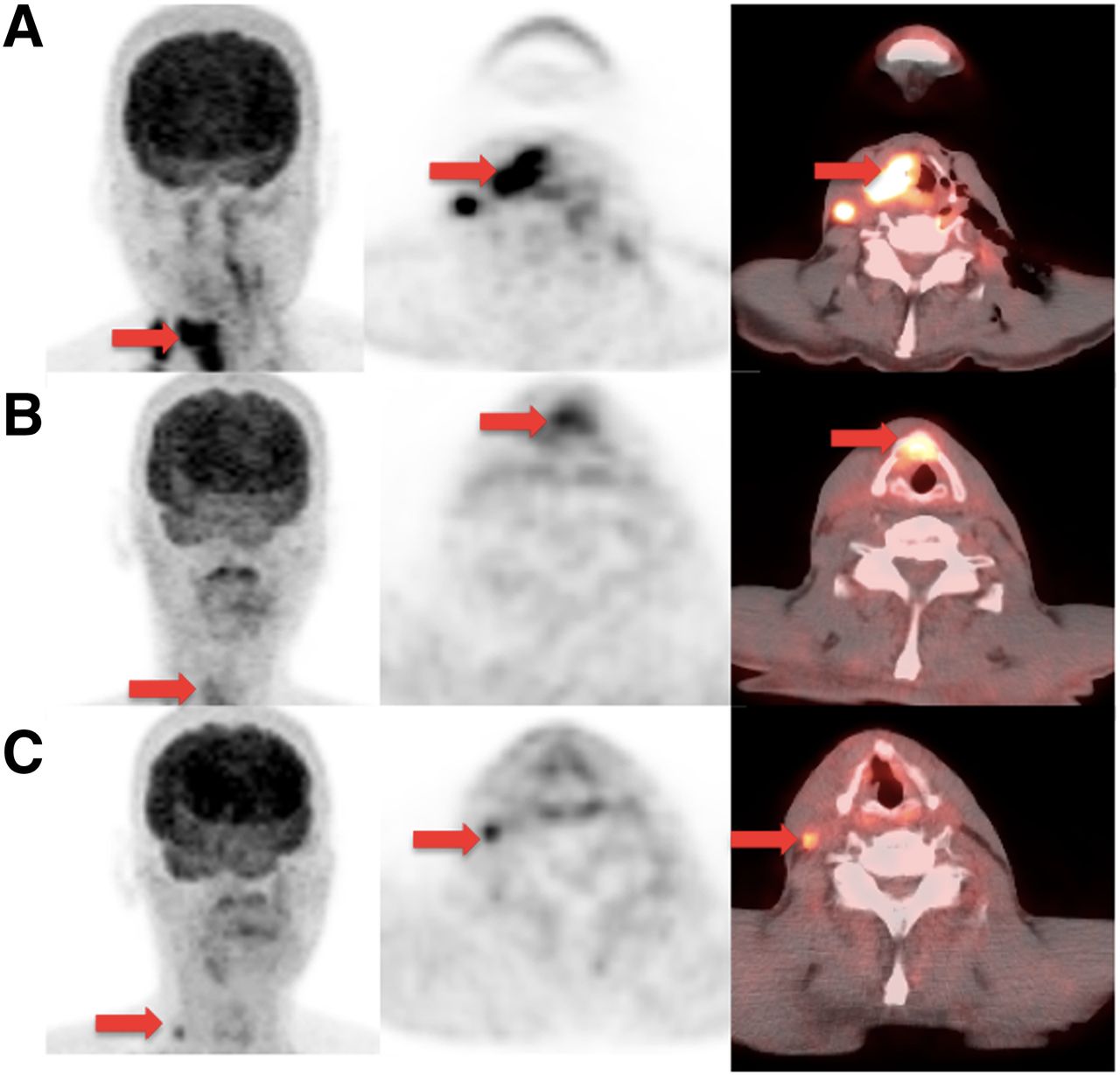
  • FIGURE 5.
    • Download figure
    • Open in new tab
    • Download powerpoint
    FIGURE 5.

    No clinical suspicion. PET/CT identified nodal recurrence at 6–12 mo of follow-up in 61-y-old man diagnosed with T3N2c squamous cell carcinoma of right supraglottic larynx. (A) Baseline coronal PET, axial PET, and fused PET/CT (from left to right) demonstrated intense 18F-FDG activity in primary lesion (arrows), with locoregional disease in right neck. He received chemoradiation to total dose of 70 Gy. (B) PET/CT scan obtained using same views 4 mo after end of therapy revealed interval decrease in size and uptake in right supraglottic laryngeal area. Although some of this uptake could be inflammatory, findings were suggestive of residual disease. Subsequently, patient underwent suspension microscopic laryngoscopy with excision of right false vocal fold irregularity. Biopsy samples were read as negative. (C) However, follow-up PET/CT scan at 10 mo after therapy revealed 18F-FDG–avid right level III lymph node (arrows), biopsy of which was positive for disease.
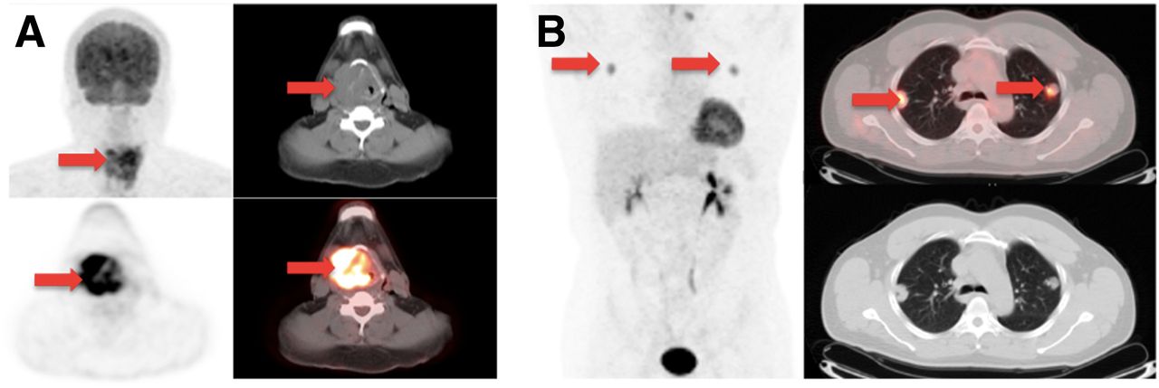
  • FIGURE 6.
    • Download figure
    • Open in new tab
    • Download powerpoint
    FIGURE 6.

    Positive PET/CT scan at 12–24 mo of follow-up in 43-y-old man with history of T4N2bM0 squamous cell carcinoma of supraglottic larynx. (A) Baseline coronal and axial PET (bottom left), axial CT (top right), and fused PET/CT (bottom left) show supraglottic mass (arrows). Patient underwent laryngectomy and right neck dissection. He then completed course of cisplatin and radiation to 66 Gy. (B) At 13 mo after therapy, follow-up PET/CT, not presented here, showed locoregional recurrence in neck; maximum intensity projection 3-dimensional representation (left), axial CT (bottom right), and fused PET/CT (top right) sections showed metastatic disease to lung upper lobes (arrows).

Tables

  • Figures
    • View popup
    TABLE 1

    Demographics of the 134 Patients Included in the Study

    DemographicCharacteristicn
    Age (y)*<4013 (9.7%)
    41–6072 (53.73%)
    >6049 (36.56%)
    SexMale98 (73%)
    Female36 (27%)
    Site of tumorOropharynx72 (53.7%)
    Oral cavity23 (17.2%)
    Larynx14 (10.44%)
    Nasopharynx11 (8.2%)
    Other14 (10.44%)
    HPV statusPositive55 (41.04%)
    Negative13 (9.7%)
    Not available14 (49.26%)
    StageI9 (6.7%)
    II12 (8.95%)
    III19 (14.17%)
    IV82 (61.2%)
    Unknown primary8 (5.79%)
    Undetermined stage4 (2.98%)
    Primary treatmentRadiotherapy7 (5.2%)
    Chemoradiotherapy75 (56%)
    Surgery52 (38.8%)
    • ↵* Mean ± SD, 57 ± 12 y.

    • HPV = human papillomavirus.

    • View popup
    TABLE 2

    Accuracy of Follow-up PET/CT Between 4 and 24 Months After Treatment

    Biopsy or 3-mo follow-up
    PET/CT findingPositiveNegativeTotal
    Positive221941
    Negative4182186
    Total26201227
    • Histopathology or 3-mo clinical follow-up was used as reference standard. Sensitivity, specificity, positive predictive value, and negative predictive value of follow-up PET/CT 4–24 months after treatment for HNSCC were 84.6%, 90.5%, 53.6%, and 97.8%, respectively.

PreviousNext
Back to top

In this issue

Journal of Nuclear Medicine: 54 (12)
Journal of Nuclear Medicine
Vol. 54, Issue 12
December 1, 2013
  • Table of Contents
  • Table of Contents (PDF)
  • About the Cover
  • Index by author
Print
Download PDF
Article Alerts
Sign In to Email Alerts with your Email Address
Email Article

Thank you for your interest in spreading the word on Journal of Nuclear Medicine.

NOTE: We only request your email address so that the person you are recommending the page to knows that you wanted them to see it, and that it is not junk mail. We do not capture any email address.

Enter multiple addresses on separate lines or separate them with commas.
Addition of 18F-FDG PET/CT to Clinical Assessment Predicts Overall Survival in HNSCC: A Retrospective Analysis with Follow-up for 12 Years
(Your Name) has sent you a message from Journal of Nuclear Medicine
(Your Name) thought you would like to see the Journal of Nuclear Medicine web site.
Citation Tools
Addition of 18F-FDG PET/CT to Clinical Assessment Predicts Overall Survival in HNSCC: A Retrospective Analysis with Follow-up for 12 Years
Vasavi Paidpally, Abdel K. Tahari, Stella Lam, Krishna Alluri, Shanthi Marur, Wayne Koch, Richard L. Wahl, Rathan M. Subramaniam
Journal of Nuclear Medicine Dec 2013, 54 (12) 2039-2045; DOI: 10.2967/jnumed.113.121285

Citation Manager Formats

  • BibTeX
  • Bookends
  • EasyBib
  • EndNote (tagged)
  • EndNote 8 (xml)
  • Medlars
  • Mendeley
  • Papers
  • RefWorks Tagged
  • Ref Manager
  • RIS
  • Zotero
Share
Addition of 18F-FDG PET/CT to Clinical Assessment Predicts Overall Survival in HNSCC: A Retrospective Analysis with Follow-up for 12 Years
Vasavi Paidpally, Abdel K. Tahari, Stella Lam, Krishna Alluri, Shanthi Marur, Wayne Koch, Richard L. Wahl, Rathan M. Subramaniam
Journal of Nuclear Medicine Dec 2013, 54 (12) 2039-2045; DOI: 10.2967/jnumed.113.121285
Twitter logo Facebook logo LinkedIn logo Mendeley logo
  • Tweet Widget
  • Facebook Like
  • Google Plus One
Bookmark this article

Jump to section

  • Article
    • Abstract
    • MATERIALS AND METHODS
    • RESULTS
    • DISCUSSION
    • CONCLUSION
    • DISCLOSURE
    • Footnotes
    • REFERENCES
  • Figures & Data
  • Info & Metrics
  • PDF

Related Articles

  • This Month in JNM
  • PubMed
  • Google Scholar

Cited By...

  • PET/CT Versus Standard Imaging for Prediction of Survival in Patients with Recurrent Head and Neck Squamous Cell Carcinoma
  • A PET/CT-Based Strategy Is a Stronger Predictor of Survival Than a Standard Imaging Strategy in Patients with Head and Neck Squamous Cell Carcinoma
  • 18F-FDG PET/CT: Therapy Response Assessment Interpretation (Hopkins Criteria) and Survival Outcomes in Lung Cancer Patients
  • 18F-FDG PET/CT and Lung Cancer: Value of Fourth and Subsequent Posttherapy Follow-up Scans for Patient Management
  • 111In-Cetuximab-F(ab')2 SPECT and 18F-FDG PET for Prediction and Response Monitoring of Combined-Modality Treatment of Human Head and Neck Carcinomas in a Mouse Model
  • Head and Neck PET/CT: Therapy Response Interpretation Criteria (Hopkins Criteria)--Interreader Reliability, Accuracy, and Survival Outcomes
  • Follow-up or Surveillance 18F-FDG PET/CT and Survival Outcome in Lung Cancer Patients
  • PET/CT Imaging and Human Papilloma Virus-Positive Oropharyngeal Squamous Cell Cancer: Evolving Clinical Imaging Paradigm
  • Google Scholar

More in this TOC Section

  • Feasibility of Ultra-Low-Activity 18F-FDG PET/CT Imaging Using a Long–Axial-Field-of-View PET/CT System
  • Cardiac Presynaptic Sympathetic Nervous Function Evaluated by Cardiac PET in Patients with Chronotropic Incompetence Without Heart Failure
  • Validation and Evaluation of a Vendor-Provided Head Motion Correction Algorithm on the uMI Panorama PET/CT System
Show more Clinical Investigations

Similar Articles

Keywords

  • oncology
  • PET/CT
  • follow-up
  • head and neck
  • squamous cell cancer
SNMMI

© 2025 SNMMI

Powered by HighWire