


Abstract
The purpose of this study was to examine whether staging with 18F-FDG PET/CT better predicts survival in patients with recurrent head and neck squamous cell carcinoma (HNSCC) than chest x-ray (CXR) plus head and neck MRI or chest CT (CCT) plus head and neck MRI. Methods: This was a prospective cohort study based on paired data. Consecutive patients with histologically verified HNSCC recurrence were enrolled from September 2013 to March 2016. All patients underwent CXR/MRI, CCT/MRI, and PET/CT on the same day and before biopsy. All imaging studies underwent masked interpretation by separate teams of experienced nuclear physicians or radiologists. Recurrent carcinomas were categorized as localized (equivalent to primary stages I–II), locally advanced (equivalent to primary stages III–IVB), or metastatic (equivalent to primary stage IVC). Discriminative abilities for each imaging strategy with respect to cancer-specific and stage-based survival were compared using Kaplan–Meier analysis, Cox proportional-hazards regression with the Harrell concordance index (C-index), and net reclassification improvement. Results: In total, 110 patients (90 men and 20 women; median age, 66 y; range, 40–87 y) were included. PET/CT significantly changed the assigned tumor stage when compared with imaging strategies based on CXR/MRI or CCT/MRI (P < 0.001 for both). Kaplan–Meier analysis of PET/CT-based staging showed progressively worsened prognosis with localized, locally advanced, or metastatic disease (log-rank test, P < 0.001), whereas CXR/MRI and CCT/MRI were unable to distinguish between these groups in terms of survival (log-rank test, P = 0.18 and P = 0.58, respectively). Overall discriminative ability in predicting cancer-specific mortality was significantly greater for PET/CT (C-index, 0.72) than for CXR/MRI (C-index, 0.55) (P = 0.001) and CCT/MRI (C-index, 0.55)(P < 0.001). The addition of PET/CT to either CXR/MRI or CCT/MRI was associated with a significantly positive net reclassification improvement (P < 0.001 for both). Conclusion: Contrary to standard imaging strategies, PET/CT-based staging in recurrent HNSCC was able to significantly discriminate among the survival courses of patients with local, locally advanced, or metastatic disease and predict their respective survival probability.
- computed tomography
- head and neck squamous cell carcinoma
- positron emission tomography
- recurrent
- staging
- survival
Head and neck squamous cell carcinoma (HNSCC) arises from the mucosal lining of the oral cavity, pharynx, or larynx. The anatomic subsites are closely related, but incidence, treatment, and prognosis of HNSCC in the 3 locations vary considerably. For instance, despite the relatively favorable prognosis of HNSCC as a whole (5-y survival of 60%), only 25% of patients with hypopharyngeal carcinoma are alive at 5 y. Conversely, glottic malignancies are cured with definitive treatment in more than 90% of cases (1–4). Treatment of primary HNSCC is often multidisciplinary and encompasses combinations of surgery, radiation, and chemotherapy. Despite aggressive treatment, up to 54% of patients suffer from recurrences, most of which occur within the first 2 y (5–7). In addition, normal tissue is often disrupted by postsurgical and radiation-induced changes causing edema, interstitial fibrosis, or necrosis, which may obscure detection and subsequent treatment of recurrent disease (8–10).
Compared with standard imaging strategies, that is, head and neck MRI or CT, 18F-FDG PET/CT has the ability to differentiate radiation-induced tissue changes from residual disease (11–14). As a consequence, PET/CT has been implemented as part of the U.S. National Comprehensive Cancer Network guidelines for evaluation of response after radiotherapy (15). However, follow-up imaging strategies in monitoring HNSCC, and evaluation of patients with recurrent disease, appear less clear in contemporary guidelines (15,16). The National Comprehensive Cancer Network guidelines recommend imaging of the primary tumor site within 6 mo of completing treatment using PET/CT, CT, or contrast-enhanced MRI, whereas further reimaging is based on symptomatology (15). The European Head and Neck Society and the European Society for Medical Oncology recommend a strategy with standard imaging for suspected recurrence (e.g., head and neck MRI plus either chest x-ray [CXR] or chest CT [CCT]). PET/CT is suggested when there are doubtful findings (17). Several studies have demonstrated high accuracy for PET/CT in detecting suspected recurrent HNSCC (18–21). However, the clinical value of PET/CT for prediction of survival in recurrent HNSCC is still unknown. Therefore, the objective of this study was to examine whether cancer restaging by PET/CT better predicts survival in patients with recurrent HNSCC than standard imaging strategies based on CXR/MRI or CCT/MRI.
MATERIALS AND METHODS
Setting and Participants
We conducted a masked prospective cohort study based on paired data. The overall cohort has previously been used for publications concerning patients with primary HNSCC (22–24). This study examined recurrent HNSCC, which constituted a different subgroup of patients from our overall cohort. We considered consecutive patients with suspected recurrent oral, pharyngeal, or laryngeal carcinoma referred to the Head and Neck Cancer Center, Odense University Hospital, from September 2013 to March 2016. At referral, an experienced head and neck cancer specialist performed a clinical examination, including nasopharyngolaryngoscopy and neck ultrasound. Criteria used for suspected recurrence with nasopharyngolaryngoscopy were morphologic changes in the mucosa such as ulceration and elevation, whereas topography, size, shape, echogenicity, flow, and delineation were used for ultrasound examination. The patient was offered inclusion in the study if suspicion of recurrent HNSCC was sustained (Fig. 1). Inclusion criteria were previous histologically verified and treated HNSCC (without distant metastasis) at a minimum of 4 mo from primary treatment. We defined a primary site recurrence as squamous cell carcinoma within 5 y at the same anatomic site (i.e., oral cavity, pharynx, or larynx).
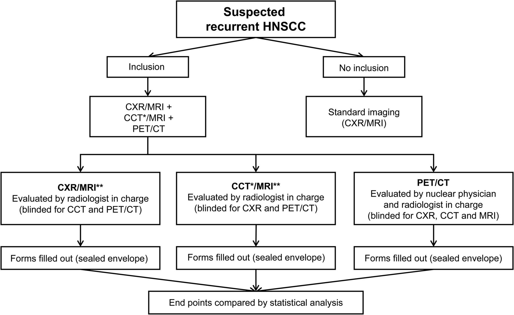
Diagram of patient inclusion and image interpretation. *Extracted from PET/CT. **Same MRI was used for staging with CXR or CCT.
Exclusion criteria included allergy or intolerance toward iodine contrast, use of high-dose systemic corticosteroids (equivalent to >50 mg of prednisone daily), impaired renal function (plasma creatinine > 90 μmol/L for women and > 105 μmol/L for men or previously diagnosed kidney disease), inability to cooperate, or blood glucose above 8 mmol/L (slightly elevated blood glucose levels were accepted).
All patients underwent upfront (before biopsy) CXR/MRI, CCT/MRI, and PET/CT on the same day. Subsequent management decisions were based on all available imaging, to ensure that patients benefited maximally from the examinations performed as part of their workup for recurrent HNSCC and after treatment. Upfront imaging is the standard procedure for all patients in the Danish Head and Neck Cancer Fast-Track Program (25). The CCT was derived from the PET/CT examinations to minimize radiation exposure. PET/CT was performed with full diagnostic-quality CT scans. The evaluation results used for both the CXR/MRI and the CCT/MRI were based on a single MRI examination. Patients with histologically verified recurrent HNSCC constituted the final study population, and their data were used for further analysis of disease extension and the discriminative ability of the 3 imaging strategies (Fig. 2).

Flowchart of patient selection. *Extracted from PET/CT. **Same MRI was used for staging with CXR or CCT.
Imaging Techniques
PET/CT data were acquired on a hybrid PET/CT scanner (Discovery 690, 710, VCT, or RX; GE Healthcare). A 4 MBq/kg dose of 18F-FDG was injected intravenously after a fasting period of at least 4 h. The PET scan was acquired using a standard whole-body protocol extending from the vertex to the thigh, and a time of 2.5 min per bed position. PET data were reconstructed into transaxial slices with a matrix size of 128 × 128 (pixel size, 5.47 mm) or 256 × 256 (pixel size, 2.73 mm) and a slice thickness of 3.27 mm using iterative 3-dimensional ordered-subset expectation maximization. A multislice, diagnostic-quality CT scan with intravenous contrast medium (Ultravist, 370 mg/mL; Bayer) was acquired after the PET scan. The CT scan was obtained with continuous shallow breathing. Data were reconstructed with a standard filter into transaxial slices with a field of view of 50 cm, a matrix size of 512 × 512 (pixel size, 0.98 mm), and a slice thickness of 3.75 mm. The scan field of view was 70 cm for both PET and CT scans. PET/CT was performed approximately 1 h after 18F-FDG administration, and imaging analysis was done on an Advantage workstation (version 4.4 or 4.3; GE Healthcare) or an AW server (version 3.1 or 3.2; GE Healthcare).
MRI was performed on Achieva, Achieva dStream, or Ingenia 1.5-T hardware (Philips) using a 20-channel (dStream; Philips) head–neck coil. The examination protocol was kept unchanged for the duration of the study and consisted of short-tau inversion recovery, turbo spin-echo T2, and turbo spin-echo T1 sequences with and without contrast enhancement, in the axial or coronal planes, with coverage from the skull base to the aortic arch using 5-mm slices. Diffusion-weighted imaging with spectral fat saturation, and apparent diffusion coefficient maps derived from b-values of 0 and 1,000 mm2/s, were done in axial 6-mm slices. Images were read on a Centricity RA1000 PACS workstation (GE Healthcare). The acquisition parameters of the MRI sequences are displayed in Supplemental Table 1 (supplemental materials are available at http://jnmt.snmjournals.org).
CXR was performed to departmental standards in full inspiration anteroposterior and lateral projections with 130–145 kV and automatic exposure control. FD-X hardware systems (Siemens Healthineers) were used, and studies were read using a Centricity RA1000 PACS workstation with dual 3-megapixel medical-grade monitors (22).
Image Interpretation
The separate imaging modalities were evaluated separately. CXR/MRI was interpreted by 2 experienced head and neck radiologists. CCT/MRI was evaluated by 1 radiologist. PET/CT was read by a team of 2 experienced radiologists and 2 nuclear physicians. Standard forms, including variables corresponding to the Union for International Cancer Control (UICC) classification of primary malignant head and neck cancer (26), were completed by each diagnostic team during imaging interpretation to define the extension of recurrent disease. The same referral text was used for each of the evaluation sessions, and the teams were masked to one another.
Discrimination of recurrent disease from benign posttreatment sequelae was evaluated at the primary site, regional lymph nodes, and distant metastases. In general, morphologic changes, altered signal intensity, contrast enhancement, changes in diffusion, and metabolic information on 18F-FDG avidity by PET were evaluated. The presence of postsurgical or radiation-induced edema and inflammation was thoroughly assessed for all modalities. In particular, increased metabolism of the oral cavity after resection or irradiation was thoughtfully recognized using the intensity of 18F-FDG uptake compared with that of the primary tumor in previous scans, as well as using other information from prior scans to help discriminate recurrence from benign posttreatment effects. As such, low-metabolism diffuse lesions would be ascribed to inflammation or treatment sequelae (or even infection) if the primary tumor was highly 18F-FDG–avid. Of course, differences in metabolic activity caused by different sizes and partial-volume effects were taken into consideration.
The 18F-FDG uptake was assessed visually and compared with the surrounding tissue and the contralateral side, when reasonable and achievable. SUVmax (g/mL) was used only as a supportive tool, and only occasionally. No cutoffs or increase in percentage was used. Non–attenuation-corrected images were reviewed in cases of motion or metal artifacts.
Characteristics that were considered for lymph nodes were enlargement, shape (round or not), consistency of hilum (fatty or nonfatty), necrosis (present or not), consistency of center (dense or not), topography of node distribution, and 18F-FDG avidity by PET.
For CT, lung lesions were labeled as distant metastases if one or more nodules were present. Small subpleural nodules on CT, particularly when calcified, were not considered metastases, unless multiple nodules were present. 18F-FDG uptake in lung nodules was considered suggestive of malignancy when the uptake level was above that of the surrounding tissue and when the pattern of the metabolically active nodules did not suggest another obvious origin, such as infection. Lung lesions in the field of view of the MR image were considered suggestive if they had a nodular or specular configuration, inconsistent with an infectious pattern. Likewise, on CXR, distant metastases were suspected when opacification was not consistent with an infectious pattern.
Bone lesions on PET/CT were considered metastatic when 18F-FDG–avid osteolytic (or osteosclerotic) lesions were present. Focally increased 18F-FDG uptake in the bone marrow, regardless of the presence of lytic or sclerotic changes, was also considered to be metastasis. Lesions close to the joints were rarely considered metastasis.
With respect to MRI, focal signal changes on T2 or short-tau inversion recovery sequences and the presence of enhancement were examined, but because of their variation, only lesions with a low T1 signal were considered suggestive of malignancy. Osteolytic or osteosclerotic changes on CXR were considered suggestive of bone metastasis.
With PET/CT, liver metastases and malignant pleural effusion were suspected when the 18F-FDG activity was above that of the normal tissue. Muscle metastases were suspected in patients with randomly distributed focal areas of increased 18F-FDG uptake in the muscles if there were corresponding morphologic changes on CXR or CCT. Finally, longitudinal muscular 18F-FDG uptake was considered to be physiologic (22).
Outcomes
The main outcome measure was the discriminative ability of cancer stages (disease extension) for survival. UICC-equivalent relapse stages were categorized as localized (stage I–II), locally advanced (stage III, IVA, or IVB), or metastatic (stage IVC) disease. Cancer-specific mortality was used as the clinical outcome to assess the discriminative ability of the 3 imaging strategies. Follow-up data on mortality were obtained from the patients’ medical records at least 6 mo (August 31, 2016) after termination of inclusion.
Statistical Methods
Continuous variables are presented as medians and ranges (minimum and maximum value), and categoric variables as counts and corresponding percentages. Overall comparisons of CXR/MRI or CCT/MRI versus PET/CT for staging of recurrent HNSCC were conducted using the McNemar test with patients stratified according to localized, locally advanced, or metastatic disease for each imaging modality. Kaplan–Meier analysis (27) including the log-rank test, and Cox proportional-hazards regression with the Harrell concordance index (C-index) (28,29), were used to compare the discriminative abilities of PET/CT versus CXR/MRI or CCT/MRI for stage-based survival. The C-index informs one of the information provided by one or several medical tests (30). Basically, C-statistics quantify the global capacity of an estimated risk score, using a fitted survival model, to discriminate among subjects, including those with different event times. The number rendered (0–1) describes the ability of a model to distinguish subjects who eventually develop an event (cases) from those who do not (controls). A high C-index means that for any case–control pair, the predicted event risk using the specified model will more likely be higher for the case.
Finally, the ability of PET/CT to enhance accurate determination of disease mortality was tested with the net reclassification improvement (NRI) (31). The NRI is a relatively novel method that allows one to quantify the predictive capabilities of a model with the introduction of a new test or marker (32,33). The values range from −2 to 2. In its classic form for dichotomous outcomes, the NRI is estimated by examining events and nonevents separately. The new test may appropriately upclassify the risk (increase the predicted probability of events) of patients who actually develop an event. Conversely, it may inappropriately downgrade the risk of patients with an event. The opposite would be true for patients who do not experience an event. The NRI is then calculated as the sum of the two; that is, it is a summary measure of the net proportion of events with increased model-based probability plus the net proportion of nonevents with decreased model-based probability.
The significance level was 5%. All analyses were performed with Stata/IC 15 (StataCorp LP).
Ethics and Disclosures
The study was conducted in accordance with the Declaration of Helsinki and contained essential elements from good clinical practice. The Regional Ethics Committee approved this study (project S_20120217), and all subjects gave written informed consent. Permission was also granted by the Danish Data Protection Agency (journal no. 12/26356). The project was implemented without the involvement of private organizations or companies.
RESULTS
In total, 110 patients were included in the study: 90 (82%) men and 20 (18%) women with a median age of 66 y (range, 40–87 y) at study entry. The most frequent initial primary tumor site was the pharynx (51%), followed by the larynx (34%) and oral cavity (15%).
Follow-up varied among patients because of the temporally spaced recruitment and because the survival data were accrued at the end of the last included patient’s follow-up period of at least 6 mo. Median follow-up from time of recurrence was 491 d, ranging from 13 to 1,505 d. The very short minimum follow-up duration recorded was due to rapid death after inclusion. The overall cancer-specific mortality rate at study termination was 57% (63/110) (Table 1).
Characteristics and Outcome of 110 Patients with Recurrent HNSCC
Table 2 presents the distribution of localized, locally advanced, and metastatic recurrent disease for each imaging strategy: 40%, 54%, and 6%, respectively, for CXR/MRI; 25%, 44%, and 31%, respectively, for CCT/MRI; and 21%, 45%, and 34%, respectively, for PET/CT. The difference in assigned tumor stage by PET/CT compared with CXR/MRI and CCT/MRI was statistically significant (P < 0.001 for both).
Distribution of UICC-Equivalent Relapse Stages According to Upfront Imaging Strategy
Hazard Risk
Figure 3 shows Kaplan–Meier plots with unadjusted survival rates for localized, locally advanced, and metastatic disease as assessed by CXR/MRI, CCT/MRI, and PET/CT.

Unadjusted Kaplan–Meier estimates of survival in 110 patients with recurrent HNSCC.
Neither CXR/MRI nor CCT/MRI was able to separate localized, locally advanced, and metastatic disease from one another, when considering stage-based differentiation of survival (log-rank test, P = 0.18 and P = 0.58, respectively). Conversely, disease extension defined by PET/CT showed a progressive worsening of the prognosis in relation to localized, locally advanced, and metastatic disease (log-rank test, P < 0.001).
The adjusted hazard ratios from the Cox regression analysis confirmed these findings. PET/CT-based staging showed a significantly different cancer-specific survival in the locally advanced and metastatic groups, compared with that in the localized disease group (Table 3).
Hazard Ratios from Age- and Sex-Adjusted Cox Regression Models for Cancer Staging According to Imaging Modality
The overall discriminative ability in predicting cancer-specific mortality was significantly greater for PET/CT (C-index, 0.72; 95% confidence interval [CI], 0.75–0.90) than for CXR/MRI (C-index, 0.55; 95% CI, 0.53–0.74) (P = 0.001) and CCT/MRI (C-index, 0.55; 95% CI, 0.48–0.69) (P < 0.001).
Reclassification
The NRI for PET/CT compared with CXR/MRI and CCT/MRI was 51% (95% CI, 30%–73%) and 73% (95% CI, 54%–92%), respectively. In other words, the addition of PET/CT to either CXR/MRI or CCT/MRI was associated with a significantly positive NRI (P < 0.001 for both).
PET/CT correctly upstaged 64% and 43% of patients with events already staged by CXR/MRI and CCT/MRI, respectively (Supplemental Tables 2 and 3). Furthermore, PET/CT correctly downstaged 43% of patients without events already staged by CCT/MRI (Supplemental Table 3).
NRI was calculated on the basis of difference in assigned tumor stage and pertained only to survival rates. Thus, “correctness” was unrelated to whether the factual cancer stage was correctly determined.
DISCUSSION
In patients with recurrent HNSCC, PET/CT provided significant changes in assigned tumor recurrence stage and was a significantly stronger predictor of cancer-specific and stage-based survival than standard imaging by CXR/MRI or CCT/MRI. PET/CT also correctly reclassified a significant proportion of patients stratified by either of these standard imaging strategies.
PET/CT is generally considered the standard evaluation technique in determining response after radiotherapy of HNSCC (with or without chemotherapy) (15–17). Indeed, PET/CT is superior to standard imaging strategies for identification of local and nodal recurrence and, importantly, distant metastasis (13,34,35). The negative predictive values of PET/CT at the primary site and at the neck have been reported to be 95% and 100%, respectively (36), whereas the sensitivity and specificity for detection of distant metastasis in patients with recurrent HNSCC were 92% and 95% (19). A recent systematic review by Jadvar et al. (14) found the current evidence for use of PET/CT for restaging and treatment response assessment of recurrent HNSCC to be appropriate.
PET/CT may also have important clinical implications for surveillance of these patients. For instance, Kim et al. (20) found a 100% 3-y survival rate in patients with a negative PET/CT result at 12 mo of follow-up. Moreover, a retrospective study by Paidpally et al. (37) demonstrated that the addition of PET/CT to routine clinical follow-up of HNSCC enhanced prediction of survival. The investigators considered 134 HNSCC patients with 227 follow-up PET/CT examinations performed at 4 and 24 mo and found a significant difference in survival for patients with a tumor-positive PET/CT scan compared with those who had a tumor-negative scan.
To our knowledge, no previous study has prospectively compared PET/CT with current guideline-recommended imaging strategies using hard clinical endpoints in patients with recurrent HNSCC. Our study was a direct head-to-head comparison of the prognostic value of PET/CT with that of CXR/MRI- and CCT/MRI-based strategies. The results suggest that a sequential imaging strategy should be avoided and that follow-up should rely solely on PET/CT. Theoretically, the more precise prognostication obtained by PET/CT may also improve patient management.
The National Comprehensive Cancer Network guidelines appear to be vague concerning recommended imaging in recurrent HNSCC (i.e., a nonspecific imaging strategy and symptom-based indications), whereas the European guidelines suggest the use of PET/CT only when the diagnosis is uncertain with standard imaging strategies (17). The British Association of Head and Neck Oncologists recommends PET/CT for patients with HNSCC recurrence who are considered candidates for active curative treatment (38). Our own Danish national guidelines, as of 2015, have no specific recommendations on imaging but do include an option for site- and tumor-type–guided imaging modalities.
Whether patients benefit from improved follow-up imaging, including PET/CT, has yet to be demonstrated, and the results of the HETeCo Trial are awaited (39). However, the present study showed that the significant effect of PET/CT, compared with CXR/MRI or CCT/MRI, in assessment of disease extension in recurrent HNSCC was not confined to identification of metastatic disease; importantly, PET/CT also correctly downstaged patients (in terms of survival), as is crucial for the selection of appropriate relapse treatment. Thus, our results show potential benefit from using PET/CT in the diagnostic work-up of all patients with recurrent HNSCC and indicate that PET/CT may stand alone for this group of patients.
This study was specifically designed to compare PET/CT with standard imaging strategies through a prospective design, each patient acting as his or her own control, and all imaging modalities being performed before biopsy and histologic evaluation on the same day and using state-of-the-art technology. Furthermore, all imaging valuations were performed by experts who were masked to the other imaging modalities. We deliberately chose a paired data design rather than a randomized one for various reasons. First, a paired data design eliminates the risk of confounding, and second, paired designs possess the possibility of early unmasking of results at the individual level (40,41).
Some limitations deserve mention. The study was performed at a single institution with that inherent limitation. The CT scan was obtained during continuous shallow breathing, whereas standard CCT is performed at breath-hold, which is more sensitive, particularly for basal lung nodules. Comparing with a whole-body modality such as PET/CT may give an unfair imbalance to the study. However, we wanted the compare contemporary imaging strategies used in most head and neck cancer centers to mimic daily routine procedures. Furthermore, the study investigated the ability of each individual imaging strategy to provide clinically meaningful separation of recurrent cancer stages, but since no comparison with a histopathologic reference standard (the gold standard) was made, the accuracy of the individual imaging modalities for correct staging per se could not be assessed. No details regarding treatment strategy were available. However, actual treatment strategies for the patients included in this study were based on a comprehensive assessment of all available imaging, including PET/CT. Therefore, the resulting predictive value for mortality may have been overestimated. No economic examination was provided in this study. Financial analysis is warranted, to decide whether PET/CT should replace the standard imaging strategies for patients with recurrent HNSCC.
A generally accepted definition of an HNSCC recurrence does not exist, other than reemergence of cancer disease after definitive treatment (42). Differentiation of a new from a previous primary carcinoma is therefore unclear. Moreover, clear distinction of residual disease from recurrence is not defined either. In this study, we included patients with a minimum 4-mo disease-free interval from primary treatment, and we defined primary site recurrences as squamous cell carcinoma at the primary site within 5 y at the same anatomic site (i.e., oral cavity, pharynx, or larynx).
CONCLUSION
In contrast to standard CXR/MRI or CCT/MRI, PET/CT-based staging in recurrent HNSCC was able to significantly discriminate among the survival courses of patients with local, locally advanced, or metastatic disease and predict their respective survival probability.
DISCLOSURE
This study was financially supported by the University of Southern Denmark, the Danish Cancer Society, and the Region of Southern Denmark and by doctoral research grants given by the University of Southern Denmark, the Danish Cancer Society, and the Region of Southern Denmark. No other potential conflict of interest relevant to this article was reported.
Footnotes
Published online Oct. 12, 2018.
- © 2019 by the Society of Nuclear Medicine and Molecular Imaging.
REFERENCES
- Received for publication July 18, 2018.
- Accepted for publication September 24, 2018.


