


Abstract
The gastrin-releasing peptide receptor (GRPR) is overexpressed in human prostate cancer. Bombesin (BBN) is a neurotransmitter of 14 amino acids and binds with selectivity and with high affinity to GRPRs. We have synthesized a NOTA-conjugated bombesin derivative, NOTA-8-Aoc-BBN(7-14)NH2, to label this analog with 18F using the new Al18F method. In this study, the GRPR-targeting potential of 18F-labeled NOTA-8-Aoc-BBN(7-14)NH2 was studied using 68Ga-NOTA-8-Aoc-BBN(7-14)NH2 as a reference. Methods: The NOTA-conjugated bombesin analog was synthesized and radiolabeled with 68Ga or 18F. For 18F labeling, we used our new 1-pot, 1-step method. The labeled product was purified by reversed-phase high-performance liquid chromatography. The log P values of the radiotracers were determined. The tumor-targeting characteristics of the compounds were assessed in mice with subcutaneously growing PC-3 xenografts. GRPR-binding specificity was studied by coinjection of an excess of unlabeled NOTA-8-Aoc-BBN(7-14)NH2. Small-animal PET/CT images were acquired. Results: NOTA-8-Aoc-BBN(7-14)NH2 could be efficiently labeled with 18F or with 68Ga. NOTA-8-Aoc-BBN(7-14)NH2 was labeled with 18F in a single step, with 50%–90% yield. Radiolabeling, including purification, was performed in 45 min and resulted in a specific activity of greater than 10 GBq/μmol. The log P values of 18F- and 68Ga-labeled NOTA-8-Aoc-BBN(7-14)NH2 were −1.47 ± 0.05 and −1.98 ± 0.03, respectively. In mice, both radiolabeled compounds cleared rapidly from the blood (<0.07 percentage injected dose per gram at 1 h after injection), mainly via the kidneys. At 1 h after injection, the uptake of 18F- and 68Ga-labeled NOTA-8-Aoc-BBN(7-14)NH2 in the PC-3 tumors was 2.15 ± 0.55 and 1.24 ± 0.26 percentage injected dose per gram, respectively. GRPR-binding specificity was demonstrated by reduced tumor uptake of radiolabeled NOTA-8-Aoc-BBN(7-14)NH2 after coinjection of a 100-fold excess of unlabeled NOTA-8-Aoc-BBN(7-14)NH2 peptide. The accumulation of 18F-NOTA-8-Aoc-BBN(7-14)NH2 in the subcutaneous PC-3 tumors could be visualized via small-animal PET. Conclusion: NOTA-8-Aoc-BBN(7-14)NH2 could be labeled rapidly and efficiently with 18F using a 1-pot, 1-step method. Radiolabeled NOTA-8-Aoc-BBN(7-14)NH2 specifically accumulated in the GRPR-expressing PC-3 tumors and should be evaluated clinically.
Prostate cancer is the third-leading cause of cancer-related deaths and the most frequently diagnosed cancer among men in the Western world (1). There is a great need for an imaging technique that provides adequate staging of patients with this disease. A promising approach to cancer diagnosis is the use of radiolabeled receptor-binding peptides. It has been shown that prostate tumors overexpress the gastrin-releasing peptide receptor (GRPR) (2–6). GRPRs are expressed in primary prostate cancer, invasive prostate carcinoma, and prostatic intraepithelial neoplasms at high density, whereas normal prostate tissue and benign prostate hyperplasia (BPH) are predominantly GRPR-negative (2). These findings suggest that the GRPR represents an ideal molecular target for radiolabeled GRPR ligands for diagnosis and staging of prostate cancer.
Bombesin (BBN) is the 14-amino acid amphibian peptide analog of the 27-amino acid mammalian gastrin-releasing peptide (GRP). BBN and GRP share a homologous 7–amino acid amidated C-terminus, Trp-Ala-Val-Gly-His-Leu-Met-NH2, which is necessary for binding to the GRPR. Because GRP suffers from poor in vivo stability, developments have focused on BBN analogs. Synthetic BBNs are truncated versions of BBNs containing the sequence or a full-length 14–amino acid sequence in which one or more amino acids have been replaced. The C-terminus of BBN is required for high-affinity binding and biologic potency (7). Therefore, the N-terminus of BBN is usually modified for labeling the analogs with radioisotopes. A variety of BBN analogs has been developed and labeled with various radionuclides, such as 99mTc and 111In, for SPECT (8) and 64Cu, 68Ga, or 86Y for PET (9,10). 18F is the most widely used radionuclide in PET. 18F has excellent characteristics for peptide-based receptor imaging studies: the half-life (109.7 min) matches the pharmacokinetics of most peptides, and the low positron energy of 635 keV results in a short penetration range in tissue and excellent imaging resolution (11). Several methods have been developed to label peptides with 18F. However, in general these methods are laborious and require a multistep synthesis. Recently, McBride et al. reported a facile method wherein 18F is first attached to aluminum as Al18F, which is then complexed in a chelating agent attached to the peptide, forming a stable Al18F–chelate peptide complex in an efficient 1-pot process (12).
Several studies have indicated that the DOTA-conjugated BBN analog, DOTA-8-Aoc-BBN(7-14)NH2, satisfies inherent in vitro and in vivo requirements for radiopharmaceutical development (13–15). However, the DOTA chelator in the studies of Rogers et al. (13) showed demetallation of 64Cu. Therefore, Prasanphanich et al. conjugated the NOTA chelator to 8-Aoc-BBN(7-14)NH2 to produce a kinetically inert BBN conjugate (16).
As shown previously, NOTA-conjugated peptides can be labeled with 18F using the Al18F method (17–19). Here, we describe the 18F labeling of NOTA-8-Aoc-BBN(7-14)NH2, using the new 1-pot, 1-step, labeling method. The in vitro affinity and the in vivo tumor targeting characteristics of 18F-labeled NOTA-8-Aoc-BBN(7-14)NH2 were determined and compared with those of the 68Ga-labeled counterpart.
MATERIALS AND METHODS
Synthesis of NOTA-Conjugated BBN Derivative 8-Aoc-BBN(7-14)NH2
The BBN derivative was synthesized using Fmoc-based solid-phase peptide synthesis as described previously (16). The structural formula of NOTA-8-Aoc-BBN(7-14)NH2 is shown in Figure 1.
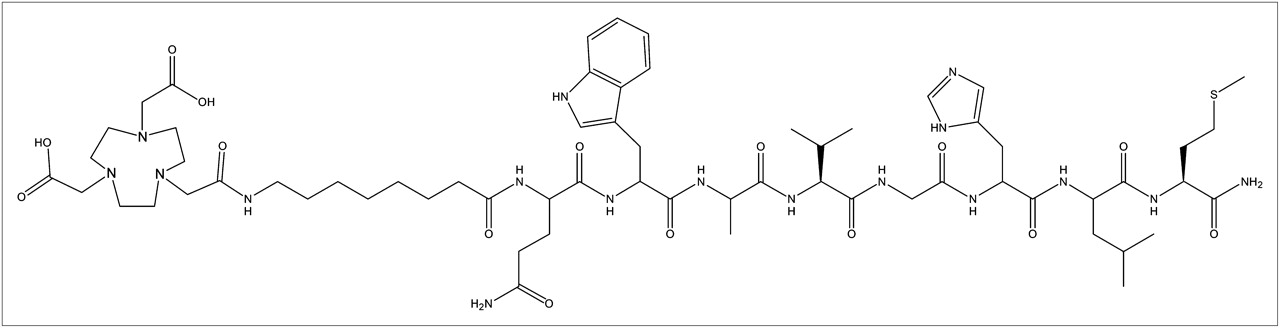
Structural formula of NOTA-conjugated BBN analog NOTA-8-Aoc-BBN(7-14)NH2.
Radiolabeling
Labeling with 18F
NOTA-8-Aoc-BBN(7-14)NH2 was radiolabeled with 18F as described by Laverman et al. (17). Briefly, a Chromafix cartridge with 2–6 GBq of 18F (BV Cyclotron VU) was washed with 3 mL of metal-free water. 18F was eluted from the cartridge with 100 μL of 0.4 M KHCO3. The pH of the eluate was adjusted to 4.1 with 5 μL of metal-free glacial acetic acid, and 2–40 μL of 2 mM AlCl3 in 0.1 M sodium acetate buffer (pH 4.1) were added. Finally, 80 μL of this Al18F solution was added to 400 μL of MeCN and 20 μL of NOTA-8-Aoc-BBN(7-14)NH2 (5 μg/μL) in 0.5 M sodium acetate, pH 4.1. The reaction mixture was heated at 100°C for 15 min. The radiolabeled peptide was purified by reversed-phase high-performance liquid chromatography (RP-HPLC) using the gradient as in the “Quality Control” section. Fractions containing 18F-NOTA-8-Aoc-BBN(7-14)NH2 were collected and diluted 10-fold with H2O and purified on an Oasis HLB 1-cm3 (10-mg) cartridge (Waters) to remove acetonitrile and trifluoroacetic acid. In brief, the fraction was applied on the cartridge, and the cartridge was washed with 3 × 1 mL of H2O. The radiolabeled peptide was eluted with 200 μL of EtOH/H2O (1:1, v/v). For injection into mice, the peptide was diluted with 0.5% bovine serum albumin in 0.9% NaCl.
Labeling with 68Ga
68Ga was obtained from a 1,850-MBq 68Ge/68Ga generator (IGG-100, Eckert & Ziegler), where 68Ge was attached to a TiO2-based column. The 68Ga was eluted with 0.1 M HCl (Ultrapure; J.T. Baker) using an Econo Pump (Bio-Rad) at 1 mL/min. Five 1-mL fractions were collected, and an aliquot of the second fraction was used for labeling the peptide.
68Ga-labeled NOTA-8-Aoc-BBN(7-14)NH2 was prepared by adding 20 μL of 2.5 M HEPES (4-(2-hydroxyethyl)piperazine-1-ethanesulfonic acid) solution to 5 μL of the peptide dissolved in 2.5 M HEPES (2 μg/μL). Then, the second fraction from the 68Ge/68Ga generator (315–365 MBq) was added. After 10 min at 95°C, the 68Ga-labeled peptide was purified on an Oasis HLB (30-mg) cartridge (Waters). After the sample was applied to the cartridge, the cartridge was washed with 3 × 1 mL of H2O and eluted with 200 μL of EtOH/H2O (1:1, v/v).
Quality Control
The radiolabeled preparations were analyzed by RP-HPLC on an Agilent 1200 system (Agilent Technologies). A C18 column (Onyx monolithic, 4.6 mm × 100 mm; Phenomenex) was used at a flow rate of 3 mL/min with the following buffer system: buffer A, 0.1% v/v trifluoroacetic acid in H2O; buffer B, 0.1% v/v trifluoroacetic acid in acetonitrile; and a gradient of 80% buffer A at 0–5 min and 80% buffer A to 76% buffer A at 5–20 min. The radioactivity of the eluate was monitored using an in-line NaI radiodetector (Raytest GmbH). Elution profiles were analyzed using Gina-star software (version 2.18; Raytest GmbH). For injection into mice, the peptide was diluted to less than 10% (v/v) ethanol with 0.5% (w/v) bovine serum albumin in 0.9% NaCl.
Octanol/Water Partition Coefficient
To an Eppendorf tube filled with 0.5 mL of the radiolabeled peptide in phosphate-buffered saline (pH 7.4), 0.5 mL of octanol was added. After the tube was vigorously stirred by a vortex mixer for 2 min at room temperature, the 2 layers were separated by centrifugation (100g, 5 min). Samples of 100 μL were taken from each layer, radioactivity was measured in a well-type γ-counter (Wallac Wizard 3″; Perkin-Elmer), and log P values were calculated (n = 3).
Competitive Cell-Binding Assay
The PC-3 human prostate cancer cell line was cultured in RPMI 1640 medium supplemented with 10% (v/v) fetal calf serum. Cells were grown in tissue culture flasks at 37°C in a humidified atmosphere containing 5% CO2 and routinely passed using 0.25% trypsin/ethylenediaminetetraacetic acid.
Binding affinities toward the GRPR for natGa-, AlnatF-, and nonlabeled NOTA-8-Aoc-BBN(7-14)NH2 were determined in a competitive binding assay on PC-3 tumor cells, using 111In-NOTA-8-Aoc-BBN(7-14)NH2 as a tracer. PC-3 cells were seeded into 6-well plates at 3 × 10⁵ cells per well and cultured until confluency. On the day of the assay, cells were washed with binding buffer (RPMI, 0.5% bovine serum albumin). Subsequently, binding buffer (1.5 mL), natGa-, AlnatF-, or nonlabeled NOTA-8-Aoc-BBN(7-14)NH2 in a range of 0.1 to 300 nM and 200 Bq of 111In-NOTA-8-Aoc-BBN(7-14)NH2 were added to each well. After incubation for 3 h, medium was removed. Then, cells were washed with binding buffer and extracted from the wells, and cell-associated radioactivity was determined in a γ-counter. For the complexation of NOTA-8-Aoc-BBN(7-14)NH2 with natGa3+, the peptide was dissolved in an aqueous solution (2.5 M HEPES; 1 μg/μL) and a 3-fold molar excess of natGa(NO3)3 (Sigma-Aldrich Chemie) in 0.1N HCl solution was added. Metal complexation was performed overnight at room temperature. AlnatF complexation of NOTA-8-Aoc-BBN(7-14)NH2 was performed by mixing an AlF solution (0.02 M AlCl3 in 0.5 M NaAc, pH 4, with 0.1 M NaF in 0.5 M NaAc, pH 4.1) with the NOTA-conjugated peptide. This reaction mixture was heated for 15 min at 100°C and then purified by RP-HPLC. GraphPad Prism software (version 5.03 for Windows [Microsoft]; GraphPad Software) was used to calculate inhibitory concentration of 50% (IC50) values.
Biodistribution Studies
Male nude BALB/c mice (6–8 wk old) were injected subcutaneously in the right flank with 0.2 mL of a PC-3 cell suspension of 1.5 × 10⁷ cells/mL (66% RPMI, 33% Matrigel [BD Biosciences]). Two weeks after inoculation of the tumor cells, mice were injected intravenously with the 18F- or 68Ga-labeled NOTA-8-Aoc-BBN(7-14)NH2 (<1 μg). Mice were killed by CO2 asphyxiation 1 h after injection (5–6 mice per group). Blood, tumor, and the major organs and tissues were collected, weighed, and counted in a γ-counter. The percentage injected dose per gram (%ID/g) was determined for each sample.
The receptor-mediated localization of the radiolabeled peptide was investigated by determining the biodistribution of the 18F- or 68Ga-labeled peptide after coinjection of an excess of unlabeled peptide (n = 3). All animal experiments were approved by the local Animal Welfare Committee in accordance with Dutch legislation and performed in accordance with their guidelines.
Small-Animal PET/CT
Mice with subcutaneous PC-3 tumors were injected intravenously with 2 MBq of 18F-labeled NOTA-8-Aoc-BBN(7-14)NH2. One hour after the injection of the radiolabeled peptide, mice were scanned on an animal PET/CT scanner (Inveon; Siemens Preclinical Solutions) with an intrinsic spatial resolution of 1.5 mm (11). The animals were placed supine in the scanner. PET emission scans were acquired over 20–25 min, followed by a CT scan for anatomic reference (spatial resolution, 113 μm; 80 kV; and 500 μA). Scans were reconstructed using Inveon Acquisition Workplace software (version 1.5; Siemens Preclinical Solutions), using a 3-dimensional ordered-subset expectation maximization/maximum a posteriori algorithm with the following parameters: matrix, 256 × 256 × 159; pixel size, 0.43 × 0.43 × 0.8 mm; and β-value, 1.5, with uniform variance
Statistical Analysis
All mean values are given with SD. Statistical analysis was performed using a Welch corrected unpaired Student t test or 1-way ANOVA using GraphPad InStat software (version 3.06; GraphPad Software). The level of significance was set at P less than 0.05.
RESULTS
Radiolabeling
RP-HPLC analysis indicated that the radiochemical purity of the 18F- or 68Ga-labeled NOTA-8-Aoc-BBN(7-14)NH2 preparations used in these experiments always exceeded 95%. Radio-HPLC chromatograms of 18F- and 68Ga-labeled NOTA-Aoc-BBN(7-14)NH2 are shown in Figure 2. Al18F-NOTA-Aoc-BBN(7-14)NH2 had a retention time of 16.3 min and eluted at 23% of buffer B. 68Ga-NOTA-Aoc-BBN(7-14)NH2 had a 12.7-min retention time and eluted at 22% of buffer B.
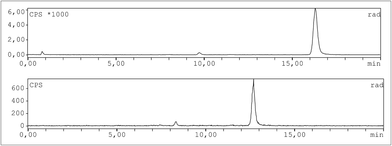
Radio-HPLC chromatograms of 18F- (top) and 68Ga-labeled (bottom) NOTA-8-Aoc-BBN(7-14)NH2.
Octanol/Water Partition Coefficient
To establish lipophilicity of the 18F- and 68Ga-labeled NOTA-8-Aoc-BBN(7-14)NH2, the octanol/water partition coefficients were determined. The log Poctanol/water value for the 18F-labeled BBN analog was −1.47 ± 0.05. The 68Ga-labeled analog was more hydrophilic, with a log Poctanol/water value of −1.98 ± 0.03.
Competitive Binding Assay
The affinity of AlnatF-, natGa-, and nonlabeled NOTA-8-Aoc-BBN(7-14)NH2 for GRPR was determined in a competitive binding assay. The results of these assays are summarized in Figure 3. Binding of 111In-labeled BBN peptide to GRPR was displaced by natGa-, AlnatF-, and nonlabeled NOTA-8-Aoc-BBN(7-14)NH2 in a concentration-dependent manner. The IC50 values of the AlnatF-, natGa-, and nonlabeled compounds were not significantly different and in the subnanomolar range (0.28 ± 0.15, 0.41 ± 0.15, and 0.37 ± 0.15 nM, respectively), indicating high binding affinity for the GRPR.

Competition of specific binding of 111In-NOTA-8-Aoc-BBN(7-14)NH2 with various NOTA-8-Aoc-BBN(7-14)NH2 analogs.
Biodistribution Studies
The results of the biodistribution studies of both 18F- and 68Ga-labeled NOTA-8-Aoc-BBN(7-14)NH2 are summarized in Figure 4. NOTA-8-Aoc-BBN(7-14)NH2 radiolabeled with either 18F or 68Ga cleared rapidly from the blood, in which levels of both tracers were below 0.07 %ID/g at 1 h after injection. Tumor uptake of 18F-labeled NOTA-8-Aoc-BBN(7-14)NH2 was significantly higher than that of 68Ga-labeled NOTA-8-Aoc-BBN(7-14)NH2 (2.15 ± 0.55 vs. 1.24 ± 0.26 %ID/g, respectively; P < 0.05). In addition to tumor uptake, 18F-labeled NOTA-8-Aoc-BBN(7-14)NH2 had significantly higher uptake in the pancreas than did the 68Ga-labeled analog (27.09 ± 12.77 %ID/g vs. 5.93 ± 2.10 %ID/g; P < 0.01).

(A) Biodistribution of 18F-NOTA-8-Aoc-BBN(7-14)NH2 at 1 h after injection in athymic mice with subcutaneous PC-3 tumors in absence (5 mice per group) or presence (2 mice per group) of excess of nonradiolabeled NOTA-8-Aoc-BBN(7-14)NH2. (B) Biodistribution of 68Ga-NOTA-8-Aoc-BBN(7-14)NH2 at 1 h after injection in athymic mice with subcutaneous PC-3 tumors in absence (6 mice per group) or presence (3 mice per group) of excess of nonradiolabeled NOTA-8-Aoc-BBN(7-14)NH2. p.i. = after injection; xs = excess.
The coinjection of an excess of unlabeled NOTA-8-Aoc-BBN(7-14)NH2 (50 μg) along with 18F- or 68Ga-labeled NOTA-8-Aoc-BBN(7-14)NH2 resulted in a significantly reduced radioactivity concentration in the tumor, indicating that the major fraction of the uptake of radiolabeled NOTA-8-Aoc-BBN(7-14)NH2 in the tumor was GRPR-mediated. Also, GRPR-specific uptake in the pancreas for 18F- and 68Ga-labeled NOTA-8-Aoc-BBN(7-14)NH2, in the presence of an excess of nonradiolabeled peptide showed significant reduction in accumulation of the tracer (0.43 ± 0.08 and 0.21 ± 0.09 %ID/g).
Small-Animal PET/CT
Fused PET and CT images are shown in Figure 5. PET scans were in line with the biodistribution data. PC-3 tumors could be clearly visualized. However, there was considerable accumulation and retention in other organs such as the kidneys, liver, and intestines. In addition, uptake in the pancreas was relatively high and appeared to be specific. As a whole, uptake in the abdominal cavity was relatively high and is not entirely unexpected.

3-dimensional volume–rendering projections of fused PET and CT scans of mice with subcutaneously growing PC-3 tumor after intravenous injection of 18F-NOTA-8-Aoc-BBN(7-14)NH2 in absence (A) or presence (B) of excess of nonradiolabeled NOTA-8-Aoc-BBN(7-14)NH2. Scans were recorded at 1 h after injection.
DISCUSSION
The diagnostic accuracy of metabolic PET tracers, such as 18F-FDG, 18F- or 11C-choline, and 11C-acetate, for imaging prostate cancer is limited, because most prostate tumor lesions are characterized by a low metabolic activity (20). The discovery of GRPR overexpression in prostate cancer disease has led to the development and application of radiolabeled BBN derivatives for imaging and staging of prostate cancer using SPECT or PET. Here, the feasibility of using an 18F-labeled BBN analog for radionuclide imaging of GRPR expression with PET was investigated. The NOTA-conjugated BBN analog was radiolabeled with 18F, using the new chelator-based method developed by McBride et al. (12). The in vitro and in vivo characteristics of this 18F-labeled peptide were directly compared with those of the 68Ga-labeled analog.
To radiolabel NOTA-conjugated BBN peptide with 18F, we used our optimized 1-pot method for labeling peptides with 18F as described recently (17). First, 18F− was complexed with Al3+, and subsequently the Al18F complex was chelated by NOTA. It is still unclear which aluminum–fluoride complex is formed, but because the formation of Al18F is performed at pH 4.1, we believe that the form in sodium acetate buffer is probably AlF2+. In addition, the molar ratio of aluminum to 18F is such that it is unlikely that any AlF2+ or AlF3 is formed (21).
The 18F-labeled peptide was slightly more lipophilic than the 68Ga-labeled BBN analog, as indicated by their log P values (−1.98 ± 0.03 and −1.47 ± 0.05, respectively). This difference could be due to the dianionic NOTA chelator, which stabilizes the +2 charge of the Al18F-complex but not the +3 charge of the 68Ga atom, resulting in an overall +1 charge for the 68Ga-NOTA complex. However, the presence of counter ions could play a role as well.
In a competitive binding assay using GRPR-expressing PC-3 cells, the IC50 values of the nonlabeled and cold labeled analogs were not significantly different and were in the subnanomolar range. Prasanphanich et al. also determined the IC50 value of NOTA-8-Aoc-BBN(7-14)NH2, and in this assay the binding affinity of their compound was somewhat lower than the binding affinity as determined in our assay (3.1 ± 0.5 vs. 0.37 ± 0.2 nM) (16). Although they also used PC-3 cells in their assay, their tracer was 125I-Tyr4-BBN, whereas in our assay the 111In-NOTA-8-Aoc-BBN(7-14)NH2 ligand was used as the displacement radioligand.
In the subcutaneous PC-3 xenograft model, the tumor uptake of 18F-labeled NOTA-8-Aoc-BBN(7-14)NH2 was significantly higher than that of 68Ga-labeled NOTA-8-Aoc-BBN(7-14)NH2 at 1 h after injection (2.15 ± 0.55 vs. 1.24 ± 0.26 %ID/g, respectively; P < 0.05). Both tracers showed specific tumor uptake in PC-3 tumors: the coinjection of an excess of unlabeled NOTA-8-Aoc-BBN(7-14)NH2 resulted in a significantly lower tumor uptake of 68Ga-NOTA-8-Aoc-BBN(7-14)NH2 and 18F-NOTA-8-Aoc-BBN(7-14)NH2 (0.26 ± 0.15 and 0.88 ± 0.12 %ID/g, respectively). Several tissues in mice express the GRPR, including the small intestine, large intestine, and pancreas (22). In our study, specific uptake of 68Ga-NOTA-8-Aoc-BBN(7-14)NH2 and 18F-NOTA-8-Aoc-BBN(7-14)NH2 was found in the colon and pancreas, indicating that the accumulation of these tracers in these tissues is at least partly GRPR-mediated. 18F- and 68Ga-labeled NOTA-8-Aoc-BBN(7-14)NH2 cleared rapidly from the blood, resulting in high tumor-to-blood ratios at 1 h after injection (49.51 ± 22.94 and 25.06 ± 10.55, respectively).
The uptake of the 64Cu-labeled NOTA-8-Aoc-BBN(7-14) conjugate in PC-3–bearing mice (3.59 ± 0.70 %ID/g, 1 h after injection) was similar to the tumor uptake for our radiolabeled compounds (2.15–1.24.%ID/g, 1 h after injection) (16). However, Lears et al. recently reported in this tumor model a tumor uptake as high as 13.0 %ID/g at 1 h after injection using 64Cu-labeled SarAr-SA-Aoc-BBN(7-14) (23). This compound has an additional linker and is labeled with another radionuclide via a different chelator, and each of these parameters could have caused the higher uptake of this tracer in the tumor. However, in our experiments we observed that the tumor uptake can vary largely from one experiment to the other. Even within the same experiment, the uptake of the radiolabeled BBN analog in the PC-3 tumor varied considerably.
To select the best ligand for clinical evaluation, Schroeder et al. performed a comparative study using 5 radiolabeled BBN analogs under identical experimental conditions (24). In previous studies in PC-3–bearing mice, 67Ga-PESIN, 177Lu-AMBA, and 99mTc-demobesin-1 showed tumor uptake of 14.8, 6.4, and 16.2 %ID/g, respectively (9,25–28). Variation in amounts of peptides, mouse strain (species, sex, and weight), tumor cells (passage number, culture conditions) used, tumor size, and vascularization of the tumor may all be factors that affect tumor uptake and tumor–to–non-tumor ratios. On the basis of these data, the authors selected 99mTc-demobesin-1 as the bombesin analog with optimal characteristics for prostate cancer imaging. This study of the group in Rotterdam demonstrated that the characteristics of radiolabeled BBN analogs can be appreciated only in a direct comparative study.
Overall, the biodistribution of 18F-NOTA-8-Aoc-BBN(7-14)NH2 in mice with subcutaneous PC-3 xenografts was similar to that of 68Ga-NOTA-8-Aoc-BBN(7-14)NH2, indicating that labeling the analog with Al18F did not affect the in vivo characteristics of the bombesin derivative.
The Al18F method is a fast (45 min) radiofluorination strategy and does not affect the pharmacokinetics of the bombesin peptide. The commonly used methodology for efficient 18F-labeling of peptides consists of 2 steps: first the preparation of an 18F-labeled synthon (prosthetic groups), which is subsequently conjugated to the peptide or the protein. In general, this fluorination is based on a nucleophilic substitution that requires laborious azeotropic drying of the 18F-fluoride–kryptofix complex. The total synthesis and formulation time for these methods ranges between 1 and 3 h, with most of the time dedicated to the HPLC purification of the labeled peptides to obtain the carrier-free tracer required for in vitro and in vivo experiments. We also had to purify our 18F-labeled peptide with HPLC to obtain a high-specific-activity preparation. Recently, McBride et al. have shown that the AlF method can be further optimized using other chelators (19,29). They developed a 1-pot method that provides a high labeling efficiency and specific activity with no more than only solid-phase extraction purification needed before formulation for injection. Because the Al18F-method is based on a chelator-derivatized peptide, this labeling method is easy and versatile.
CONCLUSION
The Al18F method combines the ease of chelator-based radiolabeling methods with the advantages of 18F, such as half-life, availability, and positron energy. The 18F-labeled NOTA-8-Aoc-BBN(7-14)NH2 could be synthesized in less than 45 min without the need to synthesize a synthon. The results of the biodistribution study of the 18F- and 68Ga-labeled BBN peptide show that the AlF method does not affect the in vivo behavior of the analog. The 18F-labeled BBN peptide is a suitable ligand for the noninvasive visualization of GRPR expression in vivo.
DISCLOSURE STATEMENT
The costs of publication of this article were defrayed in part by the payment of page charges. Therefore, and solely to indicate this fact, this article is hereby marked “advertisement” in accordance with 18 USC section 1734.
Acknowledgments
We thank Bianca Lemmers-van de Weem, Kitty Lemmens-Hermans, and Jonathan Disselhorst for their technical assistance. This study was performed within the framework of CTMM, the Center for Translational Molecular Medicine, PCMM project (grant 03O-203). Drs. McBride, D’Souza, and Goldenberg have employment and stock ownership with Immunomedics, Inc. No other potential conflict of interest relevant to this article was reported.
Footnotes
Published online May 8, 2012.
- © 2012 by the Society of Nuclear Medicine, Inc.
REFERENCES
- Received for publication November 16, 2011.
- Accepted for publication February 9, 2012.


Jump to section
Related Articles
Cited By...
- 18F-AlF-Labeled Biomolecule Conjugates as Imaging Pharmaceuticals
- Preclinical Comparison of Al18F- and 68Ga-Labeled Gastrin-Releasing Peptide Receptor Antagonists for PET Imaging of Prostate Cancer
- N-Terminal Modifications Improve the Receptor Affinity and Pharmacokinetics of Radiolabeled Peptidic Gastrin-Releasing Peptide Receptor Antagonists: Examples of 68Ga- and 64Cu-Labeled Peptides for PET Imaging
- A Comparative Study of Radiolabeled Bombesin Analogs for the PET Imaging of Prostate Cancer
- Three Methods for 18F Labeling of the HER2-Binding Affibody Molecule ZHER2:2891 Including Preclinical Assessment