


Article Figures & Data
Figures
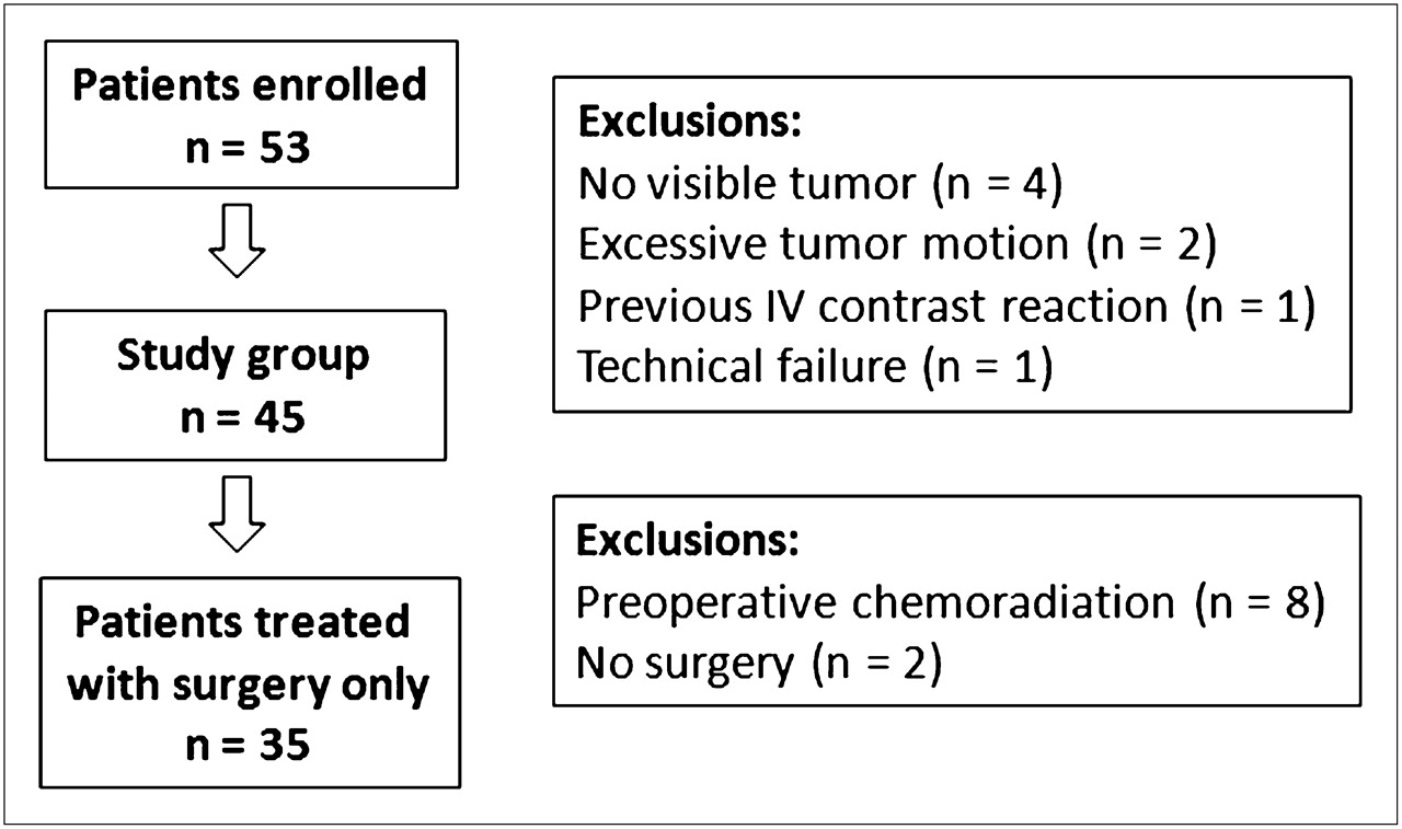
- FIGURE 1.
Flow chart of study population.
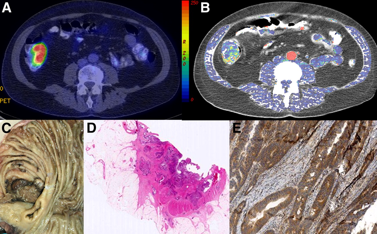
- FIGURE 2.
Representative axial images obtained from integrated 18F-FDG PET/perfusion CT study: fused anatomic CT and SUVmax image (A), fused anatomic CT and regional blood flow parametric map (B), corresponding surgical specimen (C), hematoxylin- and eosin-stained section (D), and VEGF-stained section (E).
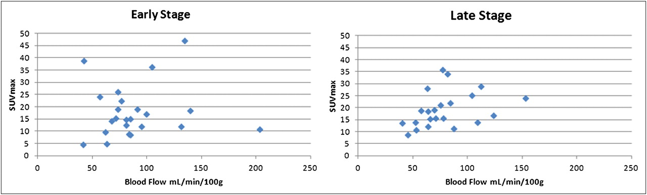
- FIGURE 3.
Scatterplot of SUVmax vs. regional blood flow for early- and late-stage tumors.

Tables
- TABLE 1
Published Phase II Trials of Neoadjuvant Chemotherapy and Chemoradiation in Rectal Cancer
Disease stage Chemotherapy agent Chemoradiation regime Author Year T3 disease and above defined by MRI or CT 5-fluorouracil (300 mg/m2) on day 1 for 12 wk; mitomycin C (7 mg/m2) intravenous bolus every 6 wk Phase I: 45 Gy in 25 fractions; phase II: 5.4- to 9-Gy boost to tumor bed; 5-fluorouracil (200 mg/m2/daily) Chau (6) 2003 T3 disease and above defined by MRI Oxaliplatin (130 mg/m2) on day 1 for 12 wk; capecitabine (1,000 mg/m2) twice daily for 14 d every 3 wk for 12 wk Phase I: 45 Gy in 25 fractions; phase II: 9-Gy boost to tumor bed; capecitabine (825 mg/m2 twice daily) Chau (7) 2006 T3 disease and above defined by MRI Oxaliplatin (130 mg/m2) on day 1 for 12 wk; capecitabine (1,000 mg/m2) twice daily for 14 d every 3 wk for 12 wk Phase I: 45 Gy in 25 fractions; phase II: 9-Gy boost to tumor bed; capecitabine (825 mg/m2 twice daily) Chua (8) 2010 T3 disease and above Oxaliplatin (130 mg/m2) on day 1 for 12 wk; capecitabine (1,000 mg/m2) twice daily for 14 d every 3 wk for 12 wk Phase I: 45 Gy in 25 fractions; phase II: 5.4-Gy boost to tumor bed; capecitabine (825 mg/m2 twice daily) Fernández-Martos (9) 2010 Characteristic TNM stage Number of patients Stage I (n = 9) T1N0M0 5 T2N0M0 4 Stage II (n = 14) T3N0M0 14 T4aN0M0/T4bN0M0 0/0 Stage III (n = 13) T2N1M0/T2N2M0 1/0 T3N1M0/T3N2M0 4/2 T4abN1M0/T4abN2M0 3/3 Stage IV (n = 9) T2N1M1 1 T3N0M1/T3N1M1/T3N2M1 0/1/4 T4N1M1 3 Moderately differentiated 36 Poorly differentiated 9 Parameter Comparison Median blood flow–to–SUVmax ratio P Tumor stage Early vs. late 5.55 vs. 3.89 0.19 Tumor size <4 cm vs. ≥4 cm 4.70 vs. 4.31 0.48 CD105 expression Low vs. high 4.94 vs. 4.00 0.63 VEGF expression Negative vs. positive 5.98 vs. 3.65 0.01* Glut-1 expression Negative vs. positive 3.90 vs. 4.59 0.56 HIF-1α expression Negative vs. positive 5.48 vs. 3.63 0.04* ↵* Significant at 5% level.
- TABLE 5
Studies That Have Assessed the Relationship Between 18F-FDG PET and Perfusion CT Parameters
Tumor type Measure Correlation Study Non–small cell lung cancer SUVmax perfusion Positive Tateshi, 2002 (22) Non–small cell lung cancer SUVmax standardized perfusion value Positive Miles, 2006 (23) Head and neck SUVmax perfusion Negative Hirasawa, 2007 (24) Head and neck SUVmax blood flow or permeability surface area product Positive Bisdas, 2008 (25) Breast SUVmax normalized perfusion Positive Groves, 2009 (26) Colorectal liver metastases SUVmax blood flow, blood volume, or mean transit time Positive Veit-Heibach, 2010 (27)


