


Article Figures & Data
Figures
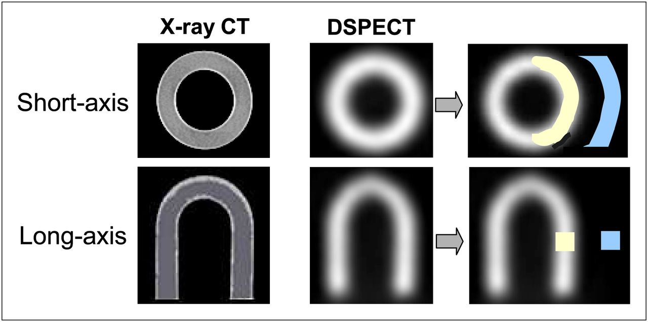
- FIGURE 1.
Images of cardiac phantom provided by CT and by DSPECT camera, as well as representation of background (blue) and myocardial (yellow) regions of interest, which were used for determining contrast-to-noise ratio.
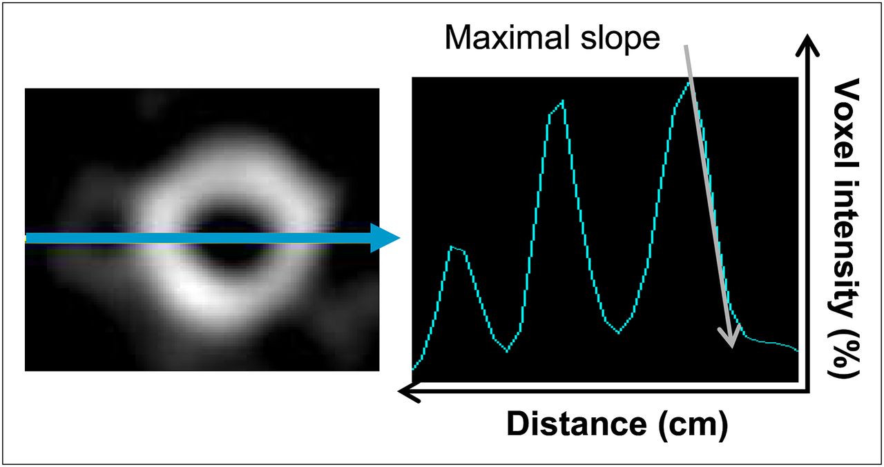
- FIGURE 2.
Example, on median short-axis slice, of determination of maximal slope of epicardial border of left lateral wall, representing sharpness index. Distance is expressed in centimeters, and voxel intensity is expressed in percentage of maximal myocardial voxel value.
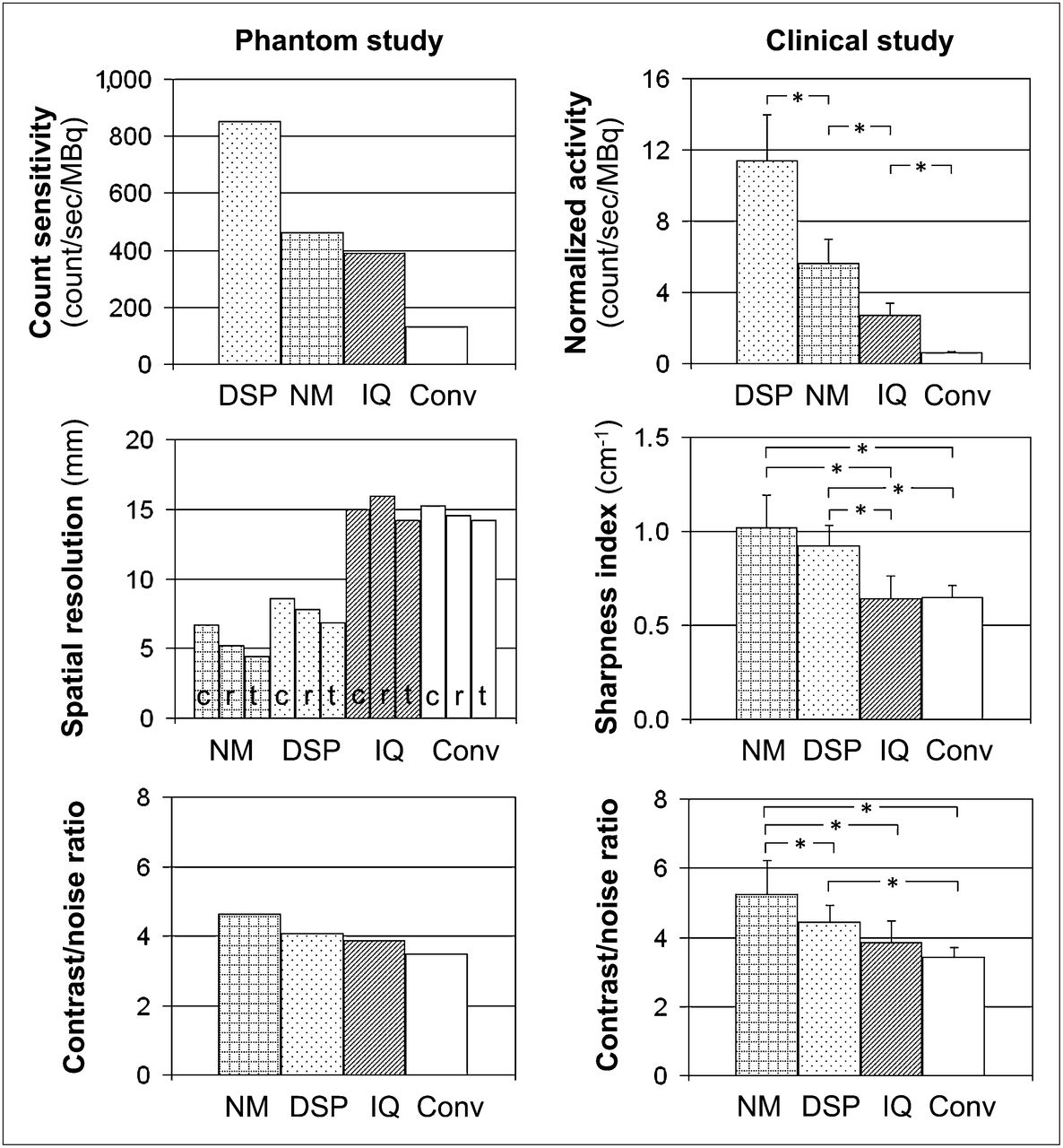
- FIGURE 3.
Comparison of the 4 cameras. Count sensitivity was determined for phantom (top left) and indirectly assessed for human (top right) SPECT images by normalizing counts to recording time and injected activity. Spatial resolution was determined for phantom insert (middle left) and indirectly assessed for human (middle right) SPECT images using sharpness index for myocardial contours. Contrast-to-noise ratio was determined for phantom (bottom left) and human (bottom right) SPECT images. Conv = conventional SPECT; DSP = DSPECT; IQ = IQ⋅SPECT; NM = Discovery NM 530c; c = central resolution; r = radial resolution; t = tangential resolution.
- FIGURE 4.
Representative SPECT images obtained with 4 different cameras: with phantom after simulation of small parietal defect (insertion of small solid cube 15 mm in length) (left) and in subjects involved in clinical part of study (right).


Tables
Parameter Discovery NM 530c DSPECT IQ⋅SPECT Conventional SPECT Recording Collimator Multipinhole Wide-angle parallel hole Astigmatic LEHR or parallel hole Energy window 140 keV ± 10% 140 keV ± 10% 140 keV ± 7.5% 140 keV ± 7.5% Number of projections 19 120 (×9 blocks) 17 (×2 heads) 16 (×2 heads) Detector angle between consecutive projections None (fixed detector) 0.4°–7°* 6° 3° Reconstruction Method Iterative 3D Iterative 3D Iterative 3D Iterative 3D Number of iterations 60 7 10 8 Number of subsets 1 32 3 4 Interiteration filter — Kernel (0.125) — — Final filter Butterworth (order, 7; cutoff, 0.37 cm−1) Normalizing filter† Gaussian (10 mm FWHM) Gaussian (10 mm FWHM) Pixel size (mm) 4.0 4.92 4.8 6.6 ↵* Angles are approximately 0.4° for projections passing through cardiac region (as defined by adjusted region of interest on prescan images) and up to 7° for other projections.
↵† Way in which normalizing filter works is proprietary information of Spectrum Dynamics.
LEHR = low-energy high-resolution; 3D = 3-dimensional; FWHM = full width at half maximum.
Characteristic Discovery NM 530c DSPECT IQ⋅SPECT Conventional SPECT Number of subjects 12 12 12 12 Age (y) 53 ± 10 56 ± 10 53 ± 8 57 ± 7 Body weight (kg) 80 ± 10 80 ± 7 84 ± 13 83 ± 13 Study protocol 1-d stress–rest 1-d stress–rest 1-d stress–rest 1-d rest–stress Tracer Sestamibi Sestamibi Sestamibi Sestamibi Activity injected at exercise (MBq)* 132 ± 22 168 ± 62 318 ± 34 1,073 ± 177 Maximal heart rate at exercise (%†) 93 ± 8 95 ± 4 91 ± 7 91 ± 4 Recording time (min) 10 6 4.5 16 Patient position Prone Semireclining Prone Supine Total myocardial counts (×103) 447 ± 96 625 ± 198 214 ± 60 1,019 ± 190


Jump to section
Related Articles
Cited By...
- SPECT/CT: Standing on the Shoulders of Giants, It Is Time to Reach for the Sky!
- Quantitative Clinical Nuclear Cardiology, Part 1: Established Applications
- Solid-State Detector SPECT Myocardial Perfusion Imaging
- Assessment of Myocardial CZT SPECT Recording in a Forward-Leaning Bikerlike Position
- Determination of the Heart-to-Mediastinum Ratio of 123I-MIBG Uptake Using Dual-Isotope (123I-MIBG/99mTc-Tetrofosmin) Multipinhole Cadmium-Zinc-Telluride SPECT in Patients with Heart Failure
- Validation of Left Ventricular Ejection Fraction with the IQ*SPECT System in Small-Heart Patients
- CZT-SPECT: Reaching its Potential
- Left Ventricular Function Assessment Using 2 Different Cadmium-Zinc-Telluride Cameras Compared with a {gamma}-Camera with Cardiofocal Collimators: Dynamic Cardiac Phantom Study and Clinical Validation
- SPECT Myocardial Perfusion Reserve in Patients with Multivessel Coronary Disease: Correlation with Angiographic Findings and Invasive Fractional Flow Reserve Measurements
- Radiation Dose and Prognosis of Ultra-Low-Dose Stress-First Myocardial Perfusion SPECT in Patients with Chest Pain Using a High-Efficiency Camera
- IQ SPECT Allows a Significant Reduction in Administered Dose and Acquisition Time for Myocardial Perfusion Imaging: Evidence from a Phantom Study
- Ischaemia testing in patients with stable angina: which test for whom?
- Comparison of Image Quality, Myocardial Perfusion, and Left Ventricular Function Between Standard Imaging and Single-Injection Ultra-Low-Dose Imaging Using a High-Efficiency SPECT Camera: The MILLISIEVERT Study