


Article Figures & Data
Figures
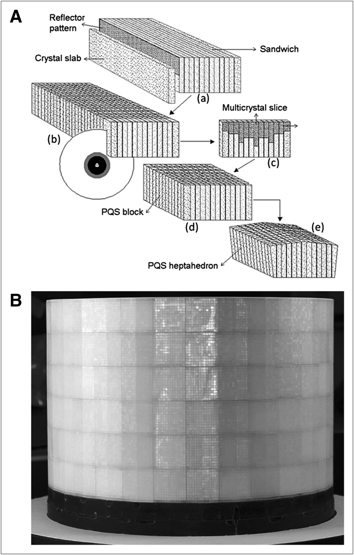
- FIGURE 1.
(A) Drawing of SSS technique for production of blocks: 13 crystal slabs and 12 film reflectors are sandwiched together using optical glue (a); sandwich is cut (b) into multicrystal slices (c); square block is made by gluing together 13 multicrystal slices and 12 film reflector patterns (d); and square block is ground on 4 sides with taper angle of 6° to produce heptahedron block (e). (B) Image of solid MuPET detector ring with 30,420 small LYSO crystals.
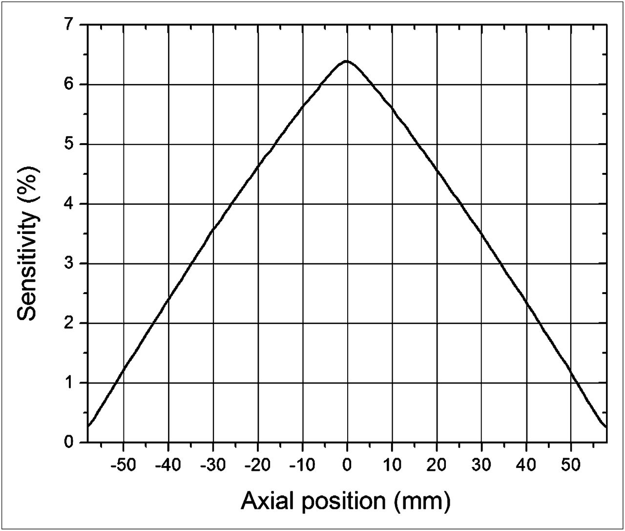
- FIGURE 2.
Axial sensitivity profile measured with 22Na point source moving along central axis of scanner.
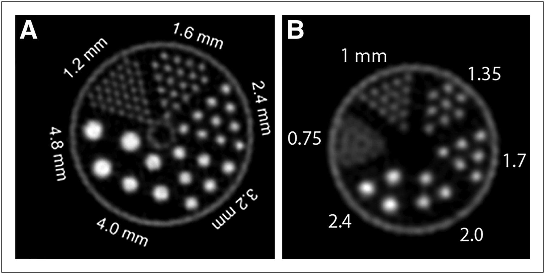
- FIGURE 3.
Counting rate performance plots as function of total activity for mouselike (A) and ratlike (B) phantoms.
- FIGURE 4.
NECR as function of total activity for mouse- and ratlike phantoms.
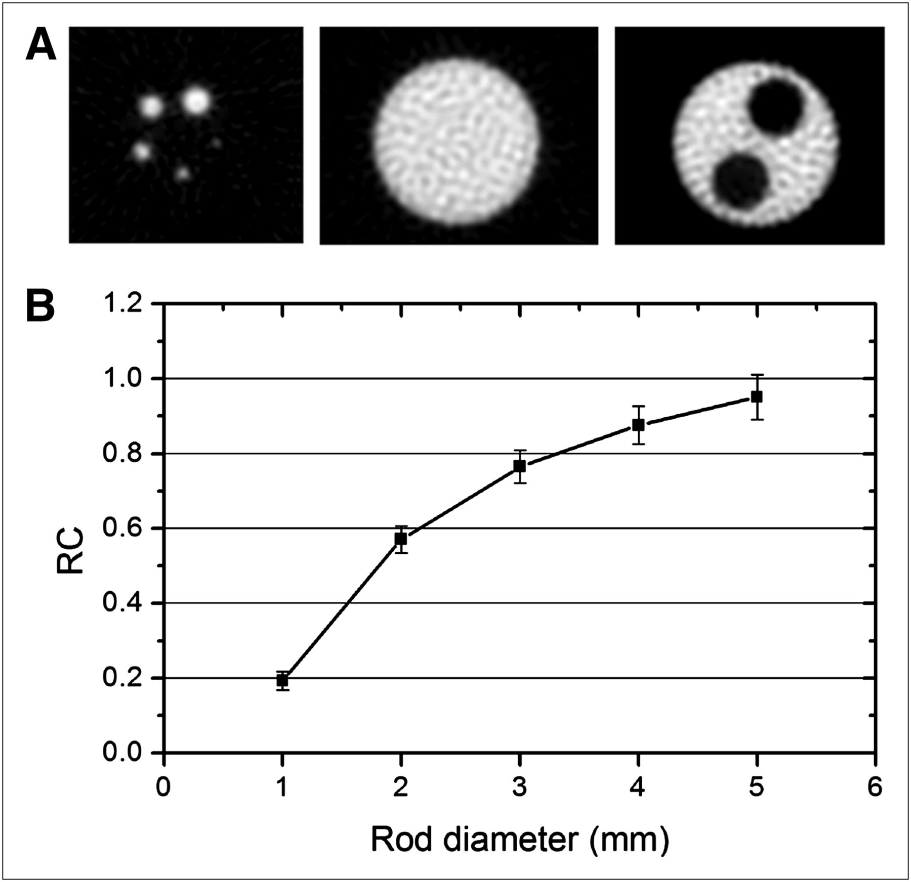
- FIGURE 5.
(A) Transverse images of NEMA NU 4-2008 image-quality phantom in 5-rod region (left), uniform region (middle), and 2-chamber region (right). (B) Recovery coefficients of 5 rods as function of rod diameter. RC = recovery coefficient.
- FIGURE 6.
Transverse images of Micro Deluxe (A) and Ultra-Micro Hot Spot (B) phantoms. Diameter of hot rods for each rod sector is indicated in figure. The 1-mm rods were well resolved.
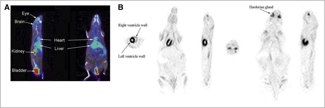
- FIGURE 7.
Nongated images of 2 healthy mice: sagittal and coronal slices of first mouse fused with CT images (A); transverse, coronal, and sagittal slices of second mouse (B) showing heart and Harderian glands. Both mice were injected with 18F-FDG. First mouse was anesthetized with ketamine–xylazine–atropine cocktail. Second mouse was anesthetized with isoflurane.


Tables
Category MuPET Inveon (10, 11) NanoPET (11) LabPET8 (12) Crystal material LYSO LSO LYSO LYSO/LGSO Photosensor PMT (19-mm diameter) 6 × 6 PSPMT 256-channel PSPMT APD No. of photosensors 210 64 24 6,144 No of signal channels 210 768 6,144 6,144 Crystal size (mm) 1.24 × 1.4 × 9.5 1.51 × 1.51 × 10 1.12 × 1.12 × 13 2 × 2 × 14 Packing fraction 95% 90% 92% No. of crystals 30,420 25,600 37,908 6,144 No. of rings 78 80 81 32 Ring diameter (cm) 16.6 16.1 18.1 16.2 Axial FOV (cm) 11.6 12.7 9.48 7.5 Transaxial FOV (cm) 10.0 10.0 12.3 10.0 LSO = lutetium oxyorthosilicate; LGSO = lutetium gadolinium oxyorthosilicate; PSPMT = position-sensitive photomultiplier tube; APD = avalanche photodiode.
Radial offset (mm) Radial resolution (mm) Tangential resolution (mm) Axial resolution (mm) FWHM FWTM FWHM FWTM FWHM FWTM 0 1.25 3.03 1.14 2.42 0.94 2.35 2 1.22 2.92 1.30 2.42 0.96 2.57 5 1.48 2.92 1.34 2.53 0.99 2.52 10 1.52 3.01 1.39 2.51 1.00 2.60 15 1.67 3.20 1.36 2.43 1.05 2.62 20 1.74 3.47 1.34 2.45 1.04 2.63 25 1.88 3.74 1.34 2.43 1.08 2.68 30 2.08 4.02 1.36 2.49 1.12 2.71 40 2.61 4.89 1.57 2.96 1.26 2.98 FWTM = full width at tenth maximum.
- TABLE 3
Radial and Tangential Spatial Resolutions for Resolution Recovery Method as Function of Radial Offset
Radial spatialresolution (mm) Tangential spatial resolution (mm) Radial offset (mm) FWHM FWTM FWHM FWTM 0 0.86 2.02 0.75 1.68 2 1.13 2.48 1.02 2.08 5 1.11 2.43 1.0 2.14 10 1.16 2.26 1.0 2.05 15 1.17 2.4 1.06 2.16 20 1.19 2.53 1.07 2.05 25 1.26 2.43 1.06 2.14 30 1.25 2.4 1.07 2.17 40 1.32 2.54 1.11 2.38 FWTM = full width at tenth maximum.


