


Article Figures & Data
Figures
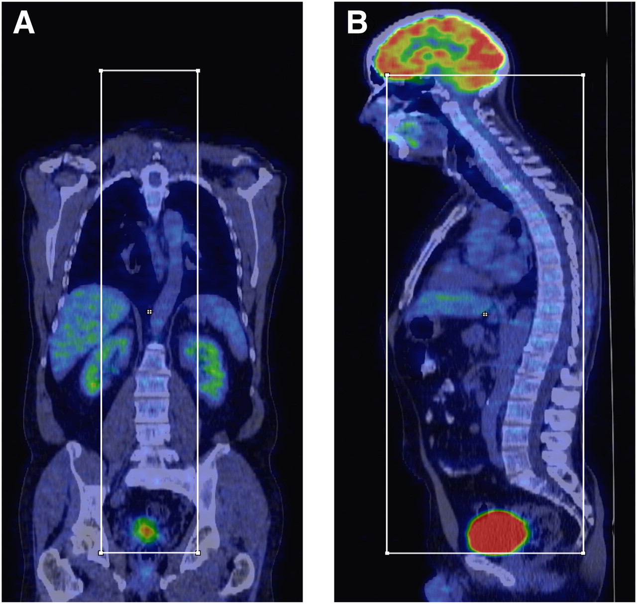
- FIGURE 1.
PET/CT image processing. Example of definition of bounding box used for segmentation of CT data: coronal 18F-FDG PET/CT image (A) and corresponding sagittal PET/CT image (B).
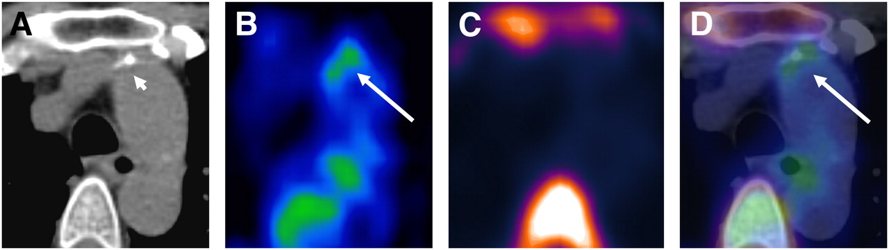
- FIGURE 2.
Transaxial PET/CT images of atherosclerotic plaque in ascending aorta: CT image (A), 18F-FDG PET image (B), 18F-fluoride PET image (C), and coregistered and fused 18F-FDG/18F-fluoride PET/CT image (D). Uptake of 18F-FDG coincides with calcification but not with 18F-fluoride accumulation. 18F-FDG activity adjacent to esophagus represents activity spillover from esophageal wall. Short arrow = calcification; long arrow = tracer uptake.
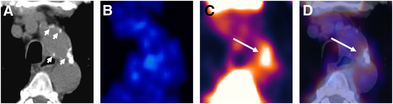
- FIGURE 3.
Transaxial PET/CT images of atherosclerotic plaque in aortic arch: CT image (A), 18F-FDG PET image (B), 18F-fluoride PET image (C), and coregistered and fused 18F-FDG/18F-fluoride PET/CT image (D). Accumulation of 18F-fluoride is colocalized with large calcification but not with 18F-FDG uptake. Short arrow = calcifications; long arrow = tracer uptake.


Tables
No. of subjects Parameter CT-positive 18F-fluoride PET–positive 18F-FDG PET–positive Total study population Subjects (n) 34 (75.6) 27 (60) 34 (75.6) 45 (100) Age at risk 28 (82.4) 24 (88.9) 27 (79.4) 29 (64.4) Male sex 16 (47.1) 12 (44.4) 15 (44.1) 21 (46.7) Body mass index > 25 14 (41.2) 9 (33.3) 13 (38.2) 17 (37.8) Cardiovascular risk factors (n) Hypertension 15 (44.1) 12 (44.4) 17 (50.0) 18 (40.0) Hypercholesterolemia 11 (32.4) 9 (33.3) 10 (29.4) 11 (24.4) Diabetes mellitus 3 (8.8) 2 (7.4) 3 (8.8) 3 (6.7) Smoking 16 (47.1) 13 (48.1) 16 (47.1) 17 (37.8) Prior vascular event 6 (17.6) 5 (18.5) 6 (17.6) 6 (13.3) Statin medication 7 (20.6) 7 (25.9) 7 (20.6) 7 (15.6) Values in parentheses are percentages.
Age at risk, 55 y or older for women or 45 y or older for men.
Parameter Left common carotid artery Right common carotid artery Ascending aorta Aortic arch Descending aorta Abdominal aorta Total No. of patients with uptake sites 11 (24.4) 12 (26.7) 10 (22.2) 16 (35.6) 4 (8.9) 17 (37.8) 27 (60) No. of uptake sites 16 (15.2) 15 (14.3) 13 (12.4) 27 (25.7) 4 (3.8) 30 (28.6) 105 (100) TBR Mean ± SD 2.3 ± 0.5 2.3 ± 0.7 2.5 ± 0.9 1.9 ± 0.5 2.1 ± 0.4 2.6 ± 0.8 2.3 ± 0.7 Range 1.6–3.3 1.5–3.6 1.2–5.0 1.4–3.4 1.8–2.5 1.5–4.7 1.2–5.0 Values in parentheses are percentages.
Parameter Left common carotid artery Right common carotid artery Ascending aorta Aortic arch Descending aorta Abdominal aorta Total No. of patients with uptake sites 4 (8.9) 10 (22.2) 21 (46.7) 18 (40.0) 14 (31.1) 29 (64.4) 34 (75.6) No. of uptake sites 5 (4.0) 11 (8.9) 24 (19.4) 19 (15.3) 17 (13.7) 48 (38.7) 124 (100) TBR Mean ± SD 1.5 ± 0.4 1.5 ± 0.4 1.6 ± 0.2 1.5 ± 0.3 1.5 ± 0.4 1.4 ± 0.3 1.5 ± 0.3 Range 1.1–2.2 1.2–2.2 1.2–2.2 1.2–2.5 1.1–2.6 0.9–2.1 0.9–2.6 Values in parentheses are percentages.
Parameter Left common carotid artery Right common carotid artery Ascending aorta Aortic arch Descending aorta Abdominal aorta Total No. of patients with calcification sites 20 (44.4) 18 (40.0) 8 (17.8) 25 (55.6) 17 (37.8) 33 (73.3) 34 (75.6) No. of calcification sites 35 (7.0) 27 (5.4) 11 (2.2) 73 (14.5) 99 (19.7) 258 (51.3) 503 (100) Lesion thickness (mm) 2.8 ± 0.9 2.7 ± 0.8 2.5 ± 0.7 3.6 ± 1.7 2.8 ± 0.8 3.0 ± 1.1 3.0 ± 1.2 Calcification score for lesions 1.6 ± 0.9 1.7 ± 0.9 1.1 ± 0.3 1.5 ± 0.8 1.2 ± 0.4 1.8 ± 1.1 1.6 ± 1.0 Values in parentheses are percentages.
- TABLE 5
18F-Sodium Fluoride Uptake, 18F-FDG Uptake, and Calcification on Per-Patient Basis
18F-Fluoride uptake (n = 27) 18F-FDG uptake (n = 34) Arterial calcification (n = 34) Total no. of patients (n = 45) PET-positive PET-positive CT-positive 25 PET-positive PET-positive CT-negative 1 PET-positive PET-negative CT-positive 1 PET-negative PET-positive CT-positive 7 PET-negative PET-positive CT-negative 1 PET-negative PET-negative CT-positive 1 PET-negative PET-negative CT-negative 9


Jump to section
Related Articles
Cited By...
- Multimodality Imaging of Aortic Valve Calcification and Function in a Murine Model of Calcific Aortic Valve Disease and Bicuspid Aortic Valve
- Multimodality Imaging of Aortic Valve Calcification and Function in a Murine Model of Calcific Aortic Valve Disease and Bicuspid Aortic Valve
- 18F-NaF PET/CT of Obese Patients on a Lutetium-Yttrium Oxyorthosilicate PET/CT System: Patient Dosimetry, Optimization of Injected Activity, and Acquisition Time
- Imaging Inflammation in Atherosclerosis with CXCR4-Directed 68Ga-Pentixafor PET/CT: Correlation with 18F-FDG PET/CT
- Nanobody-Facilitated Multiparametric PET/MRI Phenotyping of Atherosclerosis
- Three-Hour Delayed Imaging Improves Assessment of Coronary 18F-Sodium Fluoride PET
- Assessment of Physiologic Intracranial Calcification in Healthy Adults Using 18F-NaF PET/CT
- Imaging Cardiovascular Calcification
- Clinical Molecular Imaging of Chemokine Receptor CXCR4 Expression in Atherosclerotic Plaque Using 68Ga-Pentixafor PET: Correlation with Cardiovascular Risk Factors and Calcified Plaque Burden
- 18F-Flurodeoxyglucose and 18F-Sodium Fluoride Positron Emission Tomography/Computed Tomography Imaging of Arterial and Cutaneous Alterations in Pseudoxanthoma Elasticum
- 18F-Fluoride Imaging and Other Plaque-Seeking Diagnostic Strategies: Understanding the Reality and Setting the Ground Rules...
- Clinical Relevance of 18F-Sodium Fluoride Positron-Emission Tomography in Noninvasive Identification of High-Risk Plaque in Patients With Coronary Artery Disease
- Association Between Osteogenesis and Inflammation During the Progression of Calcified Plaque Evaluated by 18F-Fluoride and 18F-FDG
- Coronary Artery Calcification: From Mechanism to Molecular Imaging
- Evaluation of 68Ga-Glutamate Carboxypeptidase II Ligand Positron Emission Tomography for Clinical Molecular Imaging of Atherosclerotic Plaque Neovascularization
- Noninvasive Molecular Imaging of Disease Activity in Atherosclerosis
- Impact of Personal Characteristics and Technical Factors on Quantification of Sodium 18F-Fluoride Uptake in Human Arteries: Prospective Evaluation of Healthy Subjects
- 18F-NaF Uptake by Atherosclerotic Plaque on PET/CT Imaging: Inverse Correlation Between Calcification Density and Mineral Metabolic Activity
- 18F-Sodium Fluoride Uptake Is a Marker of Active Calcification and Disease Progression in Patients With Aortic Stenosis
- The Advancing Clinical Impact of Molecular Imaging in CVD
- Coronary Arterial 18F-Sodium Fluoride Uptake: A Novel Marker of Plaque Biology
- Feasibility of 11C-Acetate PET/CT for Imaging of Fatty Acid Synthesis in the Atherosclerotic Vessel Wall