


Article Figures & Data
Figures
- FIGURE 1.
Integrated β-camera and microfluidic chip for real-time radioassay imaging of glycolysis in small cell populations. (A) Schematic cross-section of β-camera integrated with microfluidic chip. (B) Micrograph of microfluidic chip loaded with colored dyes. Chamber array (4 × 4) allows cultured cells to be individually addressable within flow network for parametric study. (C) Micrograph of cells in chamber of microfluidic chip taken with bright-field microscope. (D) Chip operation of an 18F-FDG uptake radioassay. From left to right: Cells are allowed to adapt to closed-chamber microenvironment. Valves are opened to allow replacement of culture medium with culture medium containing 18F-FDG. Cells are incubated in 18F-FDG to start 18F-FDG uptake. Valves are opened again to wash away extracellular 18F-FDG using culture medium. Channel network is washed with culture medium to remove 18F-FDG residing outside cell chamber. 18F-FDG uptake of cells is imaged by β-camera. GND = ground; HV = high voltage; PDMS = polydimethylsiloxane.
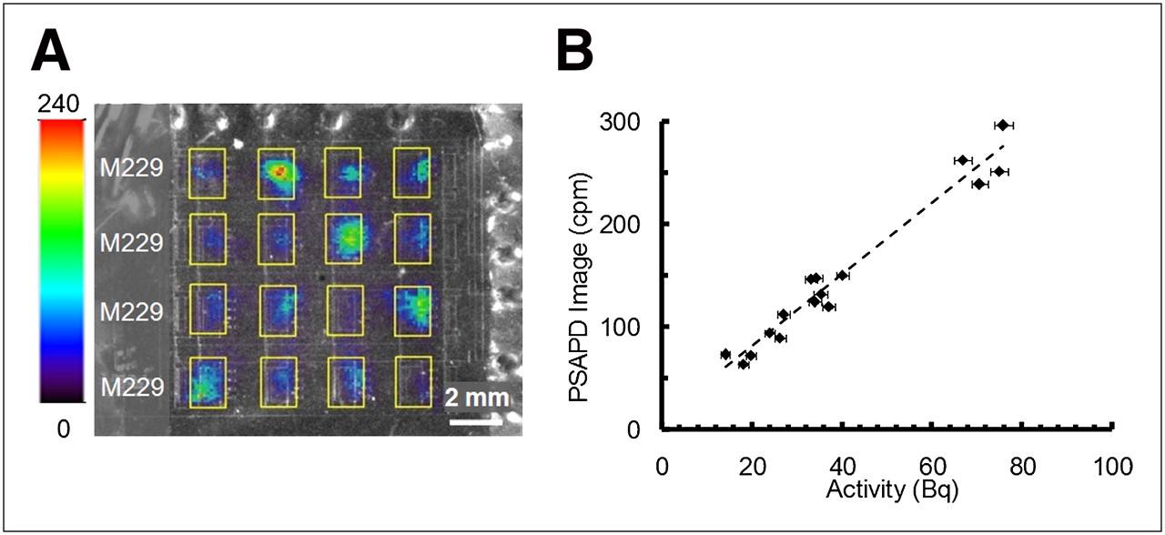
- FIGURE 2.
Overlay image of 18F-FDG uptake in melanoma primary cell line (M229) with β-camera and optical image of microfluidic chips. All images were evaluated using ROIs (yellow rectangles) of equal dimensions. Color bar represents β-camera image scale and indicates counts per minute (cpm) per square millimeter. (A) 18F-FDG uptake image of M229 cell cultures inside microfluidic chambers. (B) Calibration of β-camera image.
- FIGURE 3.
(A) Same β-camera image displayed with 2 different color-intensity scales. In each β-camera image, 18F-FDG uptake was measured for cell cultures incubated with 18F-FDG radioactivity concentrations of 0.037, 0.370, 3.700, and 37.00 MBq/mL (I, II, III, and IV, respectively). (B) 18F-FDG uptake values are given as mean (±SEM) activity normalized to number of cells in each chamber.
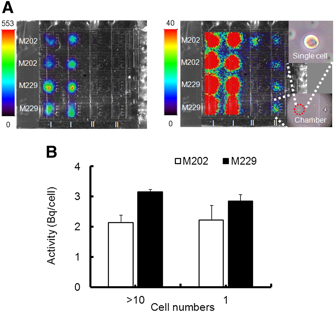
- FIGURE 4.
(A) Same β-camera image displayed with 2 different color-intensity scales. In each image, the 2 leftmost columns (I) show 18F-FDG uptake in chambers with double-digit numbers of cells. The 2 rightmost columns (II) show image of 18F-FDG uptake in chambers with single cell. Micrograph of selected chamber and zoom-in section showing same chamber that has only single cell. (B) 18F-FDG uptake values are given as mean (±SEM) activity normalized to number of cells in each chamber.
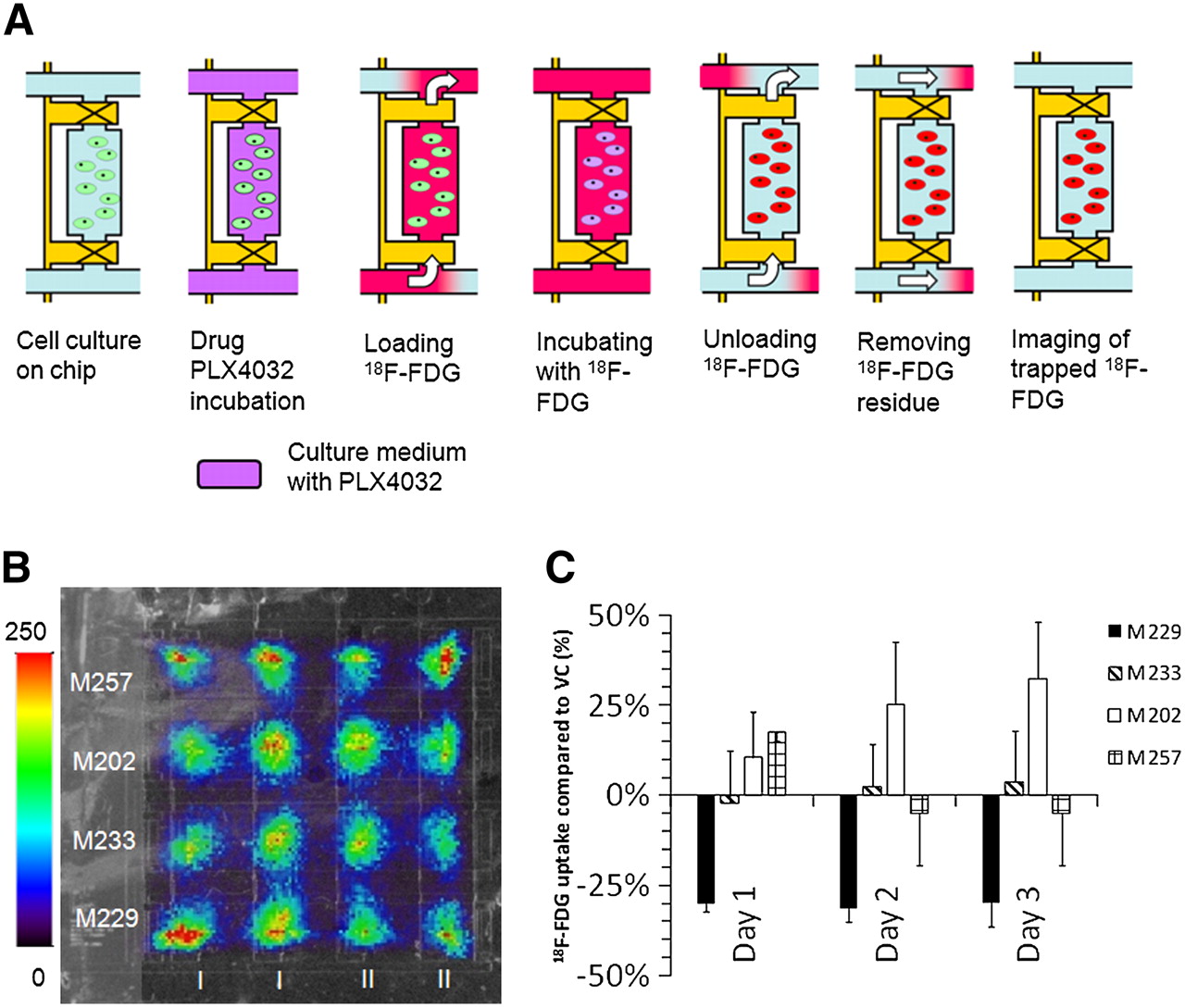
- FIGURE 5.
18F-FDG radioassay of melanoma cell lines treated with B-Raf inhibitor drug. (A) Chip operation for 18F-FDG uptake radioassay of drug-treated melanoma cells. (Left to right) Cells are allowed to adapt to closed-chamber microenvironment. Valves are opened to allow replacement of culture medium by culture medium containing PLX4032. Cells are incubated in 18F-FDG to start 18F-FDG uptake. Valves are opened again to wash away extracellular 18F-FDG using culture medium. Channel network is washed with culture medium to remove 18F-FDG residing outside cell chamber. 18F-FDG uptake of cells is imaged by β-camera. (B) Overlay image of 18F-FDG uptake with β-camera and optical image of microfluidic chip. (C) 18F-FDG uptake values are given as ratio of mean (±SEM) activity normalized to number of cells in each chamber between drug-treated cells and vehicle control.


Additional Files
Supplemental Data
Files in this Data Supplement:


Jump to section
Related Articles
Cited By...
- Development of a Lensless Radiomicroscope for Cellular-Resolution Radionuclide Imaging
- A Continuously Infused Microfluidic Radioassay System for the Characterization of Cellular Pharmacokinetics
- Fast Metabolic Response to Drug Intervention Through Analysis on a Miniaturized, Highly Integrated Molecular Imaging System
- High-Resolution Radioluminescence Microscopy of 18F-FDG Uptake by Reconstructing the {beta}-Ionization Track