
Main menu

  • Home
  • Content
    • Current
    • Ahead of print
    • Past Issues
    • JNM Supplement
    • SNMMI Annual Meeting Abstracts
    • Continuing Education
    • JNM Podcasts
  • Subscriptions
    • Subscribers
    • Institutional and Non-member
    • Rates
    • Journal Claims
    • Corporate & Special Sales
  • Authors
    • Submit to JNM
    • Information for Authors
    • Assignment of Copyright
    • AQARA requirements
  • Info
    • Reviewers
    • Permissions
    • Advertisers
  • About
    • About Us
    • Editorial Board
    • Contact Information
  • More
    • Alerts
    • Feedback
    • Help
    • SNMMI Journals
  • SNMMI
    • JNM
    • JNMT
    • SNMMI Journals
    • SNMMI

User menu

  • Subscribe
  • My alerts
  • Log in
  • My Cart

Search

  • Advanced search
Journal of Nuclear Medicine
  • SNMMI
    • JNM
    • JNMT
    • SNMMI Journals
    • SNMMI
  • Subscribe
  • My alerts
  • Log in
  • My Cart
Journal of Nuclear Medicine

Advanced Search

  • Home
  • Content
    • Current
    • Ahead of print
    • Past Issues
    • JNM Supplement
    • SNMMI Annual Meeting Abstracts
    • Continuing Education
    • JNM Podcasts
  • Subscriptions
    • Subscribers
    • Institutional and Non-member
    • Rates
    • Journal Claims
    • Corporate & Special Sales
  • Authors
    • Submit to JNM
    • Information for Authors
    • Assignment of Copyright
    • AQARA requirements
  • Info
    • Reviewers
    • Permissions
    • Advertisers
  • About
    • About Us
    • Editorial Board
    • Contact Information
  • More
    • Alerts
    • Feedback
    • Help
    • SNMMI Journals
  • View or Listen to JNM Podcast
  • Visit JNM on Facebook
  • Join JNM on LinkedIn
  • Follow JNM on Twitter
  • Subscribe to our RSS feeds
OtherBASIC SCIENCE INVESTIGATIONS

CT Hounsfield Units of Brown Adipose Tissue Increase with Activation: Preclinical and Clinical Studies

Shingo Baba, Heather A. Jacene, James M. Engles, Hiroshi Honda and Richard L. Wahl
Journal of Nuclear Medicine February 2010, 51 (2) 246-250; DOI: https://doi.org/10.2967/jnumed.109.068775
Shingo Baba
  • Find this author on Google Scholar
  • Find this author on PubMed
  • Search for this author on this site
Heather A. Jacene
  • Find this author on Google Scholar
  • Find this author on PubMed
  • Search for this author on this site
James M. Engles
  • Find this author on Google Scholar
  • Find this author on PubMed
  • Search for this author on this site
Hiroshi Honda
  • Find this author on Google Scholar
  • Find this author on PubMed
  • Search for this author on this site
Richard L. Wahl
  • Find this author on Google Scholar
  • Find this author on PubMed
  • Search for this author on this site
  • Article
  • Figures & Data
  • Info & Metrics
  • PDF
Loading

Article Figures & Data

Figures

  • Tables
  • FIGURE 1. 
    • Download figure
    • Open in new tab
    • Download powerpoint
    FIGURE 1. 

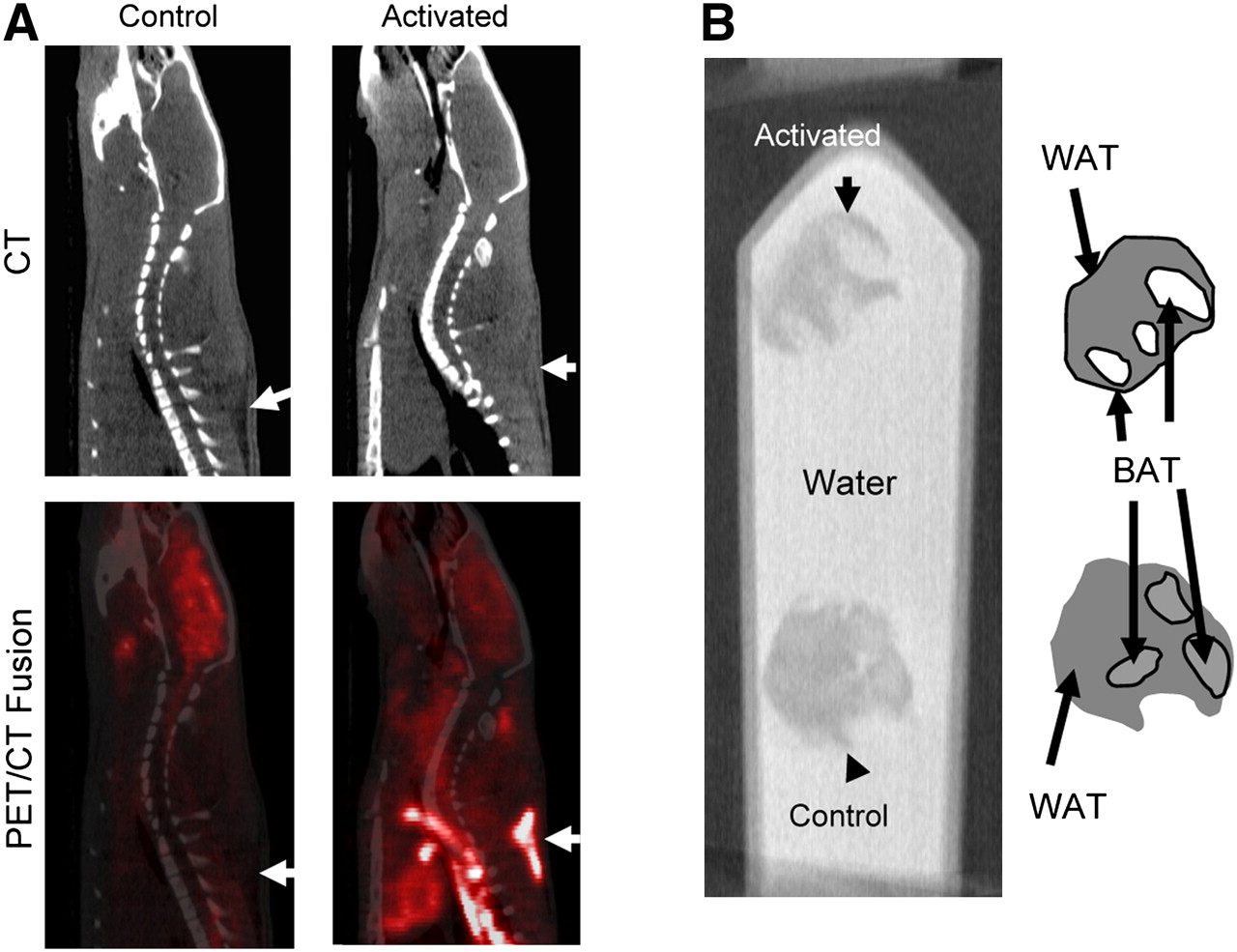
    (A) Sagittal CT HU of subcutaneous paraspinal BAT (arrow) in rodent is greater under activated conditions than under control conditions. 18F-FDG uptake of BAT is also increased under activated conditions. CT shows that BAT with increased fat density (right) fuses precisely to area of increased 18F-FDG uptake. (B) CT image of resected BAT with surrounding WAT subjected to 4 h of cold-stimulation shows greater CT density that is closer to water (top) than control kept at room temperature (bottom). Under control conditions, BAT is difficult to distinguish from surrounding WAT. On CT, higher CT HUs are shown as areas of increased whiteness, whereas low HUs are increasingly black. Areas of WAT and BAT as seen on direct visual inspection are shown in schematic at right.

  • FIGURE 2. 
    • Download figure
    • Open in new tab
    • Download powerpoint
    FIGURE 2. 

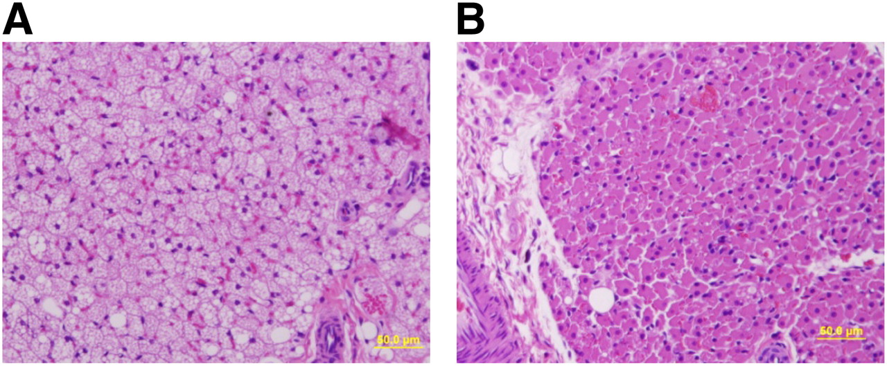
    On light microscopy, major structural changes can be seen in BAT cells stained with hematoxylin–eosin. Many large lipid vacuoles are seen in control BAT cells (A), but vacuoles are almost nonexistent in BAT cells exposed to cold (B). Lipid content is much lower in cold-stimulated BAT.

  • FIGURE 3. 
    • Download figure
    • Open in new tab
    • Download powerpoint
    FIGURE 3. 

    (A) Average CT HUs of BAT (46 regions from 23 patients) between high SUV for 18F-FDG and low SUV for 18F-FDG is shown. CT HUs in scans with high SUV were significantly higher than CT HUs in scans with low SUV (−71.6 ± 18.0 vs. −104.4 ± 16.8; P < 0.05). (B) When we plotted CT HUs against 18F-FDG uptake in all 92 ROIs, there was significant positive correlation between the 2 values (R = 0.66, P < 0.01).
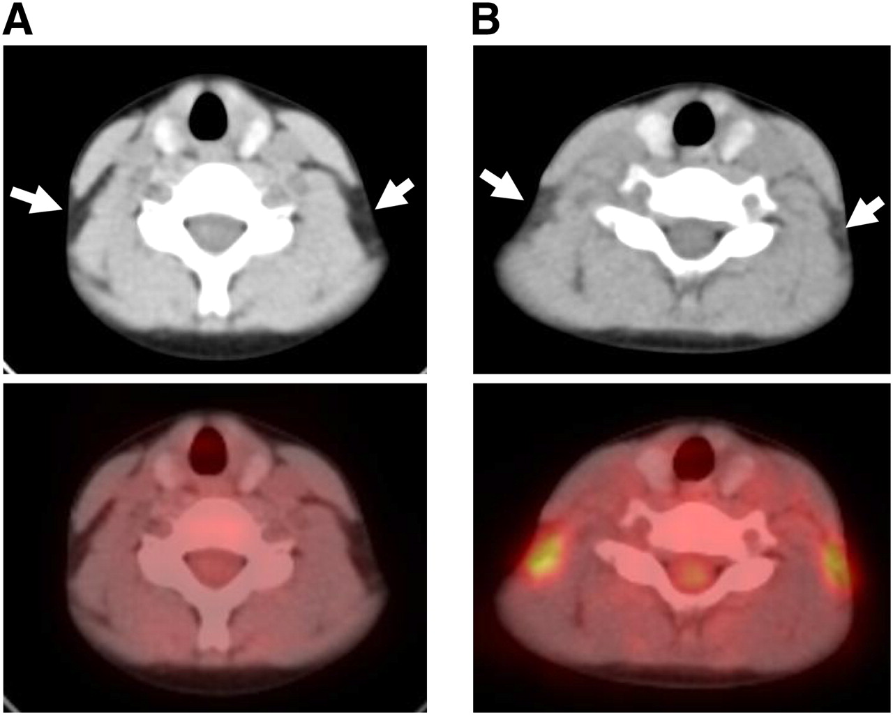
  • FIGURE 4. 
    • Download figure
    • Open in new tab
    • Download powerpoint
    FIGURE 4. 

    Representative study of patient in whom CT HU of BAT is increased in parallel with increase in 18F-FDG uptake. A 20-y-old man with malignant lymphoma displayed bilateral cloudy BAT (increased CT HUs of −53 and −56) with avid 18F-FDG uptake (SUVmax = 3.4 and 3.2) (B), whereas baseline scan showed low-attenuation BAT (CT HUs of −87 and −85) with low 18F-FDG uptake (SUVmax = 0.9 and 1.0) (A). Both CT images are displayed with same window level (−20) and narrow window width (300) of CT HU to emphasize small difference of CT density in BAT.

Tables

  • Figures
    • View popup
    TABLE 1

    Patient and Disease Characteristics (BAT-Avid Cohort)

    CharacteristicValue
    Age (y)
     Mean ± SD34 ± 17
     Range17–54
    Men/women (n)5/18
    Disease (n)
     Ovarian carcinoma5
     Non-Hodgkin lymphoma3
     Hodgkin disease2
     Thyroid carcinoma3
     Breast carcinoma2
     Malignant melanoma2
     Other6
    • View popup
    TABLE 2

    Summary of In Vivo Imaging Studies

    Study typeFirst parameterSecond parameterP
    RatCold-activatedControl
     18F-FDG uptake (Bq/cm3 of BAT)1,982.8 ± 361.3892.9 ± 357.7<0.05
     CT HU (BAT)−12.4 ± 22.4−27.9 ± 9.6<0.05
     CT HU (WAT)−69.3 ± 9.7−75.6 ± 9.0NS
     CT HU (liver)19.8 ± 4.223.6 ± 9.9NS
    HumanHigh SUVLow SUV
     18F-FDG uptake (SUVmax of BAT)6.6 ± 2.80.9 ± 0.5<0.05
     CT HU (BAT)−71.6 ± 18.0−104.4 ± 16.8<0.05
     CT HU (WAT)−101.8 ± 6.4−102.6 ± 5.1NS
     CT HU (liver)56.0 ± 7.756.2 ± 7.4NS
     Frequency during cold season*19/234/23<0.05
    • ↵* Number of scans obtained between November and March of a total of 23 human scans.

    • NS = not statistically significant.

PreviousNext
Back to top

In this issue

Journal of Nuclear Medicine: 51 (2)
Journal of Nuclear Medicine
Vol. 51, Issue 2
February 2010
  • Table of Contents
  • Table of Contents (PDF)
  • About the Cover
  • Index by author
Print
Download PDF
Article Alerts
Sign In to Email Alerts with your Email Address
Email Article

Thank you for your interest in spreading the word on Journal of Nuclear Medicine.

NOTE: We only request your email address so that the person you are recommending the page to knows that you wanted them to see it, and that it is not junk mail. We do not capture any email address.

Enter multiple addresses on separate lines or separate them with commas.
CT Hounsfield Units of Brown Adipose Tissue Increase with Activation: Preclinical and Clinical Studies
(Your Name) has sent you a message from Journal of Nuclear Medicine
(Your Name) thought you would like to see the Journal of Nuclear Medicine web site.
Citation Tools
CT Hounsfield Units of Brown Adipose Tissue Increase with Activation: Preclinical and Clinical Studies
Shingo Baba, Heather A. Jacene, James M. Engles, Hiroshi Honda, Richard L. Wahl
Journal of Nuclear Medicine Feb 2010, 51 (2) 246-250; DOI: 10.2967/jnumed.109.068775

Citation Manager Formats

  • BibTeX
  • Bookends
  • EasyBib
  • EndNote (tagged)
  • EndNote 8 (xml)
  • Medlars
  • Mendeley
  • Papers
  • RefWorks Tagged
  • Ref Manager
  • RIS
  • Zotero
Share
CT Hounsfield Units of Brown Adipose Tissue Increase with Activation: Preclinical and Clinical Studies
Shingo Baba, Heather A. Jacene, James M. Engles, Hiroshi Honda, Richard L. Wahl
Journal of Nuclear Medicine Feb 2010, 51 (2) 246-250; DOI: 10.2967/jnumed.109.068775
Twitter logo Facebook logo LinkedIn logo Mendeley logo
  • Tweet Widget
  • Facebook Like
  • Google Plus One
Bookmark this article

Jump to section

  • Article
    • Abstract
    • MATERIALS AND METHODS
    • RESULTS
    • DISCUSSION
    • CONCLUSION
    • Footnotes
    • References
  • Figures & Data
  • Info & Metrics
  • PDF

Related Articles

  • This Month in JNM
  • PubMed
  • Google Scholar

Cited By...

  • High-fructose feeding suppresses cold-stimulated brown adipose tissue glucose uptake in young men independently of changes in thermogenesis and the gut microbiome
  • Perspectives on Brown Adipose Tissue Imaging: Insights from Preclinical and Clinical Observations from the Last and Current Century
  • Recent advances in the detection of brown adipose tissue in adult humans: a review
  • Accurate quantification of brown adipose tissue mass by xenon-enhanced computed tomography
  • Association of Changes in Abdominal Fat Quantity and Quality With Incident Cardiovascular Disease Risk Factors
  • Supraclavicular Brown Adipose Tissue 18F-FDG Uptake and Cardiovascular Disease
  • Obesity-Induced Changes in Adipose Tissue Microenvironment and Their Impact on Cardiovascular Disease
  • Cross-Sectional Associations of Computed Tomography (CT)-Derived Adipose Tissue Density and Adipokines: The Framingham Heart Study
  • Sugar-Sweetened Beverage Consumption Is Associated With Change of Visceral Adipose Tissue Over 6 Years of Follow-Up
  • Selective Impairment of Glucose but Not Fatty Acid or Oxidative Metabolism in Brown Adipose Tissue of Subjects With Type 2 Diabetes
  • Detection of brown adipose tissue and thermogenic activity in mice by hyperpolarized xenon MRI
  • Association of Fat Density With Subclinical Atherosclerosis
  • Quantification of Human and Rodent Brown Adipose Tissue Function Using 99mTc-Methoxyisobutylisonitrile SPECT/CT and 18F-FDG PET/CT
  • Visceral and Subcutaneous Fat Quality and Cardiometabolic Risk
  • 15O PET Measurement of Blood Flow and Oxygen Consumption in Cold-Activated Human Brown Fat
  • Google Scholar

More in this TOC Section

  • 11C-Methionine PET of Myocardial Inflammation in a Rat Model of Experimental Autoimmune Myocarditis
  • Counting Rate Characteristics and Image Distortion in Preclinical PET Imaging During Radiopharmaceutical Therapy
  • Design and Fabrication of Kidney Phantoms for Internal Radiation Dosimetry Using 3D Printing Technology
Show more Basic Science Investigations

Similar Articles

SNMMI

© 2025 SNMMI

Powered by HighWire