
Main menu

  • Home
  • Content
    • Current
    • Ahead of print
    • Past Issues
    • JNM Supplement
    • SNMMI Annual Meeting Abstracts
    • Continuing Education
    • JNM Podcasts
  • Subscriptions
    • Subscribers
    • Institutional and Non-member
    • Rates
    • Journal Claims
    • Corporate & Special Sales
  • Authors
    • Submit to JNM
    • Information for Authors
    • Assignment of Copyright
    • AQARA requirements
  • Info
    • Reviewers
    • Permissions
    • Advertisers
  • About
    • About Us
    • Editorial Board
    • Contact Information
  • More
    • Alerts
    • Feedback
    • Help
    • SNMMI Journals
  • SNMMI
    • JNM
    • JNMT
    • SNMMI Journals
    • SNMMI

User menu

  • Subscribe
  • My alerts
  • Log in
  • My Cart

Search

  • Advanced search
Journal of Nuclear Medicine
  • SNMMI
    • JNM
    • JNMT
    • SNMMI Journals
    • SNMMI
  • Subscribe
  • My alerts
  • Log in
  • My Cart
Journal of Nuclear Medicine

Advanced Search

  • Home
  • Content
    • Current
    • Ahead of print
    • Past Issues
    • JNM Supplement
    • SNMMI Annual Meeting Abstracts
    • Continuing Education
    • JNM Podcasts
  • Subscriptions
    • Subscribers
    • Institutional and Non-member
    • Rates
    • Journal Claims
    • Corporate & Special Sales
  • Authors
    • Submit to JNM
    • Information for Authors
    • Assignment of Copyright
    • AQARA requirements
  • Info
    • Reviewers
    • Permissions
    • Advertisers
  • About
    • About Us
    • Editorial Board
    • Contact Information
  • More
    • Alerts
    • Feedback
    • Help
    • SNMMI Journals
  • View or Listen to JNM Podcast
  • Visit JNM on Facebook
  • Join JNM on LinkedIn
  • Follow JNM on Twitter
  • Subscribe to our RSS feeds
OtherThis Month in JNM

This Month in JNM

Journal of Nuclear Medicine February 2010, 51 (2) 11A-12A;
  • Article
  • Info & Metrics
  • PDF
Loading

Imaging RNA interference: Hong and colleagues offer perspective on current investigations into in vivo molecular imaging of RNA interference, a technique for regulating or silencing specific genes, with potential applications in numerous diseases.
Page 169

SUV—silly or smart metric?: Visser and colleagues provide an overview of perspective on challenges to meaningful quantitation in PET imaging and preview an article in this issue of JNM that identifies superior approaches.
Page 173

PET cost-effectiveness in HNSCC: Uyl-de Groot and colleagues compare the cost-effectiveness of whole-body 18F-FDG PET and chest CT in pretreatment screening for distant metastases in patients with head and neck squamous cell carcinoma.
Page 176

Pharmacokinetics of 18F-FDHT: Beattie and colleagues describe a clinically applicable, noninvasive method using PET and this fluorinated androgen analog to quantify changes in androgen receptor levels in patients undergoing therapy for prostate cancer.
Page 183

Imaging coronary artery macrophage activity: Rominger and colleagues correlate uptake of 68Ga-DOTATATE in the coronary arteries with the presence of calcified plaques and cardiovascular risk factors to explore a potential role for this tracer in plaque imaging.
Page 193

Figure

Vascular territories in hybrid PET/CT: Javadi and colleagues review standard techniques for assignment of vascular territories in myocardial perfusion imaging and suggest an alternative approach to correct for inaccuracies in PET/CT resulting from morphologic variability in the coronary tree.
Page 198

Figure
Figure

MPS change quantification: Prasad and colleagues analyze variables in a large group of rest–stress myocardial perfusion SPECT studies to improve optimal automatic detection of ischemia and coronary artery disease.
Page 204

Movement correction for human brain PET: Wardak and colleagues describe a retrospective image-based movement correction method and evaluate its implementation on dynamic 18F-FDDNP PET images of patients with Alzheimer disease and cognitively intact controls.
Page 210

Figure

11C-methionine uptake in normal brain: Uda and colleagues determine uptake of this increasingly useful PET tracer in normal brain and detail inter- and intraindividual variations in standardized uptake values.
Page 219

Figure

Imaging presynaptic monoaminergic system integrity: Okamura and colleagues report on the utility of 18F-AV-133, a novel radiolabeled tetrabenazine derivative, in PET assessment of the vesicular monoamine transporter type 2 in Parkinson disease patients and healthy individuals.
Page 223

Figure

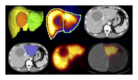
SPECT with scintigraphy for liver function: de Graaf and colleagues determine the value of the addition of SPECT to dynamic 99mTc-mebrofenin hepatobiliary scintigraphy in SPECT/CT evaluation of liver function in patients before liver resection.
Page 229

Time-of-flight PET/CT: Lois and colleagues investigate the qualitative and quantitative effects of incorporating time-of-flight data into reconstruction of clinical studies in patients with cancer.
Page 237

Figure

Assessing brown adipose tissue activation: Baba and colleague measure brown adipose tissue densities as CT Hounsfield units in a rodent model and in patients to determine whether changes in these metrics correlate with brown adipose tissue activity.
Page 246

Figure

AgRP scaffold–based PET probe: Jiang and colleagues describe experiments to determine the potential of engineered αvβ3 integrin–binding agouti-related protein peptides for use as cancer imaging agents in living subjects.
Page 251

Figure

Annexin-A5 uptake in myocardial ischemia: Kenis and colleagues explore the use of 99mTc-labeled annexin-A5 for γ-imaging of phosphatidylserine externalization as a byproduct of cardiomyocite apoptosis and report on the potential reversibility of the apoptotic process.
Page 259

Figure

Tumor volume and SUV estimation in PET: Tylski and colleagues assess the accuracy and robustness of 5 methods for tumor volume estimates in 18F-FDG PET and 10 methods for SUV estimates in a variety of configurations.
Page 268

Figure

Sincalide-stimulated cholescintigraphy: Ziessman and colleagues determine the infusion method with the least variability for calculation of gallbladder ejection fraction in sincalide-stimulated cholescintigraphy and establish reference values for the technique.
Page 277

Coin-shaped radiosynthesis microreactor: Elizarov and colleagues report on the design of an integrated microfluidic device, with a footprint equal to that of a postage stamp, and on its optimization for multistep radiosynthesis of PET tracers.
Page 282

Figure

18F-FLT fetal dosimetry: Bartlett and colleagues estimate radiation absorbed dose to the human fetus from nuclear medicine procedures with this tracer, using biodistribution data obtained with PET/CT studies in pregnant rhesus monkeys.
Page 288

Figure

Enhancing pediatric PET: Accorsi and colleagues determine quantitative injection rules for pediatric PET imaging that reduce scan time or dose while maintaining high image quality.
Page 293

90Y SIRT structural dosimetry: Gulec and colleagues describe a methodology for translating derived absorbed dose estimates to subunits of normal liver tissue into biologically relevant absorbed doses for more effective selective internal radiation treatment with 90Y microspheres.
Page 301

α-Particle emitter dosimetry: Sgouros and members of the MIRD committee review current targeted α-particle emitter therapies and provide guidance and recommendations for human α-particle emitter dosimetry in this abbreviated version of MIRD Pamphlet 23.
Page 311

Figure

ON THE COVER

Future remnant liver function is a crucial determinant of whether a patient can safely undergo major liver resection. The combination of SPECT data with measures of dynamic uptake function by planar hepatobiliary scintigraphy provides valuable information about segmental liver function and is an accurate measure of future remnant liver function.

See page 231.

Figure
PreviousNext
Back to top

In this issue

Journal of Nuclear Medicine: 51 (2)
Journal of Nuclear Medicine
Vol. 51, Issue 2
February 2010
  • Table of Contents
  • Table of Contents (PDF)
  • About the Cover
  • Index by author
Print
Download PDF
Article Alerts
Sign In to Email Alerts with your Email Address
Email Article

Thank you for your interest in spreading the word on Journal of Nuclear Medicine.

NOTE: We only request your email address so that the person you are recommending the page to knows that you wanted them to see it, and that it is not junk mail. We do not capture any email address.

Enter multiple addresses on separate lines or separate them with commas.
This Month in JNM
(Your Name) has sent you a message from Journal of Nuclear Medicine
(Your Name) thought you would like to see the Journal of Nuclear Medicine web site.
Citation Tools
This Month in JNM
Journal of Nuclear Medicine Feb 2010, 51 (2) 11A-12A;

Citation Manager Formats

  • BibTeX
  • Bookends
  • EasyBib
  • EndNote (tagged)
  • EndNote 8 (xml)
  • Medlars
  • Mendeley
  • Papers
  • RefWorks Tagged
  • Ref Manager
  • RIS
  • Zotero
Share
This Month in JNM
Journal of Nuclear Medicine Feb 2010, 51 (2) 11A-12A;
Twitter logo Facebook logo LinkedIn logo Mendeley logo
  • Tweet Widget
  • Facebook Like
  • Google Plus One
Bookmark this article

Jump to section

  • Article
    • ON THE COVER
  • Info & Metrics
  • PDF

Related Articles

  • In Vivo Imaging of RNA Interference
  • SUV: From Silly Useless Value to Smart Uptake Value
  • Chest CT and Whole-Body 18F-FDG PET Are Cost-Effective in Screening for Distant Metastases in Head and Neck Cancer Patients
  • Pharmacokinetic Assessment of the Uptake of 16β-18F-Fluoro-5α-Dihydrotestosterone (FDHT) in Prostate Tumors as Measured by PET
  • In Vivo Imaging of Macrophage Activity in the Coronary Arteries Using 68Ga-DOTATATE PET/CT: Correlation with Coronary Calcium Burden and Risk Factors
  • Definition of Vascular Territories on Myocardial Perfusion Images by Integration with True Coronary Anatomy: A Hybrid PET/CT Analysis
  • Improved Quantification and Normal Limits for Myocardial Perfusion Stress–Rest Change
  • Movement Correction Method for Human Brain PET Images: Application to Quantitative Analysis of Dynamic 18F-FDDNP Scans
  • Evaluation of the Accumulation of 11C-Methionine with Standardized Uptake Value in the Normal Brain
  • In Vivo Measurement of Vesicular Monoamine Transporter Type 2 Density in Parkinson Disease with 18F-AV-133
  • 99mTc-Mebrofenin Hepatobiliary Scintigraphy with SPECT for the Assessment of Hepatic Function and Liver Functional Volume Before Partial Hepatectomy
  • An Assessment of the Impact of Incorporating Time-of-Flight Information into Clinical PET/CT Imaging
  • CT Hounsfield Units of Brown Adipose Tissue Increase with Activation: Preclinical and Clinical Studies
  • Evaluation of a 64Cu-Labeled Cystine-Knot Peptide Based on Agouti-Related Protein for PET of Tumors Expressing αvβ3 Integrin
  • Annexin A5 Uptake in Ischemic Myocardium: Demonstration of Reversible Phosphatidylserine Externalization and Feasibility of Radionuclide Imaging
  • Comparative Assessment of Methods for Estimating Tumor Volume and Standardized Uptake Value in 18F-FDG PET
  • Sincalide-Stimulated Cholescintigraphy: A Multicenter Investigation to Determine Optimal Infusion Methodology and Gallbladder Ejection Fraction Normal Values
  • Design and Optimization of Coin-Shaped Microreactor Chips for PET Radiopharmaceutical Synthesis
  • Fetal Dose Estimates for 18F-Fluoro-l-Thymidine Using a Pregnant Monkey Model
  • Improved Dose Regimen in Pediatric PET
  • Hepatic Structural Dosimetry in 90Y Microsphere Treatment: A Monte Carlo Modeling Approach Based on Lobular Microanatomy
  • MIRD Pamphlet No. 22 (Abridged): Radiobiology and Dosimetry of α-Particle Emitters for Targeted Radionuclide Therapy
  • Google Scholar

Cited By...

  • No citing articles found.
  • Google Scholar

More in this TOC Section

  • This Month in JNM
  • This Month in JNM
  • This Month in JNM
Show more This Month in JNM

Similar Articles

SNMMI

© 2025 SNMMI

Powered by HighWire