


Article Figures & Data
Figures
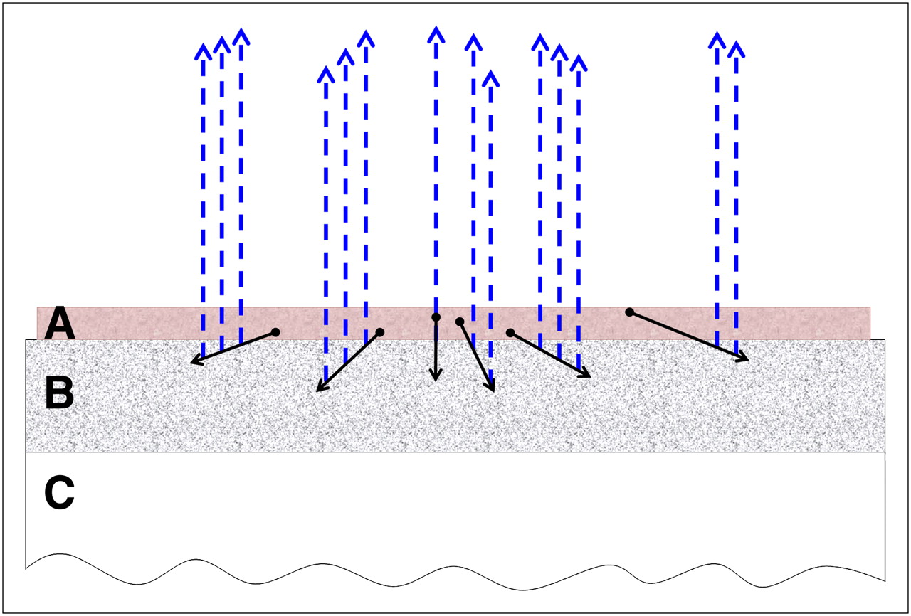
- FIGURE 1.
Schematized setup of sample configuration during imaging. Cryosections of tissues (A) were placed on scintillating layer (B) that was coated on clear polyester sheet (C). α-particles (black solid arrows) emitted in tissue hit scintillator and generate scintillation photons (blue dotted arrows), which traversed cryosection before being imaged.
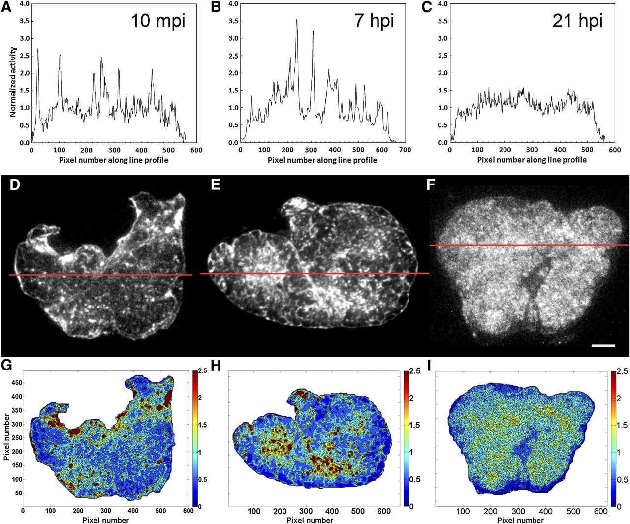
- FIGURE 2.
Activity distribution of tumor-specific 211At-MX35-F(ab′)2 in OVCAR-3 xenografts. (A–C) Pixel intensity values (normalized to line profile mean pixel intensity) of line profile plotted as function of pixel number along line. (D–F) α-Camera images taken 10 min, 7 h, and 21 h after bioconjugate injection. Red lines mark line profile positions used in A–C. (G–I) Activity distribution histograms for whole tumor. Intensity value for each pixel was divided by mean pixel intensity of whole tumor. Normalized data were divided into 10 bins between 0 and 2.5, and each level was plotted in representative color described by color-code bar. White scale bar corresponds 1,000 μm in D–F. hpi = hours after injection; mpi = minutes after injection.
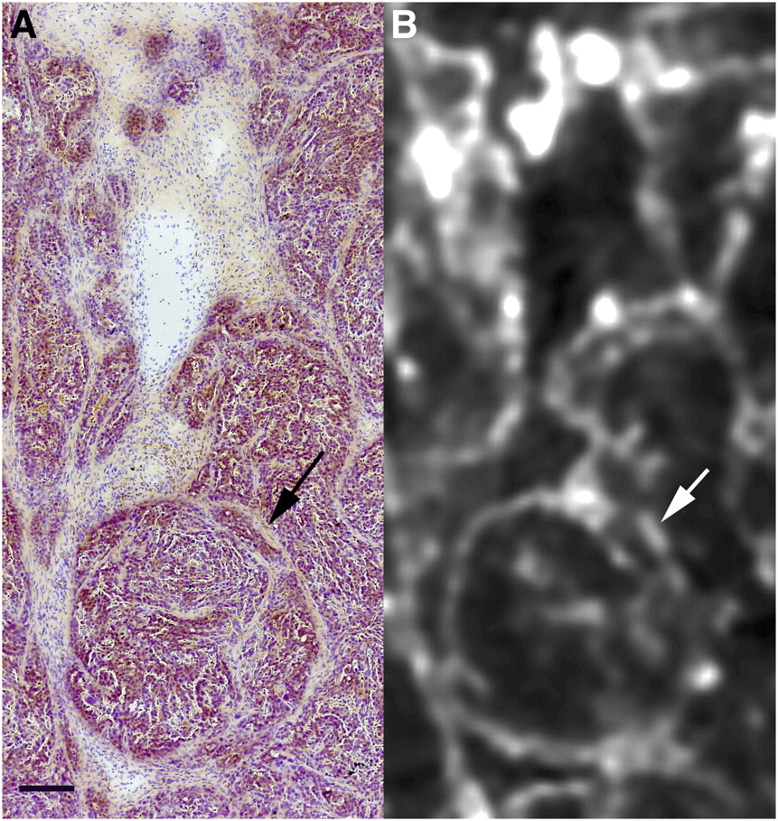
- FIGURE 3.
Overlay of α-camera image with H&E section. OVCAR-3 xenograft was imaged at 7 h after intravenous injection of tumor-specific 211At-MX35-F(ab′)2. (A) Area of H&E-stained section consecutive to α-camera images is shown in Figure 4. (B) Same area seen in α-camera image. White and black arrows indicate same region of stroma in tumor. Black bar indicates 200 μm.
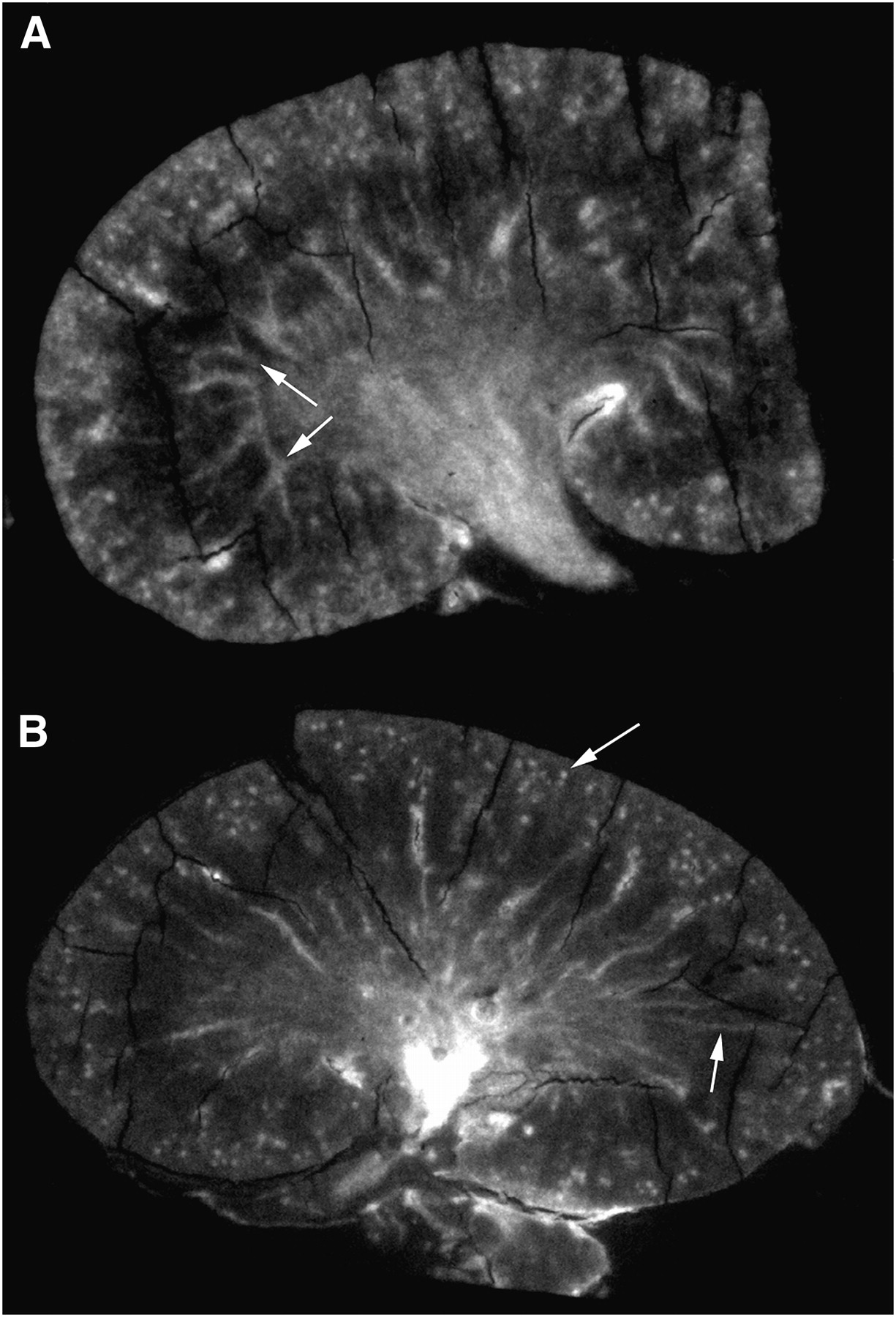
- FIGURE 4.
Cryosections of mice kidneys imaged with α-camera at 30 min (A) and 2 h (B) after intravenous injection of 211At-IgG trastuzumab. White arrows indicate vascular branches (A) and medullary rays and glomeruli (B).
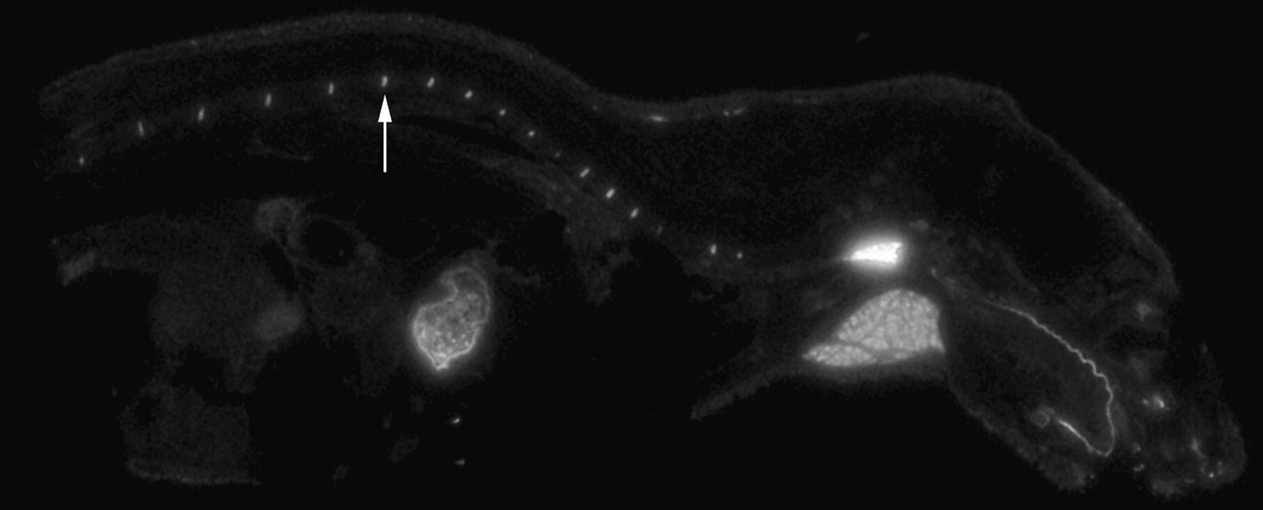
- FIGURE 5.
Whole-body mouse cryosection imaged with α-camera at 30 min after intravenous injection of free 211At. White arrow indicates cartilage in spine.

Tables
Supplemental Data
Files in this Data Supplement:


Jump to section
Related Articles
Cited By...
- Tumor Control Probability and Small-Scale Monte Carlo Dosimetry: Effects of Heterogenous Intratumoral Activity Distribution in Radiopharmaceutical Therapy
- Combination of Carriers with Complementary Intratumoral Microdistributions of Delivered {alpha}-Particles May Realize the Promise for 225Ac in Large, Solid Tumors
- Blind Image Restoration Enhances Digital Autoradiographic Imaging of Radiopharmaceutical Tissue Distribution
- Preclinical Evaluation of 213Bi- and 225Ac-Labeled Low-Molecular-Weight Compounds for Radiopharmaceutical Therapy of Prostate Cancer
- Evidence of Local Concentration of {alpha}-Particles from 211At-Labeled Antibodies in Liver Metastasis Tissue
- Cure of Human Ovarian Carcinoma Solid Xenografts by Fractionated {alpha}-Radioimmunotherapy with 211At-MX35-F(ab')2: Influence of Absorbed Tumor Dose and Effect on Long-Term Survival
- (2S)-2-(3-(1-Carboxy-5-(4-211At-Astatobenzamido)Pentyl)Ureido)-Pentanedioic Acid for PSMA-Targeted {alpha}-Particle Radiopharmaceutical Therapy
- {alpha}-Imaging Confirmed Efficient Targeting of CD45-Positive Cells After 211At-Radioimmunotherapy for Hematopoietic Cell Transplantation
- Astatine-211 conjugated to an anti-CD20 monoclonal antibody eradicates disseminated B-cell lymphoma in a mouse model
- Ex Vivo Activity Quantification in Micrometastases at the Cellular Scale Using the {alpha}-Camera Technique
- Anti-CD45 radioimmunotherapy using 211At with bone marrow transplantation prolongs survival in a disseminated murine leukemia model
- MIRD Pamphlet No. 23: Quantitative SPECT for Patient-Specific 3-Dimensional Dosimetry in Internal Radionuclide Therapy
- Anti-CD45 pretargeted radioimmunotherapy using bismuth-213: high rates of complete remission and long-term survival in a mouse myeloid leukemia xenograft model