


Article Figures & Data
Figures
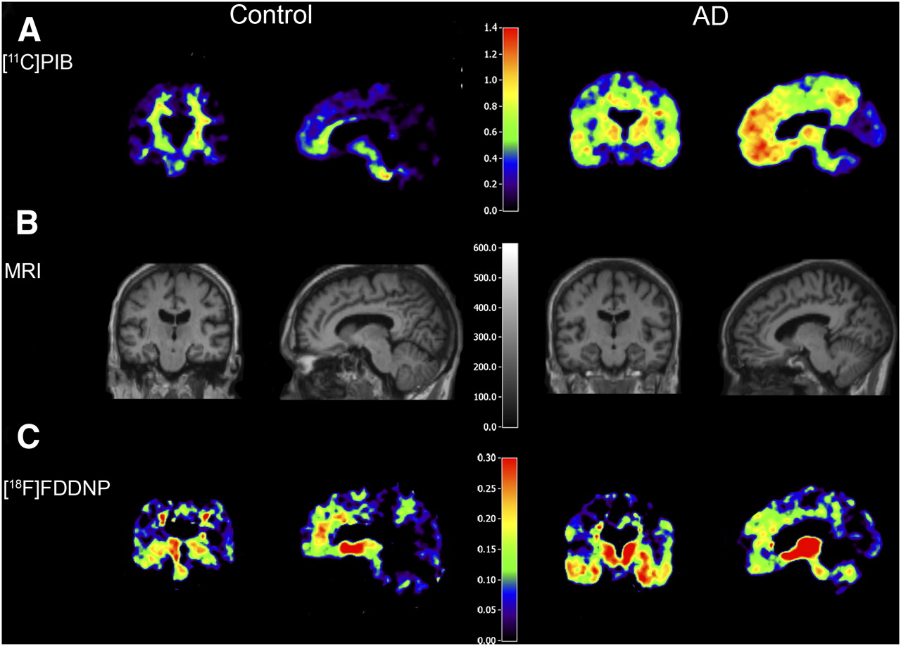
- FIGURE 1.
Examples of parametric coronal (left column) and sagittal (right column) 11C-PiB and 18F-FDDNP BPND images in control subject and AD patient with their coregistered MR image. 11C-PiB and 18F-FDDNP scans were obtained in same subjects. Corresponding CSF values for AD patient (60 y) were Aβ1–42, 504 pg/mL, and tau, 837 pg/mL. For the healthy control (61 y), CSF levels were Aβ1–42, 1,123 pg/mL, and tau, 245 pg/mL.
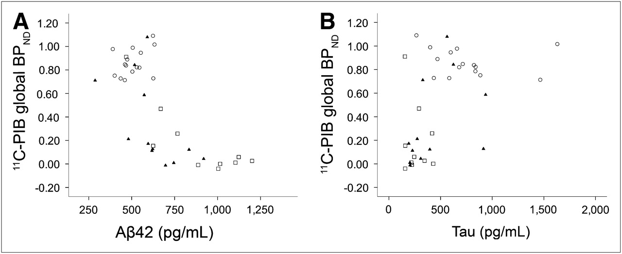
- FIGURE 2.
Scatter plots of 11C-PiB binding (global BPND) against CSF levels of Aβ1–42 (A) and tau (B). ○ are patients with AD, ▴ are patients with MCI, and □ are healthy controls. CSF values are shown as measured, whereas statistical analyses were performed on log-transformed values. Lower CSF levels of Aβ1–42 (A: r = −0.72, P < 0.001) and higher tau levels (B: r = 0.58, P < 0.001) were associated with higher global 11C-PiB binding.
- FIGURE 3.
Scatter plots of 18F-FDDNP binding (global BPND) against CSF levels of Aβ1–42 (A) and tau (B). ○ are patients with AD, ▴ are patients with MCI, and □ are healthy controls. CSF values are shown as measured, whereas statistical analyses were performed on log-transformed values. Lower CSF levels of Aβ1–42 (A: r = −0.37, P < 0.05) and higher tau levels (B: r = 0.56, P < 0.001) were associated with higher global 18F-FDDNP binding.


Tables
Characteristic Control MCI AD n 10 12 15 Age (y)* 70 ± 6 68 ± 9 63 ± 7 Sex (F/M) 3/7 2/10 6/9 MMSE† 29 ± 1 28 ± 2 24 ± 2‡§ Aβ1–42 (pg/mL)† 886 ± 244 623 ± 165§ 507 ± 78§ Tau (pg/mL)† 264 ± 104 431 ± 269 752 ± 371§|| 11C-PiB BPND Global† 0.18 ± 0.30 0.33 ± 0.37 0.86 ± 0.12‡§ Frontal 0.22 ± 0.37 0.37 ± 0.42 0.93 ± 0.11‡§ Medial temporal 0.08 ± 0.08 0.07 ± 0.06 0.15 ± 0.09||¶ Temporal 0.16 ± 0.27 0.31 ± 0.34 0.79 ± 0.11‡§ Posterior cingulate 0.17 ± 0.22 0.37 ± 0.38 0.84 ± 0.41‡§ Parietal 0.17 ± 0.30 0.34 ± 0.21 0.96 ± 0.20‡§ 18F-FDDNP BPND Global* 0.06 ± 0.03 0.08 ± 0.05 0.09 ± 0.02 Frontal* 0.05 ± 0.04 0.09 ± 0.06 0.09 ± 0.02 Medial temporal 0.12 ± 0.04 0.13 ± 0.05 0.14 ± 0.05 Temporal 0.07 ± 0.03 0.10 ± 0.05 0.10 ± 0.03 Posterior cingulate 0.04 ± 0.01 0.07 ± 0.02 0.06 ± 0.01 Parietal 0.02 ± 0.04 0.04 ± 0.04 0.06 ± 0.03 ANOVA with age as covariate:
↵* P < 0.10.
↵† P ≤ 0.001.
Post hoc least-significant difference tests:
↵‡ P ≤ 0.001, compared with MCI.
↵§ P ≤ 0.001, compared with controls.
↵|| P < 0.05, compared with MCI.
↵¶ P < 0.05, compared with controls.
Data are mean ± SD, where appropriate. Note that raw values are shown for CSF biomarkers (pg/mL) but that log-transformed values were used for statistical analysis.
MMSE = Mini-Mental State Examination.
CSF biomarker (pg/mL) Model 1 Model 2 Brain region Aβ1–42 Tau Aβ1–42 Tau 11C-PiB BPND Global 11C-PiB −0.72* 0.59* −0.50* 0.17 Frontal −0.75* 0.60* −0.56* 0.18 Medial temporal −0.42† 0.37† −0.15 0.03 Temporal −0.69* 0.57* −0.46* 0.17 Posterior cingulate −0.69* 0.52* −0.47† 0.12 Parietal −0.67* 0.57* −0.43† 0.14 18F-FDDNP BPND Global 18F-FDDNP −0.36† 0.59* −0.26 0.62† Frontal −0.36† 0.56* −0.27 0.62† Medial temporal −0.16 0.34‡ −0.07 0.33 Temporal −0.29‡ 0.56* −0.12 0.50† Posterior cingulate −0.12 0.33† −0.03 0.46† Parietal −0.36† 0.49† −0.33 0.44†


Jump to section
Related Articles
Cited By...
- Temporal Correlation of CSF and Neuroimaging in the Amyloid-Tau-Neurodegeneration Model of Alzheimer Disease
- Use of amyloid-PET to determine cutpoints for CSF markers: A multicenter study
- Detailed comparison of amyloid PET and CSF biomarkers for identifying early Alzheimer disease
- Longitudinal Change in CSF Biomarkers in Autosomal-Dominant Alzheimer's Disease
- Fluid Biomarkers in Alzheimer Disease
- CSF biomarkers for Alzheimer disease correlate with cortical brain biopsy findings
- Brain Imaging in Alzheimer Disease
- Cerebrospinal fluid biomarkers in the differential diagnosis of Alzheimer's disease from other cortical dementias
- Target-Specific PET Probes for Neurodegenerative Disorders Related to Dementia
- Differential association of [11C]PIB and [18F]FDDNP binding with cognitive impairment