


Article Figures & Data
Figures
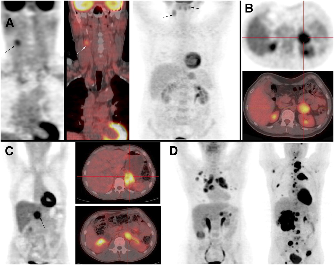
- FIGURE 1.
(A) Cervical paraganglioma (patients 13 and 12): Patient 13 is SDHB patient with right glomic paraganglioma (arrow), as seen on coronal 18F-FDG PET (left) and fusion imaging (middle). Patient 12 has bilateral cervical paraganglioma, as seen on coronal 18F-FDG PET (right). (B) Left pheochromocytoma (patient 3), as seen on axial 18F-FDG PET (top) and fusion imaging (bottom). (C) Abdominal nonmetastatic tumors (patients 7 and 10): Patient 7 has abdominal right paraganglioma, as seen on coronal 18F-FDG PET (left). Patient 10 is VHL patient with left pheochromocytoma (top right) and paraganglioma at left renal hilum (bottom right), as seen on axial fusion imaging. (D) Metastatic pheochromocytoma (patients 19 and 28): Patient 19 has recurrent pheochromocytoma, as seen on maximum-intensity-projection image (left). Patient 28 is SDHB patient with metastatic pheochromocytoma, as seen on maximum-intensity-projection image (right).
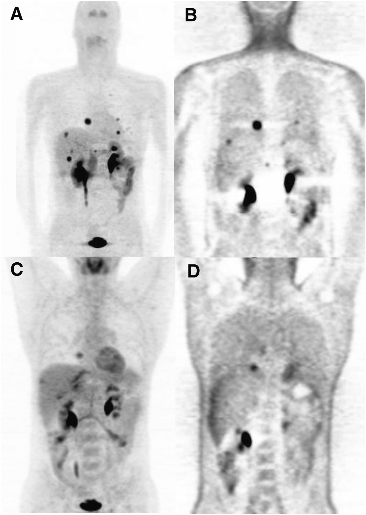
- FIGURE 2.
Metastatic SDHB-related pheochromocytoma (patient 27): multiple abdominal paraganglioma seen on axial CT (top), axial 6-18F-fluorodopa PET (middle), and axial 18F-FDG PET (bottom) (A); 6-18F-fluorodopa PET (coronal whole-body axial images) (B); and 18F-FDG PET (coronal whole-body axial images) (C). Compared with 6-18F-fluorodopa PET, 18F-FDG PET detected additional tumor sites and 18F-FDG PET avidity was higher (arrow).
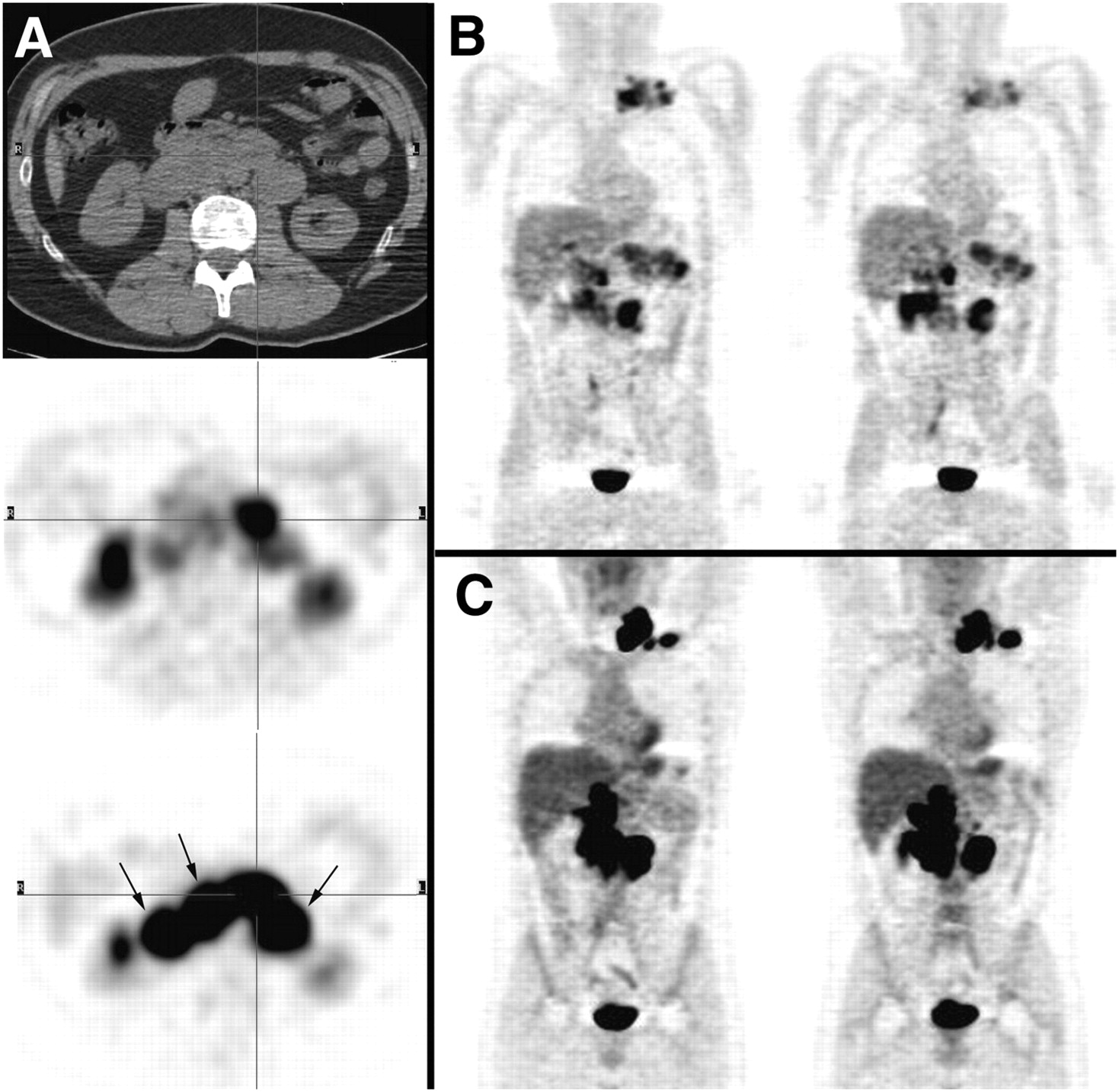
- FIGURE 3.
Metastatic sporadic pheochromocytoma (patient 23): 6-18F-fluorodopa PET maximum-intensity-projection image (A), 18F-FDG PET maximum-intensity-projection image (B), attenuation-uncorrected 6-18F-fluorodopa PET coronal image (C), and attenuation-uncorrected 18F-FDG PET coronal image (D). Compared with 6-18F-fluorodopa PET, 18F-FDG PET underestimated extent of disease.
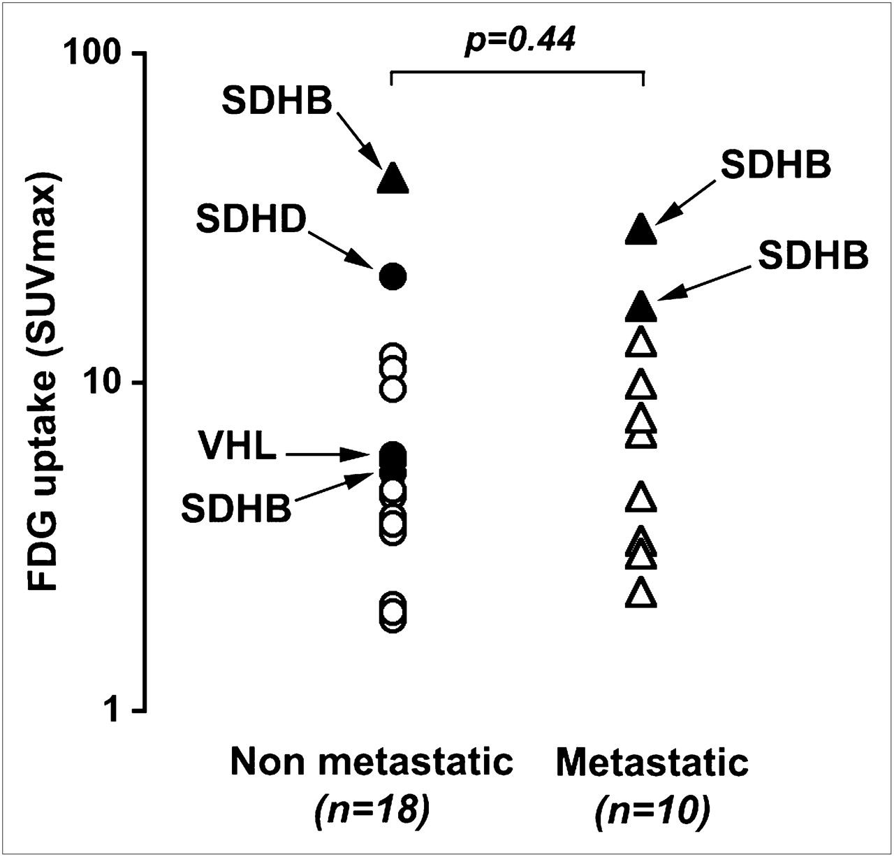
- FIGURE 4.
Comparison of SUVmax between nonmetastatic and metastatic tumors. Distributed values of SUVmax are represented. Mean SUVmax was not significantly different between tumors (P = 0.44). SDHB-related tumors were notable in being most 18F-FDG–avid tumors (SUVmax, 42, 29.3, 21, and 17).

Tables
Patient no. Initial presentation Age (y) Final status 18F-FDG PET foci Molecular imaging technique Additional foci on 18F-FDG PET, compared with…* Germline mutation Indication Sex VA SUVmax SPECT FDOPA 1 Pheo Initial staging F 58 Unifocal Adrenal Pos 3.5 MIBG No (=) ND Absent 2 Pheo Initial staging F 30 Unifocal Adrenal Pos 12 MIBG No (=) ND Absent 3 Pheo Initial staging M 38 Unifocal Adrenal Pos 11 FDOPA ND No (=) Absent 4 Pheo Initial staging M 30 Unifocal Adrenal Neg 2.1 MIBG, FDOPA No (=) No (<) Absent 5 Pheo Initial staging F 26 Unifocal Adrenal Pos 9.5 MIBG No (=) ND Absent 6 Ab PGL Initial staging M 64 Unifocal Ab PGL Pos 4.5 MIBG No (=) ND Absent 7 Ab PGL Initial staging F 52 Unifocal Ab PGL Pos 5.7 MIBG, FDOPA, SRS No (=) No (=) Absent 8 Pheo Initial staging F 30 Unifocal Adrenal Pos 3.8 FDOPA — ND Absent 9 Pheo Initial staging M 34 Unifocal Adrenal Pos 42 MIBG No (=) ND SDHB 10 Pheo Recurrence M 25 Multifocal Adrenal, ab PGL (renal pedicle) Pos 6 MIBG, FDOPA, SRS Yes No (=) VHL 11 Pheo + PGL Initial staging M 34 Multifocal Ab and ce PGL (gl) Pos 21 MIBG, FDOPA, SRS Yes Yes SDHD 12 Ce PGL Initial staging M 67 Multifocal Ce PGL (gl + vagal) Pos 3.5 SRS, FDOPA No (=) No (=) Absent 13 Pheo Recurrence M 70 Multifocal Ce PGL (gl) Pos 5.3 MIBG, FDOPA, SRS No (=) No (=) SDHB 14 Pheo + PGL Recurrence M 56 Multifocal Ab PGL Pos 3.9 MIBG Yes ND ND 15 Pheo Initial staging M 36 Multifocal Adrenal (bilateral) Pos 3.7 MIBG No (=) ND Absent 16 Pheo Initial staging M 51 Unifocal Adrenal Pos 4.7 MIBG No (=) ND NF1 17 Pheo Initial staging M 45 Unifocal Adrenal Neg 1.9 MIBG, FDOPA No (=) No (<) NF1 18 Pheo Recurrence M 35 Multifocal Adrenal Pos 2 MIBG No (=) ND RET 19 Pheo Recurrence F 61 Metastatic Lung, me, carcinosis Pos 4.5 MIBG, FDOPA Yes No (=) Absent 20 Pheo Recurrence M 75 Metastatic Adrenal, Lung, me, liver, LN Pos 13.3 MIBG, FDOPA Yes Yes Absent 21 Pheo Recurrence F 32 Metastatic Lung, liver, bone Pos 2.3 MIBG, FDOPA Yes No (<) Absent 22 Pheo Recurrence M 34 Metastatic Liver, bone Pos 9.9 MIBG, FDOPA, HMDP No (<) No (<) Absent 23 Pheo Recurrence M 43 Metastatic Lung, liver, LN Pos 3.3 MIBG, FDOPA, HMDP Yes No (<) Absent 24 PGL Recurrence M 78 Metastatic Ab PGL, bone Pos 7 MIBG, FDOPA, HMDP Yes Yes Absent 25 PGL Recurrence F 59 Metastatic Lung, LN, Bone Pos 3 MIBG, FDOPA, HMDP, SRS Yes No (<) Absent 26 Pheo Initial staging M 63 Metastatic Adrenal, Bone Pos 7.8 MIBG, FDOPA Yes No (<) ND 27 PGL Recurrence M 38 Metastatic Ab and ce PGL, LN Pos 29.3 MIBG, FDOPA Yes Yes SDHB 28 Pheo Initial staging M 34 Metastatic Adrenal, Bone Pos 17 MIBG, HMDP Yes ND SDHB VA = visual analysis; FDOPA = 6-18F-fluorodopa PET; pheo = pheochromocytoma; pos = positive; MIBG = 131I-MIBG; ND = not done; ab = abdominal; PGL = paraganglioma; SRS = somatostatin receptor scintigraphy; ce = cervical; gl = glomic; neg = negative; RET = RET protooncogene; me = mediastinal; LN = lymph node; HMDP = 99mTc-HMDP; HMDP = hydroxymethylenediphosphonate.
↵* Comparison between 18F-FDG PET and SPECT (131I-metaiodobenzylguanidine [MIBG] or SRS) and 6-18F-fluorodopa PET studies in terms of number of tumor sites. = indicates equivalent number of tumor sites on 18F-FDG PET; < indicates lower number of tumor sites on 18F-FDG PET; > indicates higher number of tumor sites on 18F-FDG PET.
In this issue


Jump to section
Related Articles
Cited By...
- Metabolic Subtyping of Pheochromocytoma and Paraganglioma by 18F-FDG Pharmacokinetics Using Dynamic PET/CT Scanning
- 18F-FLT PET/CT in the Evaluation of Pheochromocytomas and Paragangliomas: A Pilot Study
- Magnetic resonance spectroscopy of paragangliomas: new insights into in vivo metabolomics
- 15 YEARS OF PARAGANGLIOMA: Imaging and imaging-based treatment of pheochromocytoma and paraganglioma
- Current views on cell metabolism in SDHx-related pheochromocytoma and paraganglioma
- Assessment of incidental and clinically unsuspected fluorodeoxyglucose-avid foci detected on oncological positron emission tomography/CT
- Molecular and Therapeutic Advances in the Diagnosis and Management of Malignant Pheochromocytomas and Paragangliomas
- First Report of Harlequin Syndrome as the Presenting Feature of Carney Triad: A Diagnostic and Imaging Challenge
- Modern Nuclear Imaging for Paragangliomas: Beyond SPECT
- Expression of somatostatin receptors, dopamine D2 receptors, noradrenaline transporters, and vesicular monoamine transporters in 52 pheochromocytomas and paragangliomas