
Main menu

  • Home
  • Content
    • Current
    • Ahead of print
    • Past Issues
    • JNM Supplement
    • SNMMI Annual Meeting Abstracts
    • Continuing Education
    • JNM Podcasts
  • Subscriptions
    • Subscribers
    • Institutional and Non-member
    • Rates
    • Journal Claims
    • Corporate & Special Sales
  • Authors
    • Submit to JNM
    • Information for Authors
    • Assignment of Copyright
    • AQARA requirements
  • Info
    • Reviewers
    • Permissions
    • Advertisers
  • About
    • About Us
    • Editorial Board
    • Contact Information
  • More
    • Alerts
    • Feedback
    • Help
    • SNMMI Journals
  • SNMMI
    • JNM
    • JNMT
    • SNMMI Journals
    • SNMMI

User menu

  • Subscribe
  • My alerts
  • Log in
  • Log out
  • My Cart

Search

  • Advanced search
Journal of Nuclear Medicine
  • SNMMI
    • JNM
    • JNMT
    • SNMMI Journals
    • SNMMI
  • Subscribe
  • My alerts
  • Log in
  • Log out
  • My Cart
Journal of Nuclear Medicine

Advanced Search

  • Home
  • Content
    • Current
    • Ahead of print
    • Past Issues
    • JNM Supplement
    • SNMMI Annual Meeting Abstracts
    • Continuing Education
    • JNM Podcasts
  • Subscriptions
    • Subscribers
    • Institutional and Non-member
    • Rates
    • Journal Claims
    • Corporate & Special Sales
  • Authors
    • Submit to JNM
    • Information for Authors
    • Assignment of Copyright
    • AQARA requirements
  • Info
    • Reviewers
    • Permissions
    • Advertisers
  • About
    • About Us
    • Editorial Board
    • Contact Information
  • More
    • Alerts
    • Feedback
    • Help
    • SNMMI Journals
  • View or Listen to JNM Podcast
  • Visit JNM on Facebook
  • Join JNM on LinkedIn
  • Follow JNM on Twitter
  • Subscribe to our RSS feeds
OtherBASIC SCIENCE INVESTIGATIONS

PET Imaging of Prostate Cancer Xenografts with a Highly Specific Antibody against the Prostate-Specific Membrane Antigen

Ursula Elsässer-Beile, Gerald Reischl, Stefan Wiehr, Patrick Bühler, Philipp Wolf, Karen Alt, John Shively, Martin S. Judenhofer, Hans-Jürgen Machulla and Bernd J. Pichler
Journal of Nuclear Medicine April 2009, 50 (4) 606-611; DOI: https://doi.org/10.2967/jnumed.108.058487
Ursula Elsässer-Beile
  • Find this author on Google Scholar
  • Find this author on PubMed
  • Search for this author on this site
Gerald Reischl
  • Find this author on Google Scholar
  • Find this author on PubMed
  • Search for this author on this site
Stefan Wiehr
  • Find this author on Google Scholar
  • Find this author on PubMed
  • Search for this author on this site
Patrick Bühler
  • Find this author on Google Scholar
  • Find this author on PubMed
  • Search for this author on this site
Philipp Wolf
  • Find this author on Google Scholar
  • Find this author on PubMed
  • Search for this author on this site
Karen Alt
  • Find this author on Google Scholar
  • Find this author on PubMed
  • Search for this author on this site
John Shively
  • Find this author on Google Scholar
  • Find this author on PubMed
  • Search for this author on this site
Martin S. Judenhofer
  • Find this author on Google Scholar
  • Find this author on PubMed
  • Search for this author on this site
Hans-Jürgen Machulla
  • Find this author on Google Scholar
  • Find this author on PubMed
  • Search for this author on this site
Bernd J. Pichler
  • Find this author on Google Scholar
  • Find this author on PubMed
  • Search for this author on this site
  • Article
  • Figures & Data
  • Info & Metrics
  • PDF
Loading

Article Figures & Data

Figures

  • FIGURE 1. 
    • Download figure
    • Open in new tab
    • Download powerpoint
    FIGURE 1. 

    Binding of 3/A12 mAb and DOTA-3/A12 mAb, with and without serum preincubation, to PSMA-positive C4-2 cells. Cells were treated with increasing concentrations (0.15–800 nM) of first-step anti-PSMA mAb followed by incubation with saturating amount of second-step phycoerythrin-labeled goat antimouse IgG followed by cytofluorometric analysis. MFI = mean fluorescence intensity.

  • FIGURE 2. 
    • Download figure
    • Open in new tab
    • Download powerpoint
    FIGURE 2. 

    Blood immunoreactivity of 3/A12, DOTA-3/A12, and copper-DOTA-3/A12 in SCID mice after intravenous injection of 25 μg of mAb. Serum levels are given as mean fluorescence intensity (MFI) measured by flow cytometry.
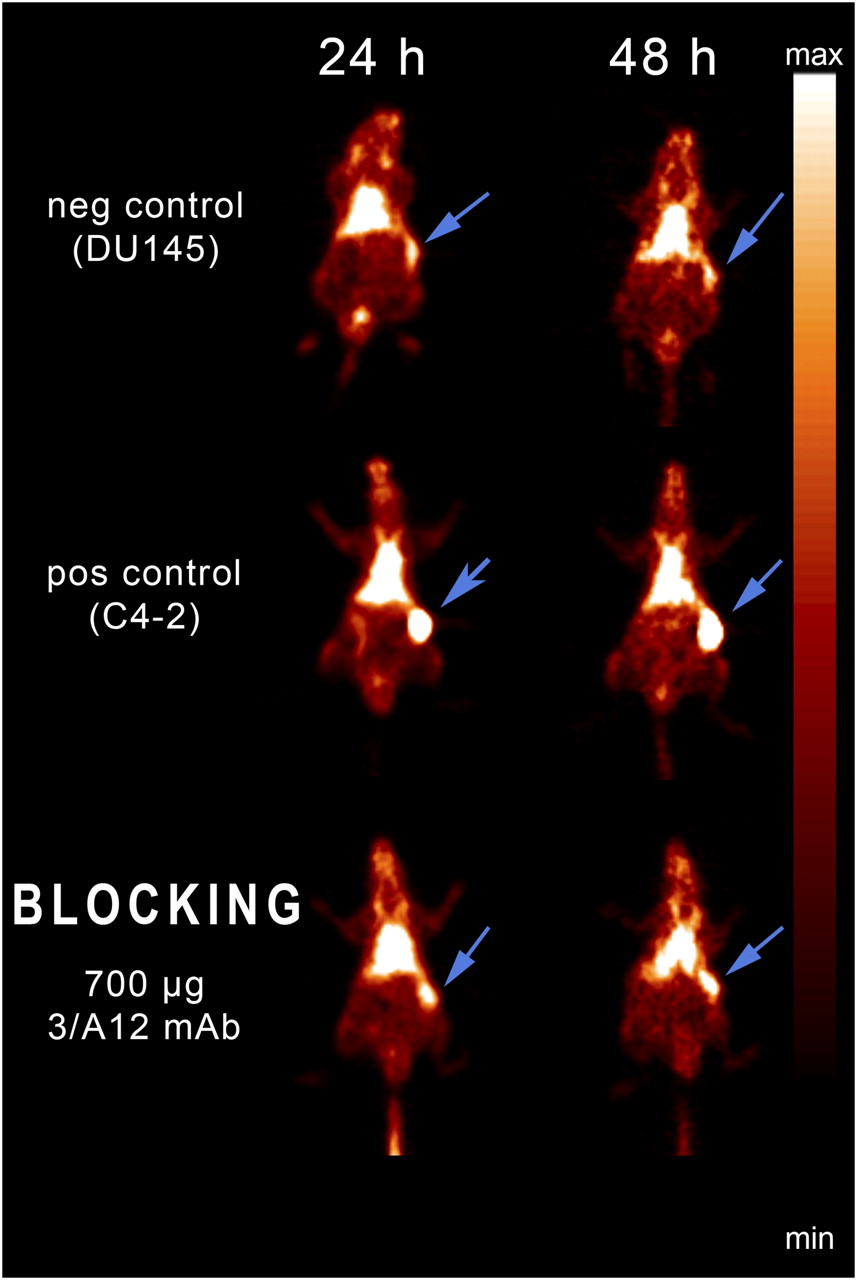
  • FIGURE 3. 
    • Download figure
    • Open in new tab
    • Download powerpoint
    FIGURE 3. 

    Small-animal PET imaging of mice bearing PSMA-positive C4-2 tumors (pos control, C4-2) and PSMA-negative DU 145 tumors (neg control, DU145) 24 and 48 h after injection of 64Cu-DOTA-3/A12 mAb. Blocking experiments were performed by injecting unlabeled 3/A12 mAb 3 h before injection of 64Cu-labeled mAb (blocking, 700 μg of 3/A12 mAb). Arrows indicate position of tumors.

  • FIGURE 4. 
    • Download figure
    • Open in new tab
    • Download powerpoint
    FIGURE 4. 

    Blocking of 64Cu-DOTA-3/A12 mAb uptake in C4-2 tumors with 250, 500, or 700 μg of nonradioactive 3/A12 mAb, given 3 h before radiotracer injection. %ID/cm3 from mice bearing DU 145 tumors (negative control) or C4-2 tumors (positive control) 3, 24, and 48 h after injection of 64Cu-DOTA-3/A12 are shown.

  • FIGURE 5. 
    • Download figure
    • Open in new tab
    • Download powerpoint
    FIGURE 5. 

    Biodistribution by γ-counting of 64Cu-DOTA-3/A12 in various organs of mice 48 h after injection.

PreviousNext
Back to top

In this issue

Journal of Nuclear Medicine: 50 (4)
Journal of Nuclear Medicine
Vol. 50, Issue 4
April 2009
  • Table of Contents
  • Table of Contents (PDF)
  • About the Cover
  • Index by author
Print
Download PDF
Article Alerts
Sign In to Email Alerts with your Email Address
Email Article

Thank you for your interest in spreading the word on Journal of Nuclear Medicine.

NOTE: We only request your email address so that the person you are recommending the page to knows that you wanted them to see it, and that it is not junk mail. We do not capture any email address.

Enter multiple addresses on separate lines or separate them with commas.
PET Imaging of Prostate Cancer Xenografts with a Highly Specific Antibody against the Prostate-Specific Membrane Antigen
(Your Name) has sent you a message from Journal of Nuclear Medicine
(Your Name) thought you would like to see the Journal of Nuclear Medicine web site.
Citation Tools
PET Imaging of Prostate Cancer Xenografts with a Highly Specific Antibody against the Prostate-Specific Membrane Antigen
Ursula Elsässer-Beile, Gerald Reischl, Stefan Wiehr, Patrick Bühler, Philipp Wolf, Karen Alt, John Shively, Martin S. Judenhofer, Hans-Jürgen Machulla, Bernd J. Pichler
Journal of Nuclear Medicine Apr 2009, 50 (4) 606-611; DOI: 10.2967/jnumed.108.058487

Citation Manager Formats

  • BibTeX
  • Bookends
  • EasyBib
  • EndNote (tagged)
  • EndNote 8 (xml)
  • Medlars
  • Mendeley
  • Papers
  • RefWorks Tagged
  • Ref Manager
  • RIS
  • Zotero
Share
PET Imaging of Prostate Cancer Xenografts with a Highly Specific Antibody against the Prostate-Specific Membrane Antigen
Ursula Elsässer-Beile, Gerald Reischl, Stefan Wiehr, Patrick Bühler, Philipp Wolf, Karen Alt, John Shively, Martin S. Judenhofer, Hans-Jürgen Machulla, Bernd J. Pichler
Journal of Nuclear Medicine Apr 2009, 50 (4) 606-611; DOI: 10.2967/jnumed.108.058487
Twitter logo Facebook logo LinkedIn logo Mendeley logo
  • Tweet Widget
  • Facebook Like
  • Google Plus One
Bookmark this article

Jump to section

  • Article
    • Abstract
    • MATERIALS AND METHODS
    • RESULTS
    • DISCUSSION
    • CONCLUSION
    • Footnotes
    • References
  • Figures & Data
  • Info & Metrics
  • PDF

Related Articles

  • This Month in JNM
  • Erratum
  • PubMed
  • Google Scholar

Cited By...

  • A Novel Fully Human Antibody targeting Extracellular Domain of PSMA Inhibits Tumor Growth in Prostate Cancer
  • Evaluation of 111In-DOTA-5D3, a Surrogate SPECT Imaging Agent for Radioimmunotherapy of Prostate-Specific Membrane Antigen
  • Phase 2 Study of 99mTc-Trofolastat SPECT/CT to Identify and Localize Prostate Cancer in Intermediate- and High-Risk Patients Undergoing Radical Prostatectomy and Extended Pelvic LN Dissection
  • Optimization of Labeling PSMAHBED with Ethanol-Postprocessed 68Ga and Its Quality Control Systems
  • ImmunoPET/MR imaging allows specific detection of Aspergillus fumigatus lung infection in vivo
  • In Vivo Evaluation of 11C-DASB for Quantitative SERT Imaging in Rats and Mice
  • Biodistribution, Tumor Detection, and Radiation Dosimetry of 18F-DCFBC, a Low-Molecular-Weight Inhibitor of Prostate-Specific Membrane Antigen, in Patients with Metastatic Prostate Cancer
  • Advances in Immuno-Positron Emission Tomography: Antibodies for Molecular Imaging in Oncology
  • 2-(3-{1-Carboxy-5-[(6-[18F]Fluoro-Pyridine-3-Carbonyl)-Amino]-Pentyl}-Ureido)-Pentanedioic Acid, [18F]DCFPyL, a PSMA-Based PET Imaging Agent for Prostate Cancer
  • Targeting Prostate Cancer Cells In Vivo Using a Rapidly Internalizing Novel Human Single-Chain Antibody Fragment
  • New Agents and Techniques for Imaging Prostate Cancer
  • Google Scholar

More in this TOC Section

  • Design and Fabrication of Kidney Phantoms for Internal Radiation Dosimetry Using 3D Printing Technology
  • Synthesis and Biologic Evaluation of Novel 18F-Labeled Probes Targeting Prostate-Specific Membrane Antigen for PET of Prostate Cancer
  • Tumor-Specific Binding of Radiolabeled PEGylated GIRLRG Peptide: A Novel Agent for Targeting Cancers
Show more Basic Science Investigations

Similar Articles

SNMMI

© 2025 SNMMI

Powered by HighWire