


Article Figures & Data
Figures
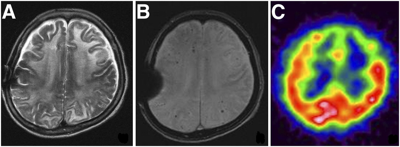
- FIGURE 1.
A 68-y-old woman with progressive cognitive decline. (A) Diffuse high signal intensities are noted along subcortical white matter of bilateral frontoparietal lobes on T2-weighted images. (B) GRE sequence revealed multiple petechic hypointense signal intensities along with cortical–subcortical distribution. Focal susceptibility artifact is shown at location of right parietal skull due to previous surgery. (C) Focal perfusion defects are demonstrated in anterior portions of both frontal lobes using 99mTc-ECD SPECT.
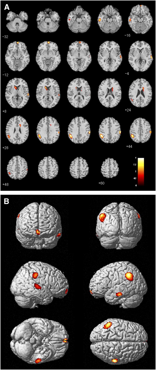
- FIGURE 2.
Results from group comparisons of patients with CAA and healthy controls: areas with decreased regional CBF. (A) Transaxial MR fused images. (B) Three-dimensional-rendered images.
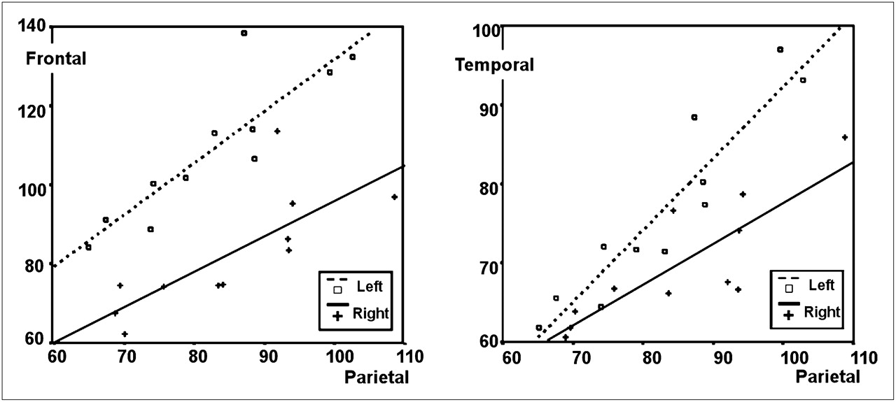
- FIGURE 3.
Correlation of regional blood flow between parietal and frontal cortices and between parietal and temporal cortices. Numbers of counts per voxel by SPAM are compared between bilateral frontal and parietal cortices and between bilateral temporal and parietal cortices and plotted separately. Both scatter plots demonstrate positive correlation (frontal and parietal regions, r = 0.83; temporal and parietal regions, r = 0.90).

Tables
Patient no. Sex Age (y) Symptoms on admission MMSE score Vessel stenosis Number of MBs Final diagnosis 1 F 60 Gait disturbance 26 — 15 CAA 2 F 76 Memory impairment 19 20% (L MCA M1) 30 CAA with vascular dementia 3 M 75 Mental confusion 25 — 45 CAA with seizure disorder 4 M 58 Headache 29 — 200 CAA with lacunar infarct 5 M 78 Motor weakness 24 10% (R MCA M1), 20% (L PCA P2) 22 CAA 6 M 70 Headache 24 — 10 CAA with lacunar infarct 7 F 63 Speaking difficulty 22 — 336 CAA with seizure disorder 8 F 78 Memory impairment 25 — 18 CAA with Alzheimer disease 9 F 68 Motor weakness, visual change 24 20% (L MCA M2) 13 CAA 10 F 71 Headache, motor weakness 24 — 98 CAA 11 M 73 Headache 25 — 104 CAA MMSE = Mini-Mental State Examination; MB = microbleed; MCA = middle cerebral artery; PCA = posterior cerebral artery.
- TABLE 2
Areas with Significantly Decreased Regional CBF in CAA Patients, Compared with Controls
Expected voxels per cluster t Z x y z Brain areas 672 6.44 4.58 −48 −50 40 Left parietal lobe, inferior parietal lobule, BA 40 672 5.13 3.98 −50 −60 24 Left temporal lobe, middle temporal gyrus, BA 39 246 6.32 4.53 62 −28 38 Right parietal lobe, postcentral gyrus 246 5.21 4.02 56 −36 44 Right parietal lobe, inferior parietal lobule, BA 40 172 6.14 4.45 62 −18 2 Right temporal lobe, superior temporal gyrus, BA 22 187 6.03 4.41 6 60 −10 Right frontal lobe, superior frontal gyrus, BA 10 193 5.72 4.26 −62 −22 −18 Left temporal lobe, inferior temporal gyrus, BA 20 203 4.91 3.85 14 0 20 Right caudate body 167 4.74 3.77 −8 0 12 Left caudate body Height threshold: t = 4.65, P = 0.0001; extent threshold: k = 100 voxels.


Jump to section
Related Articles
Cited By...
- Loss of spontaneous vasomotion precedes impaired cerebrovascular reactivity and microbleeds in a mouse model of cerebral amyloid angiopathy
- Atrophy patterns in cerebral amyloid angiopathy with and without cortical superficial siderosis
- Chronic cerebral hypoperfusion: a key mechanism leading to vascular cognitive impairment and dementia. Closing the translational gap between rodent models and human vascular cognitive impairment and dementia