
Main menu

  • Home
  • Content
    • Current
    • Ahead of print
    • Past Issues
    • JNM Supplement
    • SNMMI Annual Meeting Abstracts
    • Continuing Education
    • JNM Podcasts
  • Subscriptions
    • Subscribers
    • Institutional and Non-member
    • Rates
    • Journal Claims
    • Corporate & Special Sales
  • Authors
    • Submit to JNM
    • Information for Authors
    • Assignment of Copyright
    • AQARA requirements
  • Info
    • Reviewers
    • Permissions
    • Advertisers
  • About
    • About Us
    • Editorial Board
    • Contact Information
  • More
    • Alerts
    • Feedback
    • Help
    • SNMMI Journals
  • SNMMI
    • JNM
    • JNMT
    • SNMMI Journals
    • SNMMI

User menu

  • Subscribe
  • My alerts
  • Log in
  • My Cart

Search

  • Advanced search
Journal of Nuclear Medicine
  • SNMMI
    • JNM
    • JNMT
    • SNMMI Journals
    • SNMMI
  • Subscribe
  • My alerts
  • Log in
  • My Cart
Journal of Nuclear Medicine

Advanced Search

  • Home
  • Content
    • Current
    • Ahead of print
    • Past Issues
    • JNM Supplement
    • SNMMI Annual Meeting Abstracts
    • Continuing Education
    • JNM Podcasts
  • Subscriptions
    • Subscribers
    • Institutional and Non-member
    • Rates
    • Journal Claims
    • Corporate & Special Sales
  • Authors
    • Submit to JNM
    • Information for Authors
    • Assignment of Copyright
    • AQARA requirements
  • Info
    • Reviewers
    • Permissions
    • Advertisers
  • About
    • About Us
    • Editorial Board
    • Contact Information
  • More
    • Alerts
    • Feedback
    • Help
    • SNMMI Journals
  • View or Listen to JNM Podcast
  • Visit JNM on Facebook
  • Join JNM on LinkedIn
  • Follow JNM on Twitter
  • Subscribe to our RSS feeds
Research ArticleCLINICAL INVESTIGATIONS

In Vivo Characterization of Proliferation for Discriminating Cancer from Pancreatic Pseudotumors

Ken Herrmann, Florian Eckel, Stefan Schmidt, Klemens Scheidhauer, Bernd Joachim Krause, Joerg Kleeff, Tibor Schuster, Hans-Juergen Wester, Helmut Friess, Roland M. Schmid, Markus Schwaiger and Andreas K. Buck
Journal of Nuclear Medicine September 2008, 49 (9) 1437-1444; DOI: https://doi.org/10.2967/jnumed.108.052027
Ken Herrmann
  • Find this author on Google Scholar
  • Find this author on PubMed
  • Search for this author on this site
Florian Eckel
  • Find this author on Google Scholar
  • Find this author on PubMed
  • Search for this author on this site
Stefan Schmidt
  • Find this author on Google Scholar
  • Find this author on PubMed
  • Search for this author on this site
Klemens Scheidhauer
  • Find this author on Google Scholar
  • Find this author on PubMed
  • Search for this author on this site
Bernd Joachim Krause
  • Find this author on Google Scholar
  • Find this author on PubMed
  • Search for this author on this site
Joerg Kleeff
  • Find this author on Google Scholar
  • Find this author on PubMed
  • Search for this author on this site
Tibor Schuster
  • Find this author on Google Scholar
  • Find this author on PubMed
  • Search for this author on this site
Hans-Juergen Wester
  • Find this author on Google Scholar
  • Find this author on PubMed
  • Search for this author on this site
Helmut Friess
  • Find this author on Google Scholar
  • Find this author on PubMed
  • Search for this author on this site
Roland M. Schmid
  • Find this author on Google Scholar
  • Find this author on PubMed
  • Search for this author on this site
Markus Schwaiger
  • Find this author on Google Scholar
  • Find this author on PubMed
  • Search for this author on this site
Andreas K. Buck
  • Find this author on Google Scholar
  • Find this author on PubMed
  • Search for this author on this site
  • Article
  • Figures & Data
  • Info & Metrics
  • PDF
Loading

Article Figures & Data

Figures

  • Tables
  • FIGURE 1. 
    • Download figure
    • Open in new tab
    • Download powerpoint
    FIGURE 1. 

    Scatter plot of mean 18F-FLT SUV in pancreatic cancer with focal 18F-FLT uptake (PET-positive), in pancreatic cancer negative on visual interpretation (PET-negative), and in benign pancreatic lesions (PET-negative). MFP = mass-forming pancreatitis; PC = pancreatic cancer.
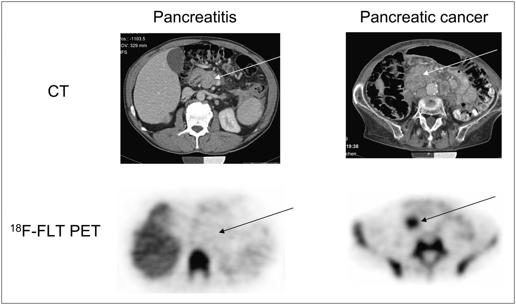
  • FIGURE 2. 
    • Download figure
    • Open in new tab
    • Download powerpoint
    FIGURE 2. 

    Spiral CT and 18F-FLT PET of patient 21, with CP, and patient 23, with pancreatic cancer. Physiologically increased 18F-FLT uptake is seen in bone marrow and liver, but only background activity of 18F-FLT is seen in area of inflammatory pancreatic mass (true-negative). Focal 18F-FLT uptake is seen in pancreatic carcinoma (true-positive).
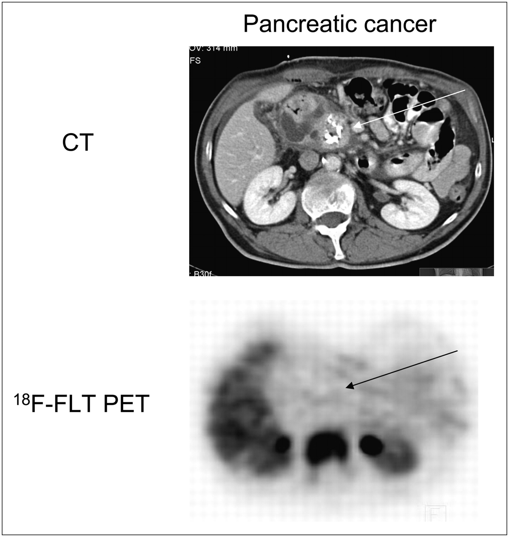
  • FIGURE 3. 
    • Download figure
    • Open in new tab
    • Download powerpoint
    FIGURE 3. 

    Spiral CT and 18F-FLT PET of patient 18, with pancreatic cancer, and false-negative findings on 18F-FLT PET. Physiologic 18F-FLT uptake is seen in bone marrow and liver. Histology indicated T1 adenocarcinoma.
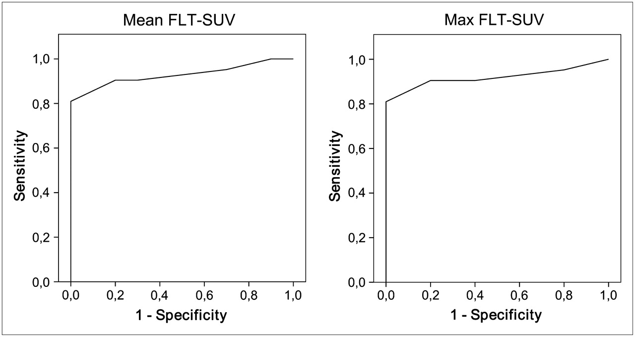
  • FIGURE 4. 
    • Download figure
    • Open in new tab
    • Download powerpoint
    FIGURE 4. 

    Receiver-operating-characteristic analysis of 18F-FLT PET for discriminating cancer from benign pancreatic lesions. Area under curve is 0.93 using cutoff of 1.8 for mean SUV or 0.92 using cutoff of 2.1 for maximal SUV.

Tables

  • Figures
    • View popup
    TABLE 1

    Tumor Characteristics, Lesion Location, 18F-FLT PET Findings, Reference Method, and Clinical Consensus

    Patient no.Lesion location18F-FLT mean SUV18F-FLT maximal SUV18F-FLT PET visual scoreReference methodClinical consensus
    1Head3.64.21Met, CFU1
    2Head1.21.50Benign pancreatic tissue (C), CFU0
    3Tail3.23.51Adenocarcinoma (H)1
    4Head1.61.80Benign pancreatic tissue (C), CFU0
    5Head3.74.41Adenocarcinoma (H)1
    6Tail2.83.51Adenocarcinoma (H)1
    7Head1.72.00Benign pancreatic tissue (H)0
    8Head/corpus1.41.70Benign pancreatic tissue (H)0
    9Head1.41.60Benign pancreatic tissue (C), CFU0
    10Head6.57.31Squamous cell carcinoma (H)1
    11Head2.63.11Adenocarcinoma (H)1
    12Head8.59.81Undifferentiated adenocarcinoma (H)1
    13Head1.31.60Benign pancreatic tissue (H)0
    14Head2.42.61Neuroendocrine carcinoma (H)1
    15Head2.83.81Adenocarcinoma (H)1
    16Corpus1.72.00Adenocarcinoma (H)1
    17Head2.02.20Cystadenocarcinoma (H)1
    18Head1.31.50Adenocarcinoma (H)1
    19Head3.43.91Adenocarcinoma (H)1
    20Corpus1.92.30Adenocarcinoma (H)1
    21Corpus1.31.50CFU0
    22Head1.41.60Benign pancreatic tissue (H)0
    23Head4.95.11Adenocarcinoma (H)1
    24Head1.71.90Benign pancreatic tissue (C), CFU0
    25Head1.41.60Benign pancreatic tissue (C), CFU0
    26Corpus2.63.01Adenocarcinoma (H)1
    27Head3.03.41Adenocarcinoma (H)1
    28Head2.14.11Adenocarcinoma (H)1
    29Head1.71.90Adenocarcinoma (H)1
    30Corpus1.41.60Met, CFU1
    31Head3.64.21Adenocarcinoma (H)1
    • 0 = benign; 1 = malignant; met = liver mets at MRI/CT; CFU = clinical follow-up; C = cytology; H = histology.

    • Nineteen of 21 malignant and 4 of 10 benign lesions were verified histologically. Cytology or clinical follow-up served as reference in remaining patients.

    • View popup
    TABLE 2

    18F-FLT PET Findings, Using Visual Interpretation, Compared with Clinical Consensus

    Clinical consensus
    18F-FLT PET findingPancreatic cancerBenign pancreatic lesionTotal
    Negative61016
    Positive15015
    Total211031
    • Clinical consensus is based on histologic verification in 23 patients, clinical follow-up/cytologic analysis in 7 patients, and evidence of metastatic disease on CT and MRI in 1 patient. Sensitivity of 18F-FLT PET is 71%; specificity, 100%.

    • View popup
    TABLE 3

    18F-FLT PET Findings, Using Cutoff of 1.8 for Mean SUV or 2.1 for Maximal SUV, Compared with Clinical Consensus

    Clinical consensus
    18F-FLT PET findingPancreatic cancerBenign pancreatic lesionTotal
    Negative41014
    Positive17017
    Total211031
    • Clinical consensus is based on histologic verification in 23 patients, clinical follow-up/cytologic analysis in 7 patients, and evidence of metastatic disease on CT and MRI in 1 patient. Sensitivity of 18F-FLT PET is 81%; specificity, 100%.

PreviousNext
Back to top

In this issue

Journal of Nuclear Medicine: 49 (9)
Journal of Nuclear Medicine
Vol. 49, Issue 9
September 2008
  • Table of Contents
  • Table of Contents (PDF)
  • About the Cover
  • Index by author
Print
Download PDF
Article Alerts
Sign In to Email Alerts with your Email Address
Email Article

Thank you for your interest in spreading the word on Journal of Nuclear Medicine.

NOTE: We only request your email address so that the person you are recommending the page to knows that you wanted them to see it, and that it is not junk mail. We do not capture any email address.

Enter multiple addresses on separate lines or separate them with commas.
In Vivo Characterization of Proliferation for Discriminating Cancer from Pancreatic Pseudotumors
(Your Name) has sent you a message from Journal of Nuclear Medicine
(Your Name) thought you would like to see the Journal of Nuclear Medicine web site.
Citation Tools
In Vivo Characterization of Proliferation for Discriminating Cancer from Pancreatic Pseudotumors
Ken Herrmann, Florian Eckel, Stefan Schmidt, Klemens Scheidhauer, Bernd Joachim Krause, Joerg Kleeff, Tibor Schuster, Hans-Juergen Wester, Helmut Friess, Roland M. Schmid, Markus Schwaiger, Andreas K. Buck
Journal of Nuclear Medicine Sep 2008, 49 (9) 1437-1444; DOI: 10.2967/jnumed.108.052027

Citation Manager Formats

  • BibTeX
  • Bookends
  • EasyBib
  • EndNote (tagged)
  • EndNote 8 (xml)
  • Medlars
  • Mendeley
  • Papers
  • RefWorks Tagged
  • Ref Manager
  • RIS
  • Zotero
Share
In Vivo Characterization of Proliferation for Discriminating Cancer from Pancreatic Pseudotumors
Ken Herrmann, Florian Eckel, Stefan Schmidt, Klemens Scheidhauer, Bernd Joachim Krause, Joerg Kleeff, Tibor Schuster, Hans-Juergen Wester, Helmut Friess, Roland M. Schmid, Markus Schwaiger, Andreas K. Buck
Journal of Nuclear Medicine Sep 2008, 49 (9) 1437-1444; DOI: 10.2967/jnumed.108.052027
Twitter logo Facebook logo LinkedIn logo Mendeley logo
  • Tweet Widget
  • Facebook Like
  • Google Plus One
Bookmark this article

Jump to section

  • Article
    • Abstract
    • MATERIALS AND METHODS
    • RESULTS
    • DISCUSSION
    • CONCLUSION
    • Acknowledgments
    • Footnotes
    • References
  • Figures & Data
  • Info & Metrics
  • PDF

Related Articles

  • This Month in JNM
  • PubMed
  • Google Scholar

Cited By...

  • PET Imaging of Proliferation with Pyrimidines
  • Genetically engineered mouse models of pancreatic cancer: unravelling tumour biology and progressing translational oncology
  • Imaging of Proliferation in Hepatocellular Carcinoma with the In Vivo Marker 18F-Fluorothymidine
  • Google Scholar

More in this TOC Section

  • Feasibility of Ultra-Low-Activity 18F-FDG PET/CT Imaging Using a Long–Axial-Field-of-View PET/CT System
  • Cardiac Presynaptic Sympathetic Nervous Function Evaluated by Cardiac PET in Patients with Chronotropic Incompetence Without Heart Failure
  • Validation and Evaluation of a Vendor-Provided Head Motion Correction Algorithm on the uMI Panorama PET/CT System
Show more Clinical Investigations

Similar Articles

SNMMI

© 2025 SNMMI

Powered by HighWire