


Article Figures & Data
Figures
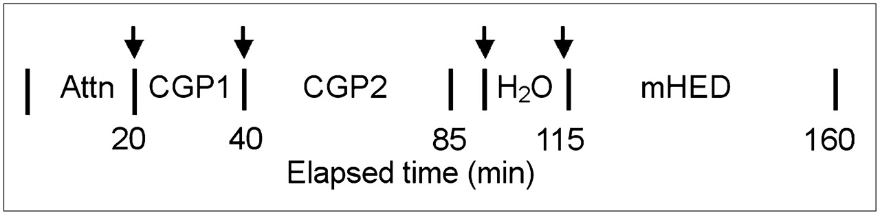
- FIGURE 1.
Time line for radiotracer injection (arrows) and PET image acquisition. Attn = transmission image acquisition; CGP1 = high-specific-activity 11C-CGP; CGP2 = low-specific-activity 11C-CGP; H2O = 15O-water.
- FIGURE 2.
Decay-corrected 11C-mHED time–activity curves from myocardial and metabolite left atrial cavity (LA Cav) ROIs in CHF patient with our model fit to myocardial time–activity curves. (Left) Location of ROI 8 (arrowhead), a visually “normal” region, and the corresponding myocardial time–activity curve. (Right) Location of ROI 3, an abnormal mHED accumulation, is shown. ROIs are bounded by inside and outside arcs within each of 8 radial lines. Parameter estimates are given for PSnt, PSves (11C-mHED release by vesicles), V′nt (virtual volume of nerve terminal), and Gseq (rate vesicular storage of mHED). MBF and the retention fraction (RF) of 11C-mHED for the 2 ROIs are also shown.
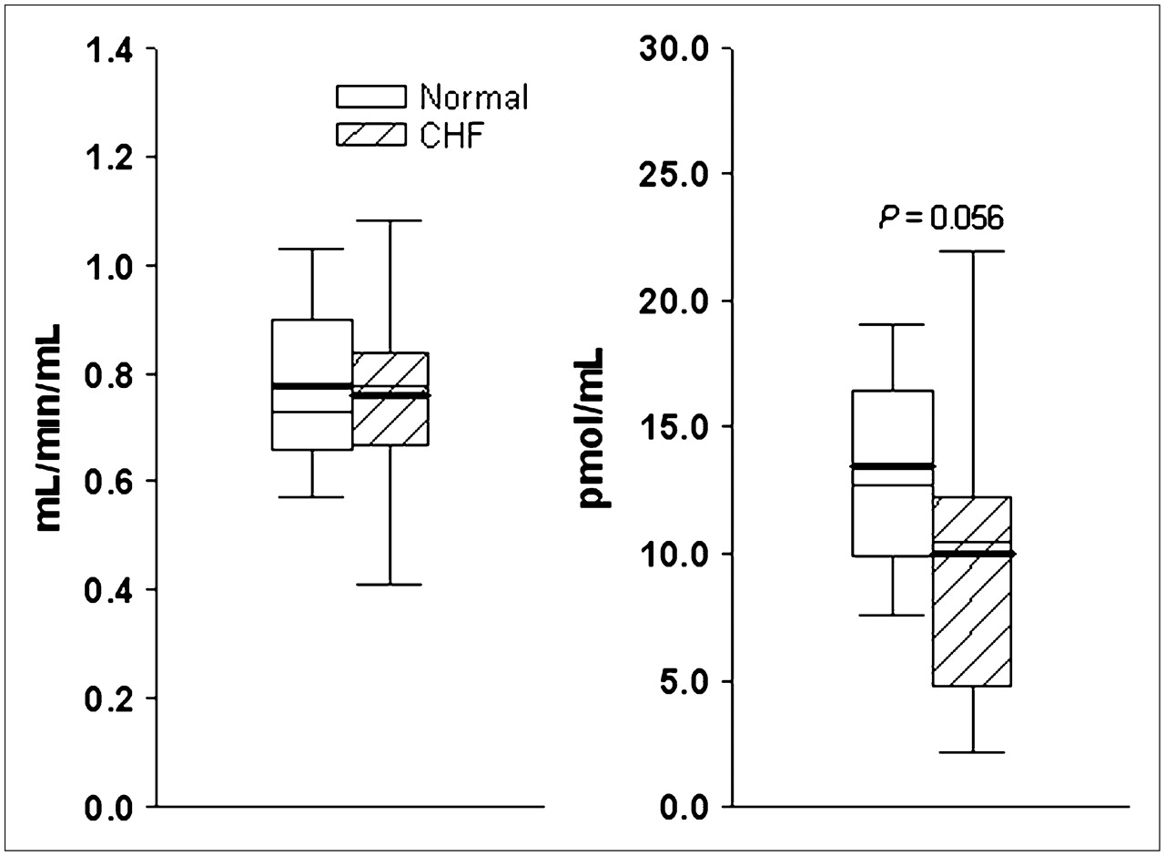
- FIGURE 3.
Box-and-whisker plots for global values for MBF (left) and B′max (right) for healthy subjects (normal) and CHF patients. Box represents 25%–75% of data; whiskers represent 5%–95%; heavy and thin solid lines are mean and median values, respectively.
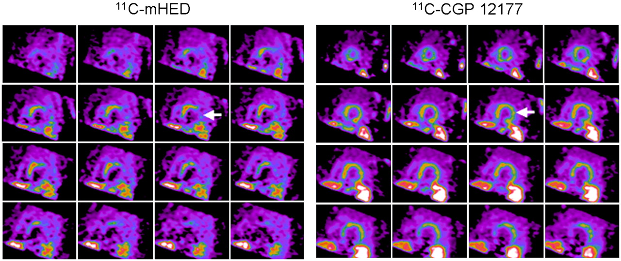
- FIGURE 4.
Short-axis PET images of 11C-mHED (35- to 45-min sum) and 11C-CGP (10- to 20-min sum from injection 1) in CHF patient. Apical slices are at upper left and basal slices are at lower right of each panel. Arrows indicate extensive mismatch between 11C-mHED and 11C-CGP.
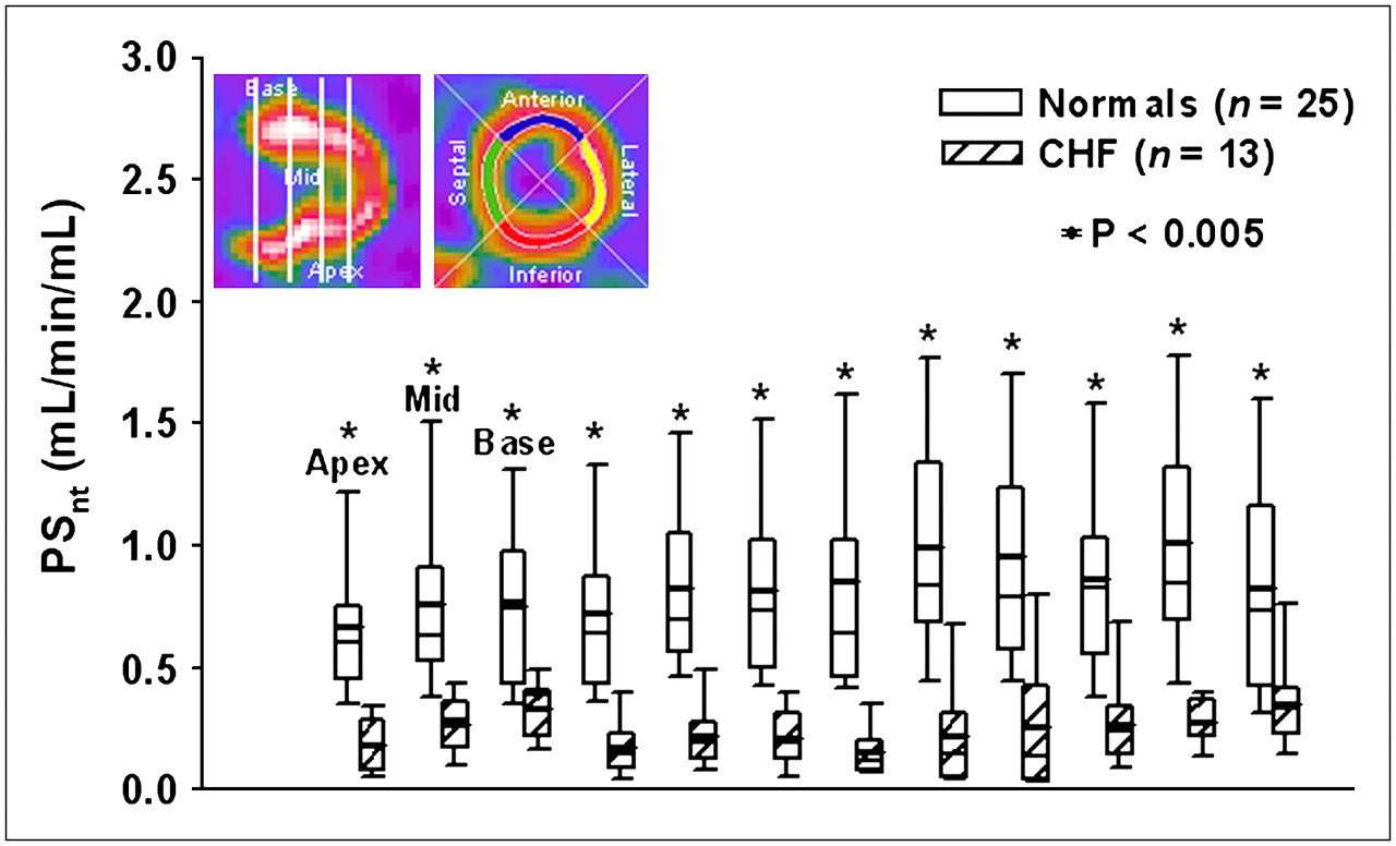
- FIGURE 5.
Box-and-whisker plots for regional NE transport (PSnt) for 12 LV regions per subject. Locations of apical, middle, and basal slices lie between the white vertical bars on the long-axis image at upper left. Locations of large sectors—anterior, lateral, inferior, septal—are shown on short-axis image. Normals = healthy subjects.
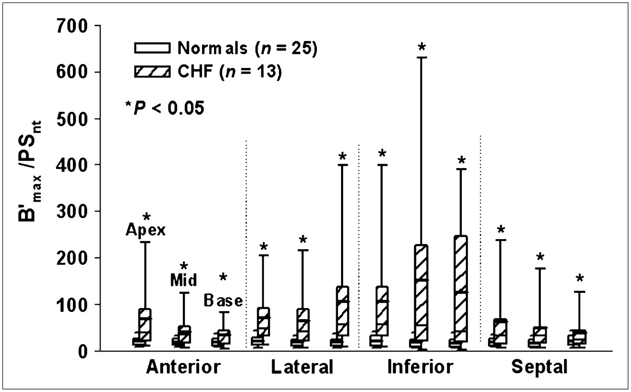
- FIGURE 6.
Box-and-whisker plots of mismatch score, which is the postsynaptic-to-presynaptic ratio (B′max:PSnt) for the same 12 LV regions as in Figure 5. Normals = healthy subjects.
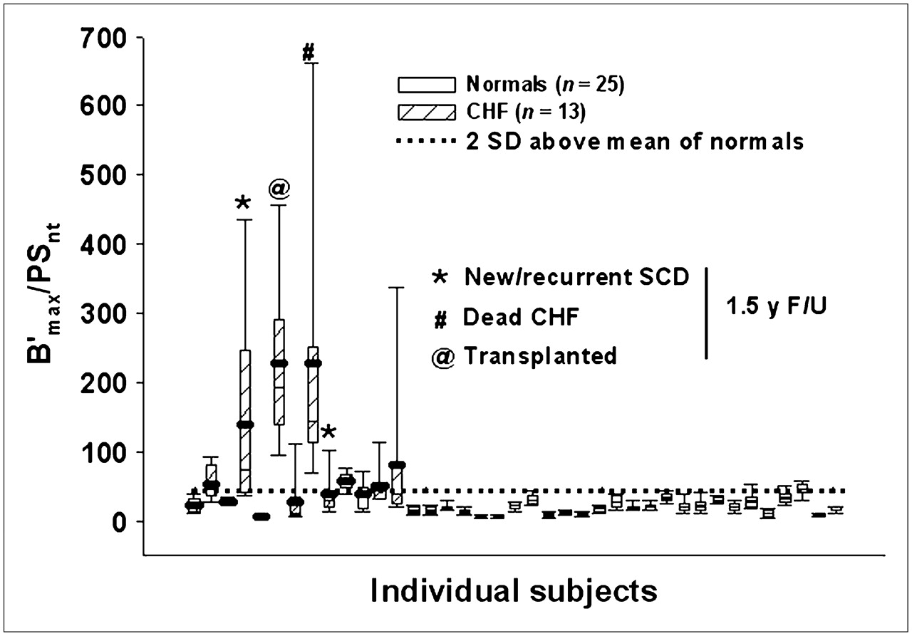
- FIGURE 7.
B′max:PSnt from the 12 ROIs per individual subject are displayed as box-and-whisker plots (mean = heavy solid line). The 5%–95% whiskers indicate within-subject B′max:PSnt heterogeneity. Horizontal dotted line indicates 2 SD above the mean B′max:PSnt of healthy subjects (normals). Patients with an adverse outcome at 1.5 y of follow-up are indicated by the symbols shown.


Tables
Age (y) EF CAD MI NYHA class ICD Amiodarone β-Blocker ACEI or ARB Event 75 0.35 1vd 3 X X X X 76 0.40 3vd 3 X X X 75 0.45 I 3 X X X 67 0.12 3vd 3 X X SCD 61 0.35 3vd 2 X 67 0.21 A 4 X X X Tx 73 0.34 I 2 X X 66 0.30 I 4 X X X CHF 77 0.35 A 2 X X SCD 67 0.23 A 4 X X 61 0.36 2vd A, L 2 X X 76 0.40 2vd 3 X X 68 0.29 3vd I 2 X X X EF = ejection fraction; CAD = coronary artery stenosis of >50% diameter stenosis; MI = electrographic myocardial infarction (I = inferior, A = anterior, L = lateral); NYHA = New York Heart Association; ICD = implanted defibrillator; ACEI or ARB = angiotensin-converting enzyme inhibitor or angiotensin-receptor blocker; 1vd, 2vd, and 3vd = number of diseased vessels; SCD = new or recurrent ventricular fibrillation/ventricular tachycardia; Tx = transplanted; CHF = death from progressive CHF.
11C-CGP1 Radiotracer injection Healthy subjects CHF patients HR* SBP* DBP* HR* SBP* DBP* Before 63.2 ± 8.0 147 ± 15 65.0 ± 10.7 74.7 ± 14† 139 ± 30 75.9 ± 11.3† After 60.9 ± 8.9‡ 151 ± 13‡ 65.9 ± 8.8 69.2 ± 13.5† 141 ± 28 75.8 ± 13.2† 11C-CGP2 Before 63.4 ± 8.7 146 ± 13 63.9 ± 9.5 72.0 ± 10.5† 137 ± 29 73.2 ± 11.4† After 60.4 ± 8.3‡ 151 ± 12‡ 66.2 ± 9.3‡ 68.1 ± 9.2† 140 ± 30 71.6 ± 12.4 11C-mHED Before 62.2 ± 8.6 148 ± 12 65.0 ± 10.2 64.8 ± 8.7§ 135 ± 33 68.2 ± 14§ After 61.3 ± 8.4 152 ± 12‡ 66.2 ± 10.1 66.9 ± 8.6 138 ± 34‡ 73.1 ± 15.5‡


Jump to section
Related Articles
Cited By...
- Radiotracers to Address Unmet Clinical Needs in Cardiovascular Imaging, Part 1: Technical Considerations and Perfusion and Neuronal Imaging
- Ventricular Tachycardia (VT) Substrate Characteristics: Insights from Multimodality Structural and Functional Imaging of the VT Substrate Using Cardiac MRI Scar, 123I-Metaiodobenzylguanidine SPECT Innervation, and Bipolar Voltage
- 123I-MIBG Scintigraphy in the Subacute State of Takotsubo Cardiomyopathy
- Global and Regional Myocardial Innervation Before and After Ablation of Drug-Refractory Ventricular Tachycardia Assessed with 123I-MIBG
- Introduction to Cardiac Neuronal Imaging: A Clinical Perspective
- Three-Dimensional 123I-Meta-Iodobenzylguanidine Cardiac Innervation Maps to Assess Substrate and Successful Ablation Sites for Ventricular Tachycardia: Feasibility Study for a Novel Paradigm of Innervation Imaging
- Noninvasive Imaging in the Assessment of the Cardiopulmonary Vascular Unit
- Altered Cardiac Innervation Predisposes to Ventricular Arrhythmia: Targeted Positron Emission Tomography Identifies Risk in Ischemic Cardiomyopathy
- Acute Myocardial Infarction Inhibits the Neurogenic Tachycardic and Vasopressor Response in Rats via Presynaptic Cannabinoid Type 1 Receptor
- Molecular Imaging of Thrombus: Technology in Evolution
- Imaging Targets of the Sympathetic Nervous System of the Heart: Translational Considerations
- Advances in SPECT and PET for the management of heart failure
- Iodine-123 Metaiodobenzylguanidine Imaging and Carbon-11 Hydroxyephedrine Positron Emission Tomography Compared in Patients With Left Ventricular Dysfunction
- Cardiac Autonomic Neuropathy in Diabetes: A clinical perspective
- Cardiac Sympathetic Imaging With mIBG in Heart Failure
- Will Imaging Assist in the Selection of Patients With Heart Failure for an ICD?
- Imaging Left Ventricular Muscarinic Receptor Heterogeneity: A Tool to Evaluate Individuals at Risk for Sudden Death?
- The Future of Cardiovascular Imaging in the Diagnosis and Management of Heart Failure, Part 1: Tasks and Tools