
Main menu

  • Home
  • Content
    • Current
    • Ahead of print
    • Past Issues
    • JNM Supplement
    • SNMMI Annual Meeting Abstracts
    • Continuing Education
    • JNM Podcasts
  • Subscriptions
    • Subscribers
    • Institutional and Non-member
    • Rates
    • Journal Claims
    • Corporate & Special Sales
  • Authors
    • Submit to JNM
    • Information for Authors
    • Assignment of Copyright
    • AQARA requirements
  • Info
    • Reviewers
    • Permissions
    • Advertisers
  • About
    • About Us
    • Editorial Board
    • Contact Information
  • More
    • Alerts
    • Feedback
    • Help
    • SNMMI Journals
  • SNMMI
    • JNM
    • JNMT
    • SNMMI Journals
    • SNMMI

User menu

  • Subscribe
  • My alerts
  • Log in
  • My Cart

Search

  • Advanced search
Journal of Nuclear Medicine
  • SNMMI
    • JNM
    • JNMT
    • SNMMI Journals
    • SNMMI
  • Subscribe
  • My alerts
  • Log in
  • My Cart
Journal of Nuclear Medicine

Advanced Search

  • Home
  • Content
    • Current
    • Ahead of print
    • Past Issues
    • JNM Supplement
    • SNMMI Annual Meeting Abstracts
    • Continuing Education
    • JNM Podcasts
  • Subscriptions
    • Subscribers
    • Institutional and Non-member
    • Rates
    • Journal Claims
    • Corporate & Special Sales
  • Authors
    • Submit to JNM
    • Information for Authors
    • Assignment of Copyright
    • AQARA requirements
  • Info
    • Reviewers
    • Permissions
    • Advertisers
  • About
    • About Us
    • Editorial Board
    • Contact Information
  • More
    • Alerts
    • Feedback
    • Help
    • SNMMI Journals
  • View or Listen to JNM Podcast
  • Visit JNM on Facebook
  • Join JNM on LinkedIn
  • Follow JNM on Twitter
  • Subscribe to our RSS feeds
Research ArticleCLINICAL INVESTIGATIONS

18F-FDG PET/CT, 99mTc-MIBI, and MRI in Evaluation of Patients with Multiple Myeloma

Rosa Fonti, Barbara Salvatore, Mario Quarantelli, Cesare Sirignano, Sabrina Segreto, Fara Petruzziello, Lucio Catalano, Raffaele Liuzzi, Bruno Rotoli, Silvana Del Vecchio, Leonardo Pace and Marco Salvatore
Journal of Nuclear Medicine February 2008, 49 (2) 195-200; DOI: https://doi.org/10.2967/jnumed.107.045641
Rosa Fonti
  • Find this author on Google Scholar
  • Find this author on PubMed
  • Search for this author on this site
Barbara Salvatore
  • Find this author on Google Scholar
  • Find this author on PubMed
  • Search for this author on this site
Mario Quarantelli
  • Find this author on Google Scholar
  • Find this author on PubMed
  • Search for this author on this site
Cesare Sirignano
  • Find this author on Google Scholar
  • Find this author on PubMed
  • Search for this author on this site
Sabrina Segreto
  • Find this author on Google Scholar
  • Find this author on PubMed
  • Search for this author on this site
Fara Petruzziello
  • Find this author on Google Scholar
  • Find this author on PubMed
  • Search for this author on this site
Lucio Catalano
  • Find this author on Google Scholar
  • Find this author on PubMed
  • Search for this author on this site
Raffaele Liuzzi
  • Find this author on Google Scholar
  • Find this author on PubMed
  • Search for this author on this site
Bruno Rotoli
  • Find this author on Google Scholar
  • Find this author on PubMed
  • Search for this author on this site
Silvana Del Vecchio
  • Find this author on Google Scholar
  • Find this author on PubMed
  • Search for this author on this site
Leonardo Pace
  • Find this author on Google Scholar
  • Find this author on PubMed
  • Search for this author on this site
Marco Salvatore
  • Find this author on Google Scholar
  • Find this author on PubMed
  • Search for this author on this site
  • Article
  • Figures & Data
  • Info & Metrics
  • PDF
Loading

Article Figures & Data

Figures

  • Tables
  • FIGURE 1. 
    • Download figure
    • Open in new tab
    • Download powerpoint
    FIGURE 1. 

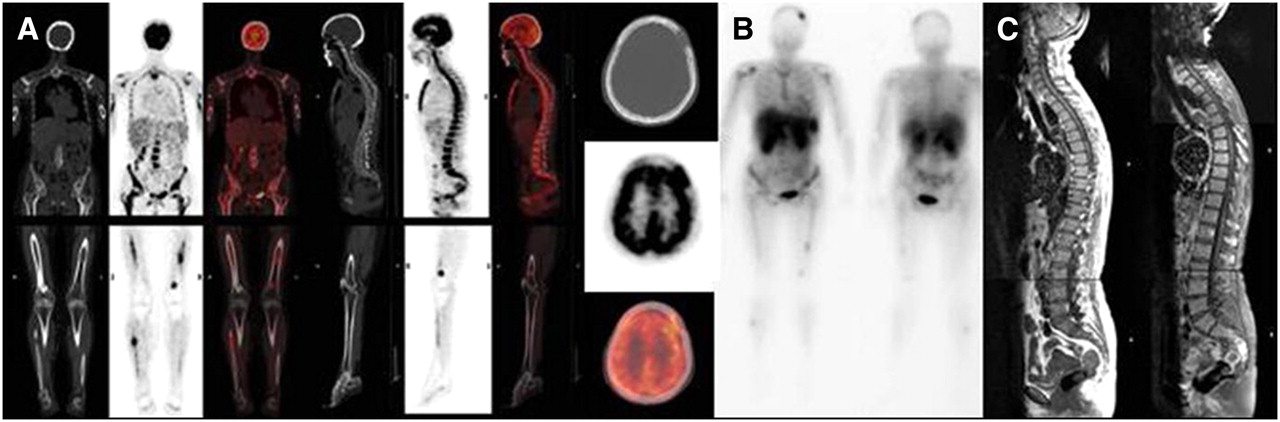
    Coronal, sagittal, and transaxial CT, PET, and fused images of whole-body 18F-FDG PET/CT (A), anterior and posterior whole-body 99mTc-MIBI (B), and sagittal T1- and T1-weighted fat-saturated with gadolinium MR images of spine (C). All 3 imaging modalities, performed on the same MM patient, showed a focal + diffuse pattern of distribution. In particular, focal lesions were shown in skull, humeri, pelvis, left femur, and right fibula by whole-body 18F-FDG PET/CT; in skull, right humerus, pelvis, left femur, and right fibula by whole-body 99mTc-MIBI; and in coccygeal bone by MRI of spine and pelvis.

Tables

  • Figures
    • View popup
    TABLE 1

    Whole-Body 18F-FDG PET/CT, Whole-Body 99mTc-MIBI, and MRI of Spine and Pelvis Performed on 33 MM Patients at Diagnosis: Comparison According to Presence of Normal, Diffuse, or Focal and Focal + Diffuse Pattern of Bone Marrow Involvement

    Imaging modalityNDF-FD
    Whole-body 18F-FDG PET/CT1 (3)3 (9)29 (88)
    Whole-body 99mTc-MIBI3 (9)11 (33)19 (57)
    MRI of spine and pelvis6 (18)13 (39)14 (42)
    • N = normal; D = diffuse; F-FD = focal and focal + diffuse.

    • Values in parentheses are percentages (2-sided Fisher exact test; P < 0.005).

    • View popup
    TABLE 2

    Comparison of Number of Focal Lesions per Patient Detected by Whole-Body 18F-FDG PET/CT, Whole-Body 99mTc-MIBI, and MRI of Spine and Pelvis

    Focal lesions per patient (n)
    Whole-body 18F-FDG PET/CTWhole-body 99mTc-MIBIMRI spine and pelvisKendall's W test
    5.94 ± 9.29*†1.91 ± 4.451.54 ± 2.45P < 0.001
    • ↵* P < 0.001, 18F-FDG PET/CT vs. 99mTc-MIBI.

    • ↵† P < 0.005, 18F-FDG PET/CT vs. MRI.

    • View popup
    TABLE 3

    18F-FDG PET/CT, 99mTc-MIBI, and MRI of Spinal and Pelvic District Performed on 33 MM Patients at Diagnosis: Comparison According to Presence of Normal, Diffuse, or Focal and Focal + Diffuse Pattern of Bone Marrow Involvement

    Imaging modalityNDF-FD
    Whole-body 18F-FDG PET/CT of spine and pelvis12 (36)6 (18)15 (45)
    Whole-body 99mTc-MIBI of spine and pelvis8 (24)18 (54)7 (21)
    MRI of spine and pelvis6 (18)13 (39)14 (42)
    • N = normal; D = diffuse; F-FD = focal and focal + diffuse.

    • Values in parentheses are percentages (2-sided Fisher exact test; P < 0.005).

    • View popup
    TABLE 4

    Comparison of Number of Focal Lesions per Patient Detected by 18F-FDG PET/CT, 99mTc-MIBI, and MRI of Spinal and Pelvic District

    Focal lesions per patient (n)
    18F-FDG PET/CT spine and pelvis99mTc-MIBI spine and pelvisMRI spine and pelvisKendall's W test
    2.27 ± 4.64*0.30 ± 0.681.54 ± 2.45†P < 0.005
    • ↵* P < 0.001, 18F-FDG PET/CT vs. 99mTc-MIBI.

    • ↵† P < 0.01, MRI vs. 99mTc-MIBI.

PreviousNext
Back to top

In this issue

Journal of Nuclear Medicine: 49 (2)
Journal of Nuclear Medicine
Vol. 49, Issue 2
February 2008
  • Table of Contents
  • Table of Contents (PDF)
  • About the Cover
  • Index by author
Print
Download PDF
Article Alerts
Sign In to Email Alerts with your Email Address
Email Article

Thank you for your interest in spreading the word on Journal of Nuclear Medicine.

NOTE: We only request your email address so that the person you are recommending the page to knows that you wanted them to see it, and that it is not junk mail. We do not capture any email address.

Enter multiple addresses on separate lines or separate them with commas.
18F-FDG PET/CT, 99mTc-MIBI, and MRI in Evaluation of Patients with Multiple Myeloma
(Your Name) has sent you a message from Journal of Nuclear Medicine
(Your Name) thought you would like to see the Journal of Nuclear Medicine web site.
Citation Tools
18F-FDG PET/CT, 99mTc-MIBI, and MRI in Evaluation of Patients with Multiple Myeloma
Rosa Fonti, Barbara Salvatore, Mario Quarantelli, Cesare Sirignano, Sabrina Segreto, Fara Petruzziello, Lucio Catalano, Raffaele Liuzzi, Bruno Rotoli, Silvana Del Vecchio, Leonardo Pace, Marco Salvatore
Journal of Nuclear Medicine Feb 2008, 49 (2) 195-200; DOI: 10.2967/jnumed.107.045641

Citation Manager Formats

  • BibTeX
  • Bookends
  • EasyBib
  • EndNote (tagged)
  • EndNote 8 (xml)
  • Medlars
  • Mendeley
  • Papers
  • RefWorks Tagged
  • Ref Manager
  • RIS
  • Zotero
Share
18F-FDG PET/CT, 99mTc-MIBI, and MRI in Evaluation of Patients with Multiple Myeloma
Rosa Fonti, Barbara Salvatore, Mario Quarantelli, Cesare Sirignano, Sabrina Segreto, Fara Petruzziello, Lucio Catalano, Raffaele Liuzzi, Bruno Rotoli, Silvana Del Vecchio, Leonardo Pace, Marco Salvatore
Journal of Nuclear Medicine Feb 2008, 49 (2) 195-200; DOI: 10.2967/jnumed.107.045641
Twitter logo Facebook logo LinkedIn logo Mendeley logo
  • Tweet Widget
  • Facebook Like
  • Google Plus One
Bookmark this article

Jump to section

  • Article
    • Abstract
    • MATERIALS AND METHODS
    • RESULTS
    • DISCUSSION
    • CONCLUSION
    • Acknowledgments
    • Footnotes
    • References
  • Figures & Data
  • Info & Metrics
  • PDF

Related Articles

  • This Month in JNM
  • PubMed
  • Google Scholar

Cited By...

  • New Developments in Myeloma Treatment and Response Assessment
  • The role of positron emission tomography-computed tomography and magnetic resonance imaging in diagnosis and follow up of multiple myeloma
  • 99mTc-Sestamibi Scintigraphy to Monitor the Long-Term Efficacy of Enzyme Replacement Therapy on Bone Marrow Infiltration in Patients with Gaucher Disease
  • Assessment of whole body MRI and sestamibi technetium-99m bone marrow scan in prediction of multiple myeloma disease progression and outcome: a prospective comparative study
  • Plasma cell disorders
  • Metabolic Tumor Volume Assessed by 18F-FDG PET/CT for the Prediction of Outcome in Patients with Multiple Myeloma
  • MRI for the detection of prostate cancer origin vertebral metastases in the preosteoblastic phase
  • Imaging of Multiple Myeloma and Related Plasma Cell Dyscrasias
  • Can multiple myeloma become a curable disease?
  • How I treat multiple myeloma in younger patients
  • A new pet for myeloma
  • NCCN Task Force: Clinical Utility of PET in a Variety of Tumor Types
  • Tissue Classification as a Potential Approach for Attenuation Correction in Whole-Body PET/MRI: Evaluation with PET/CT Data
  • Incidental Detection of Concurrent Extramedullary Plasmacytoma and Amyloidoma of the Nasopharynx on [18F]Fluorodeoxyglucose Positron Emission Tomography/Computed Tomography
  • Google Scholar

More in this TOC Section

  • Cardiac Presynaptic Sympathetic Nervous Function Evaluated by Cardiac PET in Patients with Chronotropic Incompetence Without Heart Failure
  • Validation and Evaluation of a Vendor-Provided Head Motion Correction Algorithm on the uMI Panorama PET/CT System
  • Prognostic Role of 68Ga-PSMA11 PET–Based Response in Patients with Prostate Cancer Undergoing Taxane-Based Chemotherapy
Show more Clinical Investigations

Similar Articles

SNMMI

© 2025 SNMMI

Powered by HighWire