


Article Figures & Data
Figures
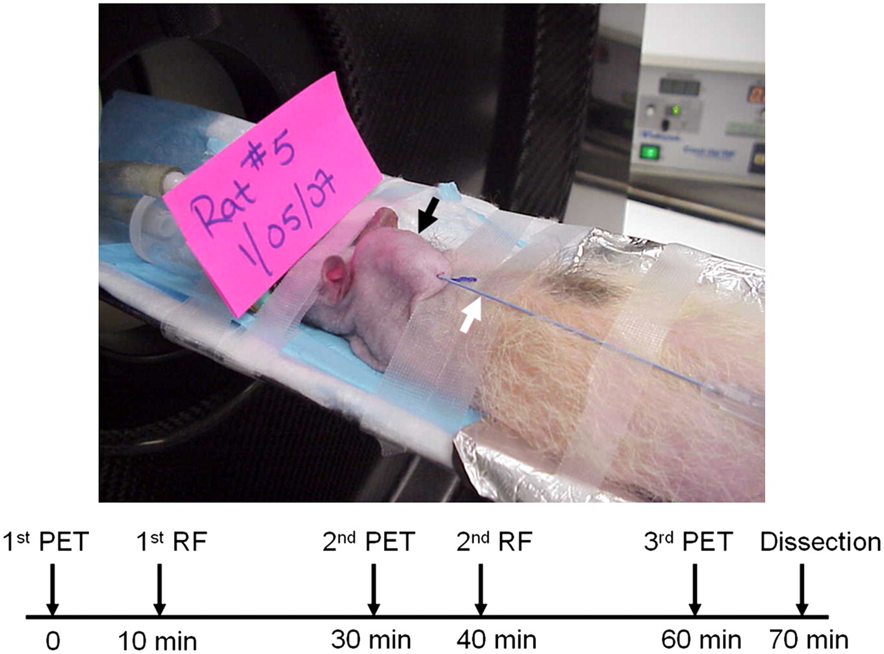
- FIGURE 1.
Experimental setup for real-time monitoring of RF ablation with 15O-water PET. RF ablation catheter (white arrow) was inserted into tumor xenograft located on back of rat's neck (black arrow). Ground electrode covered in aluminum foil was placed underneath rat. Flow chart indicates approximate times from 0 to 70 min and sequence for performing 15O-water PET, RF ablation, and tumor dissection of each animal.
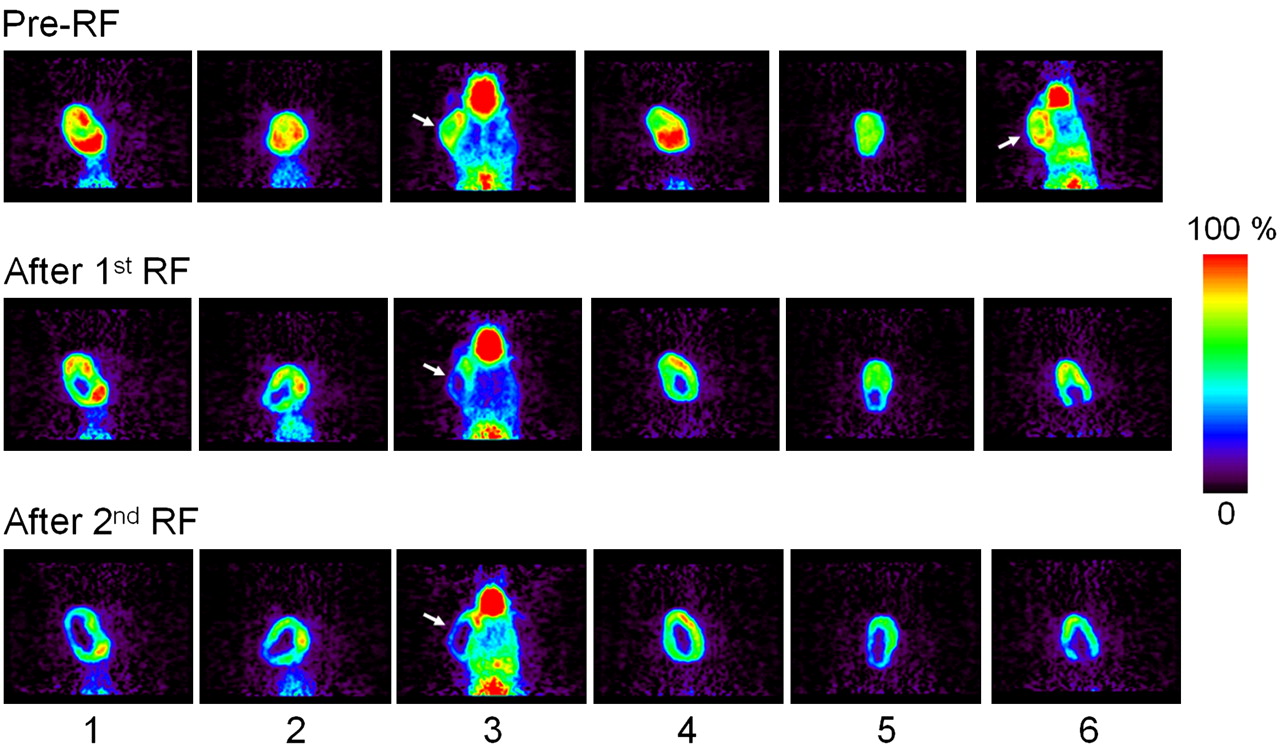
- FIGURE 2.
15O-water PET images of all 6 rats bearing head and neck SCC xenografts (coronal images) at time points of before RF ablation, after first RF ablation, and after second RF ablation. Image slices are mainly focused on tumors located in center of neck for rats 1, 2, 4, and 5 or slightly on side of neck for rats 3 and 6 (arrows). Because of motility of subcutaneous tumor xenograft, geometry of tumor in rat 6 was different before and after each RF ablation.
- FIGURE 3.
Representative 15O-water PET images of rat bearing head and neck SCC xenograft (left, axial images; middle, coronal images; right, sagittal images) at time points of before RF ablation, after first RF ablation, and after second RF ablation. Arrows point to tumor.
- FIGURE 4.
Relative pixel-value (average ± SD) time curves determined by ROI analysis of dynamic 15O-water PET images with 10 s/frame before and after RF ablation of tumors (n = 6). For pre–RF ablation analysis (A), ablated region represents ROI drawn in tumor center, whereas unablated region represents ROI drawn at tumor margin. For first RF ablation (B) and second RF ablation (C), ablated region represents area in tumor center after RF ablation and unablated region represents area in tumor margin that was not ablated (*P < 0.05, **P < 0.01, and ***P < 0.001 compare ablated and unablated regions at same time points).
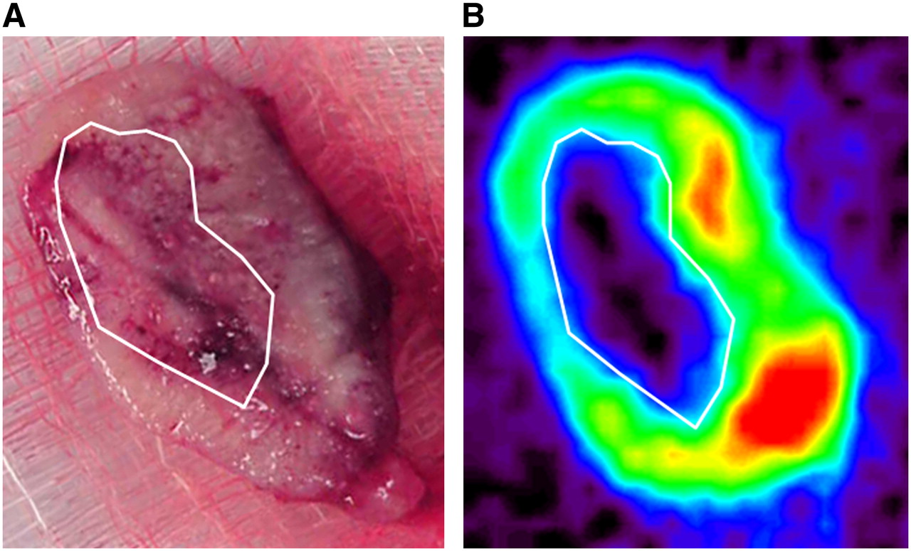
- FIGURE 5.
Comparison between coagulation zone shown in freshly dissected tumor (A) and ablated region in 15O-water PET image (B). Coagulation zone (marked area) has similar shape and size corresponding to ablated region in 15O-water PET image.
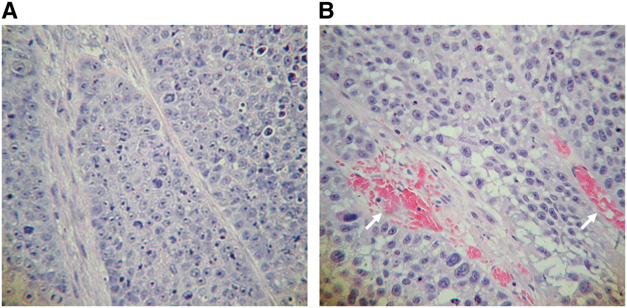
- FIGURE 6.
H&E-stained tumor section that was not RF ablated (A) and was RF ablated (B) (20× magnification for both tumor sections). Red color component (arrows) in RF-ablated tumor tissue is because of coagulated and infiltrated blood cells.


