
Main menu

  • Home
  • Content
    • Current
    • Ahead of print
    • Past Issues
    • JNM Supplement
    • SNMMI Annual Meeting Abstracts
    • Continuing Education
    • JNM Podcasts
  • Subscriptions
    • Subscribers
    • Institutional and Non-member
    • Rates
    • Journal Claims
    • Corporate & Special Sales
  • Authors
    • Submit to JNM
    • Information for Authors
    • Assignment of Copyright
    • AQARA requirements
  • Info
    • Reviewers
    • Permissions
    • Advertisers
  • About
    • About Us
    • Editorial Board
    • Contact Information
  • More
    • Alerts
    • Feedback
    • Help
    • SNMMI Journals
  • SNMMI
    • JNM
    • JNMT
    • SNMMI Journals
    • SNMMI

User menu

  • Subscribe
  • My alerts
  • Log in
  • My Cart

Search

  • Advanced search
Journal of Nuclear Medicine
  • SNMMI
    • JNM
    • JNMT
    • SNMMI Journals
    • SNMMI
  • Subscribe
  • My alerts
  • Log in
  • My Cart
Journal of Nuclear Medicine

Advanced Search

  • Home
  • Content
    • Current
    • Ahead of print
    • Past Issues
    • JNM Supplement
    • SNMMI Annual Meeting Abstracts
    • Continuing Education
    • JNM Podcasts
  • Subscriptions
    • Subscribers
    • Institutional and Non-member
    • Rates
    • Journal Claims
    • Corporate & Special Sales
  • Authors
    • Submit to JNM
    • Information for Authors
    • Assignment of Copyright
    • AQARA requirements
  • Info
    • Reviewers
    • Permissions
    • Advertisers
  • About
    • About Us
    • Editorial Board
    • Contact Information
  • More
    • Alerts
    • Feedback
    • Help
    • SNMMI Journals
  • View or Listen to JNM Podcast
  • Visit JNM on Facebook
  • Join JNM on LinkedIn
  • Follow JNM on Twitter
  • Subscribe to our RSS feeds
OtherBASIC SCIENCE INVESTIGATIONS

Performance Measurement of the microPET Focus 120 Scanner

Jin Su Kim, Jae Sung Lee, Ki Chun Im, Su Jin Kim, Seog-Young Kim, Dong Soo Lee and Dae Hyuk Moon
Journal of Nuclear Medicine September 2007, 48 (9) 1527-1535; DOI: https://doi.org/10.2967/jnumed.107.040550
Jin Su Kim
  • Find this author on Google Scholar
  • Find this author on PubMed
  • Search for this author on this site
Jae Sung Lee
  • Find this author on Google Scholar
  • Find this author on PubMed
  • Search for this author on this site
Ki Chun Im
  • Find this author on Google Scholar
  • Find this author on PubMed
  • Search for this author on this site
Su Jin Kim
  • Find this author on Google Scholar
  • Find this author on PubMed
  • Search for this author on this site
Seog-Young Kim
  • Find this author on Google Scholar
  • Find this author on PubMed
  • Search for this author on this site
Dong Soo Lee
  • Find this author on Google Scholar
  • Find this author on PubMed
  • Search for this author on this site
Dae Hyuk Moon
  • Find this author on Google Scholar
  • Find this author on PubMed
  • Search for this author on this site
  • Article
  • Figures & Data
  • Info & Metrics
  • PDF
Loading

Article Figures & Data

Figures

  • Tables
  • FIGURE 1. 
    • Download figure
    • Open in new tab
    • Download powerpoint
    FIGURE 1. 

    Spatial resolution profile measured using a 22Na point source. (A) Transaxial (radial and tangential). (B) Axial with or without oversampling. (C) Volume resolution with or without oversampling.

  • FIGURE 2. 
    • Download figure
    • Open in new tab
    • Download powerpoint
    FIGURE 2. 

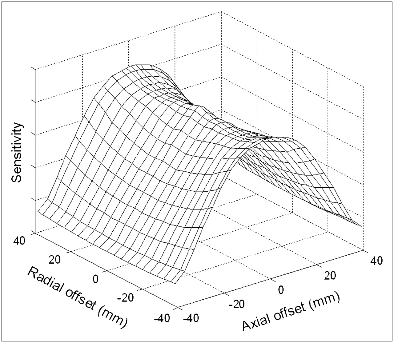
    3D sensitivity profile over a FOV.

  • FIGURE 3. 
    • Download figure
    • Open in new tab
    • Download powerpoint
    FIGURE 3. 

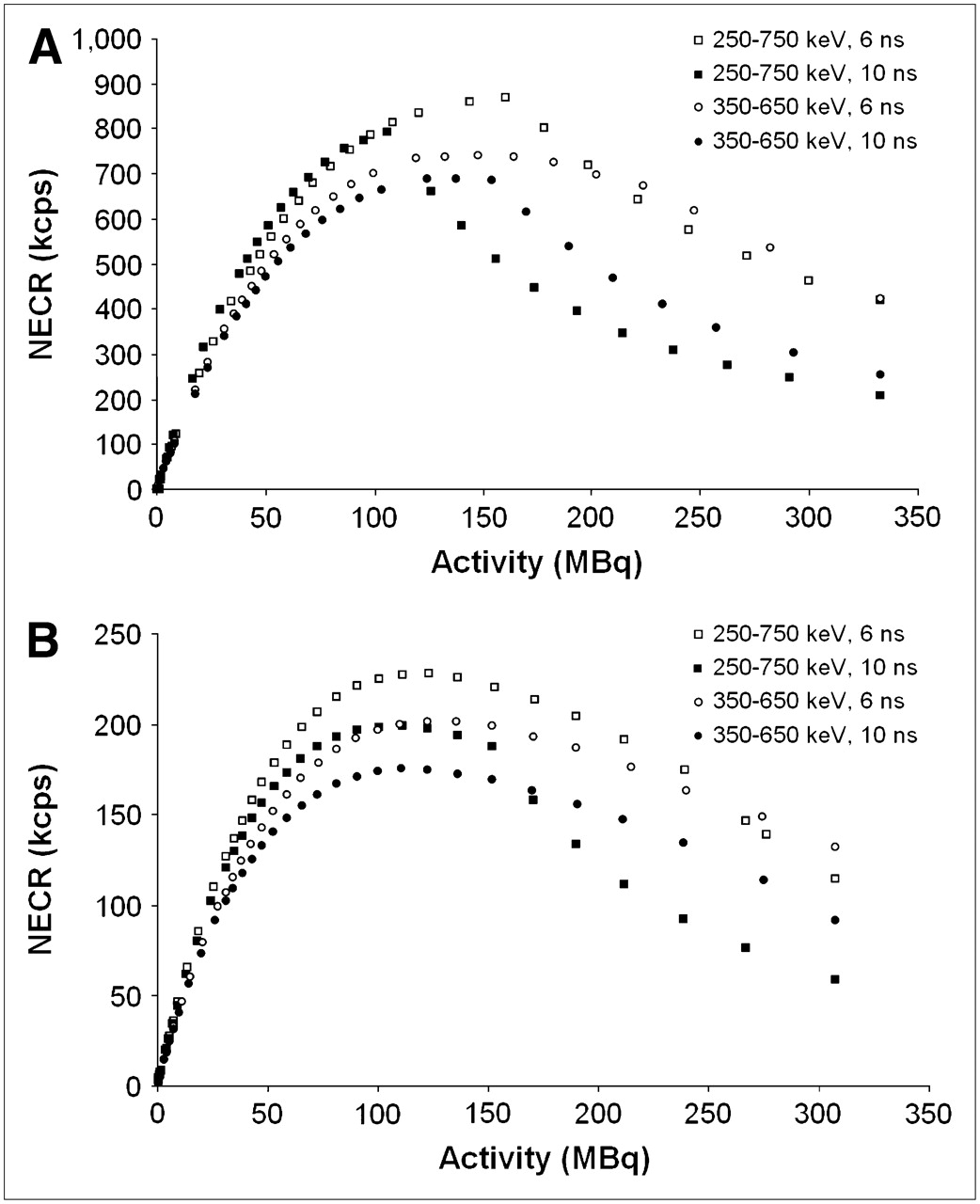
    NECR vs. total radioactivity for mouse (A) and rat (B) phantoms.

  • FIGURE 4. 
    • Download figure
    • Open in new tab
    • Download powerpoint
    FIGURE 4. 

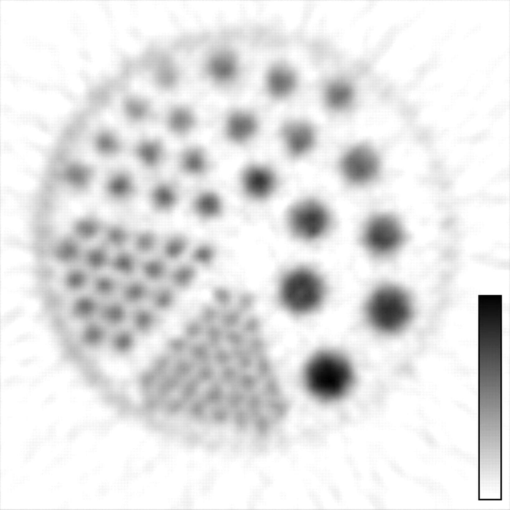
    Image of Micro Deluxe phantom with hot-rod inserts—reconstructed using the 3DRP algorithm.

  • FIGURE 5. 
    • Download figure
    • Open in new tab
    • Download powerpoint
    FIGURE 5. 

    Maximum-intensity projection (MIP) images of mouse bone 18F PET reconstructed using different algorithms: (A) FBP, (B) 3DRP, (C) EM, and (D) MAP.

  • FIGURE 6. 
    • Download figure
    • Open in new tab
    • Download powerpoint
    FIGURE 6. 

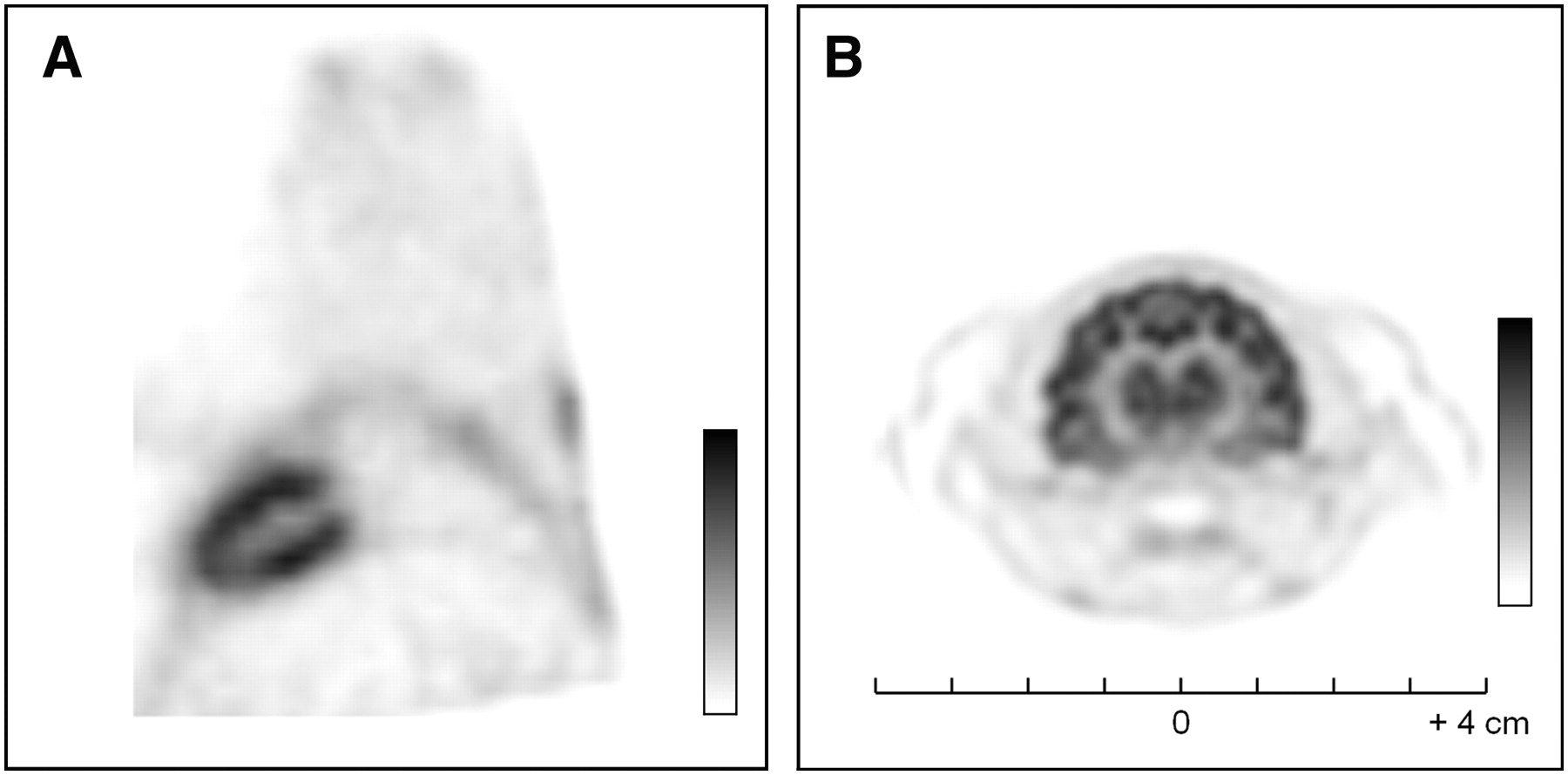
    Selected animal imaging studies. (A) Myocardial PET image of rat in end-diastolic phase. (B) Cat brain PET image acquired using 18F-FDG and reconstructed using the MAP algorithm.

Tables

  • Figures
    • View popup
    TABLE 1

    Specification Comparison of microPET Systems Related to Focus 120

    CategorySpecificationFocus 120R4*Focus 220†
    DetectorCrystal materialLSOLSOLSO
    Crystal element size (mm3)1.5 × 1.5 × 102.1 × 2.1 × 101.5 × 1.5 × 10
    Crystal pitch (mm)1.592.451.59
    Crystal array12 × 128 × 812 × 12
    Packing fraction (%)928092
    SystemNumber of detector blocks9696168
    Total number of crystal elements13,8246,14424,192
    Number of rings483648
    Ring diameter (cm)151526
    Bore size (cm)121222
    Transaxial FOV (cm)101019
    Axial FOV (cm)7.67.87.6
    Solid angle/4π0.450.460.28
    • ↵* Data published by Knoess et al. (19).

    • ↵† Data published by Tai et al. (12).

    • View popup
    TABLE 2

    Absolute Sensitivity for a Point Source Centered in Axial FOV

    Radial distance (mm)Energy window (keV)Coincidence window (ns)Sensitivity (%)
    0250–750107.0
    66.7
    350–650104.0
    63.8
    20250–750107.1
    66.9
    350–650103.0
    62.9
    • View popup
    TABLE 3

    Scatter Fraction and Counting Rate Performance

    PhantomEnergy window (keV)Coincidence window (ns)Scatter fraction (%)Peak true (kcps at kBq/mL)Peak NECR (kcps at kBq/mL)
    Mouse250∼750615.91,296 at 3,242869 at 3,242
    1015.01,218 at 2,136793 at 2,136
    350∼650611.61,088 at 3,684741 at 2,989
    1011.01,092 at 3,108689 at 2,781
    Rat250∼750635.0582 at 448228 at 290
    1034.9585 at 358199 at 262
    350∼650622.9449 at 506201 at 289
    1023.1466 at 498175 at 260
    • View popup
    TABLE 4

    Comparison of Performance Characteristics in microPET Scanners

    Performance characteristicFocus 120 (this study)Focus 120 (Laforest et al.)*R4†Focus 220‡
    Energy resolution (%)18.3N/A2318.5
    Volumetric resolution (μL)§Center1.922.875.12.5¶
    1-cm radial offset6.41N/A15.6N/A
    2-cm radial offset8.148.23∼19‖6.3¶
    Absolute sensitivity for point source at center (%)250∼750 keV, 10 ns7.07.1N/A3.4
    250∼750 keV, 6 ns6.76.7**4.373.0
    350∼650 keV, 10 ns4.04.7**N/AN/A
    350∼650 keV, 6 ns3.84.4**2.452.1
    Scatter fraction (%)††Mouse phantom15.912.332.019.0
    (3-cm Ø, 7-cm L)(2.5-cm Ø, 7-cm L)(4.3-cm Ø, 7.6-cm L)(3-cm Ø, 7-cm L)
    Rat phantom35.026.338.036.4
    (6-cm Ø, 15-cm L)(5-cm Ø, 15-cm L)(6-cm Ø, 9.6-cm L)(6-cm Ø, 15-cm L)
    Peak NECR (kcps)††Mouse phantom869 at 160.58 MBq809 at 88.8 MBq174 at 76.96 MBq645 at 146.52 MBq
    (4.34 mCi)(2.4 mCi)(2.08 mCi)(3.96 mCi)
    (3-cm Ø, 7-cm L)(2.5-cm Ø, 7-cm L)(4.3-cm Ø, 7.6-cm L)(3-cm Ø, 7-cm L)
    Rat phantom228 at 122.84 MBq300 at 149.85 MBq94 at 59.94 MBq177 at 143.19 MBq
    (3.32 mCi)(4.05 mCi)(1.62 mCi)(3.87 mCi)
    (6-cm Ø, 15-cm L)(5-cm Ø, 15-cm L)(6-cm Ø, 9.6-cm L)(6-cm Ø, 15-cm L)
    • ↵* Data published by Laforest et al. (13).

    • ↵† Data published by Knoess et al. (19).

    • ↵‡ Data published by Tai et al. (12).

    • ↵§ Data were not corrected for source dimension, positron range, or acolinearity of positron annihilation and were estimated from nonoversampled axial profiles except for Focus 220.

    • ↵¶ Axial resolution was estimated from oversampled axial profiles (Tai et al.) (12).

    • ↵‖ Estimated from Figure 5 in Knoess et al. (19).

    • ↵** Estimated from Figure 1 in Laforest et al. (13).

    • ↵†† Data were measured using same energy window (250–750 keV) and coincidence window (6 ns) but the phantom sizes (Ø, diameter; L, length) were not identical.

    • N/A = not available.

PreviousNext
Back to top

In this issue

Journal of Nuclear Medicine: 48 (9)
Journal of Nuclear Medicine
Vol. 48, Issue 9
September 2007
  • Table of Contents
  • Table of Contents (PDF)
  • About the Cover
  • Index by author
Print
Download PDF
Article Alerts
Sign In to Email Alerts with your Email Address
Email Article

Thank you for your interest in spreading the word on Journal of Nuclear Medicine.

NOTE: We only request your email address so that the person you are recommending the page to knows that you wanted them to see it, and that it is not junk mail. We do not capture any email address.

Enter multiple addresses on separate lines or separate them with commas.
Performance Measurement of the microPET Focus 120 Scanner
(Your Name) has sent you a message from Journal of Nuclear Medicine
(Your Name) thought you would like to see the Journal of Nuclear Medicine web site.
Citation Tools
Performance Measurement of the microPET Focus 120 Scanner
Jin Su Kim, Jae Sung Lee, Ki Chun Im, Su Jin Kim, Seog-Young Kim, Dong Soo Lee, Dae Hyuk Moon
Journal of Nuclear Medicine Sep 2007, 48 (9) 1527-1535; DOI: 10.2967/jnumed.107.040550

Citation Manager Formats

  • BibTeX
  • Bookends
  • EasyBib
  • EndNote (tagged)
  • EndNote 8 (xml)
  • Medlars
  • Mendeley
  • Papers
  • RefWorks Tagged
  • Ref Manager
  • RIS
  • Zotero
Share
Performance Measurement of the microPET Focus 120 Scanner
Jin Su Kim, Jae Sung Lee, Ki Chun Im, Su Jin Kim, Seog-Young Kim, Dong Soo Lee, Dae Hyuk Moon
Journal of Nuclear Medicine Sep 2007, 48 (9) 1527-1535; DOI: 10.2967/jnumed.107.040550
Twitter logo Facebook logo LinkedIn logo Mendeley logo
  • Tweet Widget
  • Facebook Like
  • Google Plus One
Bookmark this article

Jump to section

  • Article
    • Abstract
    • MATERIALS AND METHODS
    • RESULTS
    • DISCUSSION
    • CONCLUSION
    • Acknowledgments
    • Footnotes
    • References
  • Figures & Data
  • Info & Metrics
  • PDF

Related Articles

  • This Month in JNM
  • PubMed
  • Google Scholar

Cited By...

  • Immuno-PET Imaging of CD30-Positive Lymphoma Using 89Zr-Desferrioxamine-Labeled CD30-Specific AC-10 Antibody
  • Performance Assessment of a Preclinical PET Scanner with Pinhole Collimation by Comparison to a Coincidence-Based Small-Animal PET Scanner
  • Performance Evaluation of a New Dedicated Breast PET Scanner Using NEMA NU4-2008 Standards
  • 3'-Deoxy-3'-[18F]Fluorothymidine Positron Emission Tomography Imaging of Thymidine Kinase 1 Activity After 5-Fluorouracil Treatment in a Mouse Tumor Model
  • In Vivo PET/CT in a Human Glioblastoma Chicken Chorioallantoic Membrane Model: A New Tool for Oncology and Radiotracer Development
  • NEMA NU 4-2008 Comparison of Preclinical PET Imaging Systems
  • In Vivo Dopamine Transporter Imaging in a Unilateral 6-Hydroxydopamine Rat Model of Parkinson Disease Using 11C-Methylphenidate PET
  • Preclinical Pharmacology of AZD5363, an Inhibitor of AKT: Pharmacodynamics, Antitumor Activity, and Correlation of Monotherapy Activity with Genetic Background
  • Development of Small-Animal PET Prototype Using Silicon Photomultiplier (SiPM): Initial Results of Phantom and Animal Imaging Studies
  • Performance Evaluation of the FLEX Triumph X-PET Scanner Using the National Electrical Manufacturers Association NU-4 Standards
  • 89Zr-DFO-J591 for ImmunoPET of Prostate-Specific Membrane Antigen Expression In Vivo
  • Cerenkov Luminescence Imaging of Medical Isotopes
  • NEMA NU4-2008 Image Quality Performance Report for the microPET Focus 120 and for Various Transmission and Reconstruction Methods
  • Dipeptidyl Peptidase IV Inhibition With MK0431 Improves Islet Graft Survival in Diabetic NOD Mice Partially via T-Cell Modulation
  • Performance Evaluation of the Inveon Dedicated PET Preclinical Tomograph Based on the NEMA NU-4 Standards
  • Spatial Resolution and Sensitivity of the Inveon Small-Animal PET Scanner
  • Kinetic Modeling of 3'-Deoxy-3'-18F-Fluorothymidine for Quantitative Cell Proliferation Imaging in Subcutaneous Tumor Models in Mice
  • Impact of Contamination from Scattered Photons in Singles-Mode Transmission Data on Quantitative Small-Animal PET Imaging
  • Molecular-Genetic Imaging Based on Reporter Gene Expression
  • Inhibition of Dipeptidyl Peptidase IV With Sitagliptin (MK0431) Prolongs Islet Graft Survival in Streptozotocin-Induced Diabetic Mice
  • Google Scholar

More in this TOC Section

  • How Sensitive Is the Upper Gastrointestinal Tract to 90Y Radioembolization? A Histologic and Dosimetric Analysis in a Porcine Model
  • 11C-Methionine PET of Myocardial Inflammation in a Rat Model of Experimental Autoimmune Myocarditis
  • Counting Rate Characteristics and Image Distortion in Preclinical PET Imaging During Radiopharmaceutical Therapy
Show more Basic Science Investigations

Similar Articles

SNMMI

© 2025 SNMMI

Powered by HighWire