


Article Figures & Data
Figures
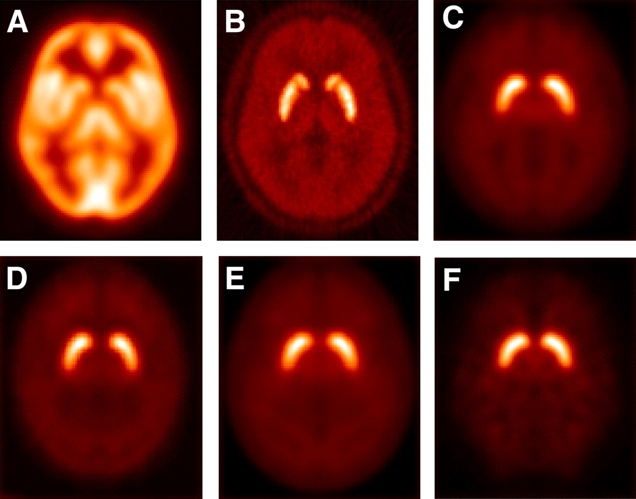
- FIGURE 1.
Templates: (Top) Regional CBF template (A) available in SPM software package; 11C-raclopride template (B) created in Orsay PET Center from images of healthy subjects; reference template of 123I-FP-CIT (C) created in center A (Template A1). Raclopride template has high specific striatal and low cortical uptake similar to the profile of 123I-FP-CIT images. (Bottom) Three other templates of 123I-FP-CIT (D = template A2, E = template B, and F = template C) constructed with images obtained from different γ-cameras or data-processing schemes.
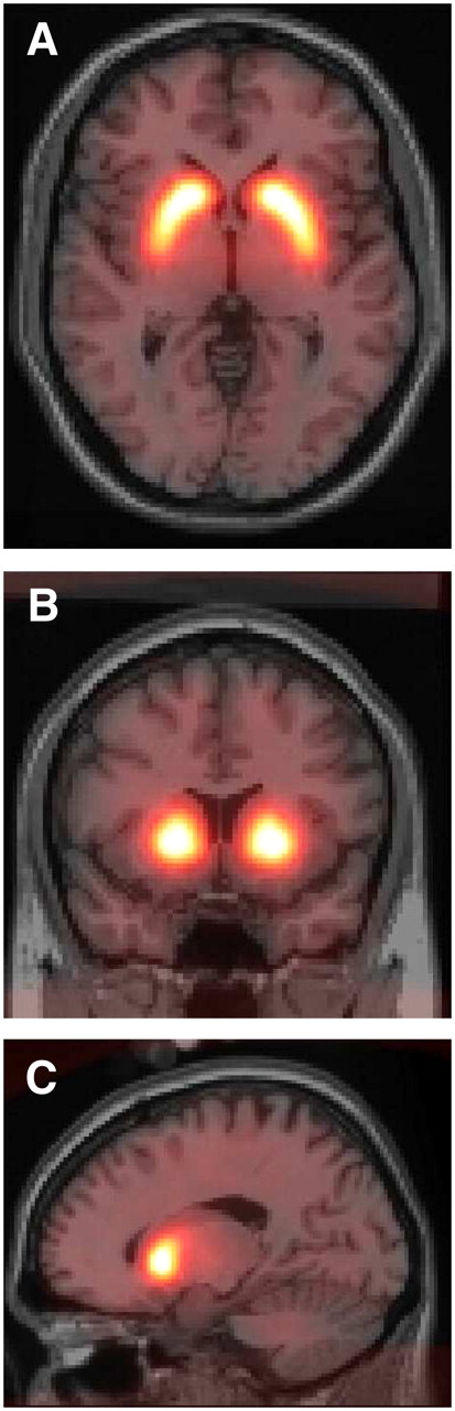
- FIGURE 2.
Template A1 overlaid on single-subject brain MRI available in SPM in 3 orthogonal views: A, axial; B, coronal; and C, sagittal. Normalized striatal 123I-FP-CIT binding is accurately superimposed onto striatum.
- FIGURE 3.
Difference against average of putaminal ratios (pBR) obtained with template A1 and 3 other templates, with 95% limits of agreement. Solid line shows mean difference score. The 95% limits of agreement (dashed lines) represent 1.96 SDs above and below the mean difference score. Each graph compares the reproducibility between the method using templates A2, B, and C and template A1.
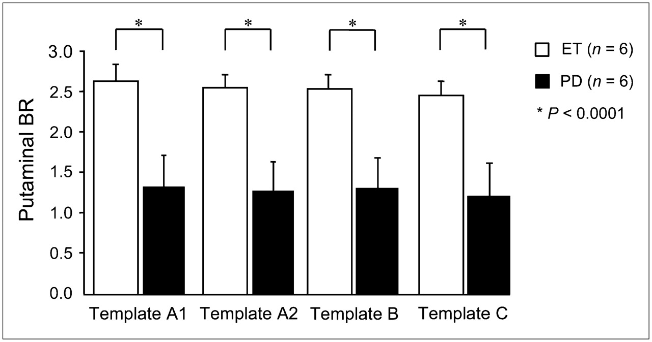
- FIGURE 4.
Mean BR ± SD values measured with VOI in putamen of subjects with ET (□) and PD (▪), after each normalization. Right and left putamen values have been averaged. Mean decrease in PD group compared with ET group was −49.6%, −49.8%, −47.4%, and −49.7% for templates A1, A2, B, and C, respectively (Student t test, all P < 0.0001).
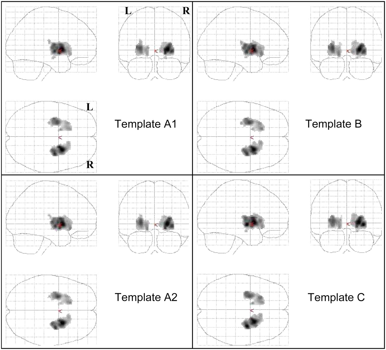
- FIGURE 5.
SPM t maps (Puncorrected < 0.001) obtained by comparing ET patients with PD patients (6 PD < 6 ET) after normalization to each template. Four statistical maps reveal similar significant differences between the 2 groups for 4 different templates. R = right; L = left.


Tables
- TABLE 1
Characteristics of Image Acquisition and Data Processing Used to Create 4 Templates
Template A1 A2 B C Center Toulouse, France Créteil, France Paris, France Camera Triple-head IRIX 3 (Picker) Dual-head AXIS (Philips) Triple-head IRIX 3 (Picker) Collimators LE-HR parallel LE-HR parallel LE-HR parallel Controls ET ET Healthy volunteers No. of controls 15 10 5 Acquisition parameters 120 projections/360°, 1282 120 projections/360°, 1282 120 projections/360°, 1282 Reconstruction parameters (iteration, subsets) OSEM (12–6) OSEM (6–4) OSEM (4) OSEM (12–6) Software/workstation Hermes Segami-Mirage Odyssey Hermes Homogeneous attenuation correction μ = 0.15 No μ = 0.15 μ = 0.15 Scatter correction No No No Yes Variable collimator response correction No No No Yes 3D postfiltering (order, cut-off frequency) Butterworth, 5, 1.7 cycles·cm−1 Wiener Butterworth, 4, 0.35 cycle/pixel Butterworth, 5, 1.7 cycles·cm−1 LE-HR = low energy, high resolution; OSEM = ordered-subsets expectation maximization; μ = attenuation coefficient (cm−1); 3D = 3-dimensional.
- TABLE 2
Evaluation of Reproducibility of BR Values Obtained from Normalization Procedure Across Templates
Template Variability in % (mean ± SD) ICC [95% CI] 95% limits of agreement A2 vs. A1 3.69 ± 3.04 0.99 [0.98−0.99] −0.11 to 0.24 B vs. A1 4.45 ± 3.10 0.99 [0.98−0.99] −0.13 to 0.23 C vs. A1 7.01 ± 3.75 0.98 [0.96−0.99] −0.02 to 0.28 - TABLE 3
SPM Results in ET Subjects Compared with PD Subjects According to Template Used for Normalization
Cluster level Voxel level MNI coordinates (mm) Pcorrected KE Puncorrected Pcorrected Z score Puncorrected t x y z Location ET > PD with template A1 0.000 1,415 0.000 0.001 5.30 0.000 14.97 24 6 2 R putamen 0.004 4.96 0.000 12.13 32 −2 −8 0.009 4.79 0.000 10.94 28 −8 10 0.000 1,074 0.000 0.014 4.71 0.000 10.45 −28 −2 4 L putamen 0.024 4.60 0.000 9.79 −34 −8 6 0.164 4.06 0.000 7.23 −14 4 4 ET > PD with template A2 0.000 1,506 0.000 0.000 5.40 0.000 15.91 24 8 0 R putamen 0.003 5.02 0.000 12.59 20 2 −8 0.003 5.01 0.000 12.50 28 4 10 0.000 1,095 0.000 0.005 4.90 0.000 11.70 −26 −2 2 L putamen 0.057 4.40 0.000 8.74 −14 22 −8 0.154 4.06 0.000 7.22 −26 16 4 ET > PD with template B 0.000 1,478 0.000 0.000 5.39 0.000 15.79 24 6 0 R putamen 0.003 4.99 0.000 12.32 32 −6 −2 0.010 4.76 0.000 10.79 28 −10 8 0.000 1,151 0.000 0.005 4.90 0.000 11.73 −26 −2 4 L putamen 0.033 4.52 0.000 9.37 −34 −8 6 0.056 4.41 0.000 8.77 −16 22 −6 ET > PD with template C 0.000 1,573 0.000 0.001 5.25 0.000 14.49 32 −6 −2 R putamen 0.001 5.23 0.000 14.34 22 6 2 0.004 4.95 0.000 12.04 26 4 10 0.000 1,115 0.000 0.017 4.66 0.000 10.17 −26 −2 2 L putamen 0.034 4.52 0.000 9.33 −32 −8 6 0.047 4.44 0.000 8.97 −22 18 4 KE = number of voxels per cluster; R = right; L = left.


Jump to section
Related Articles
Cited By...
- Generation of Structural MR Images from Amyloid PET: Application to MR-Less Quantification
- An Anthropomorphic Phantom Study of Brain Dopamine Transporter SPECT Images Obtained Using Different SPECT/CT Devices and Collimators
- Segmentation-Based MR Attenuation Correction Including Bones Also Affects Quantitation in Brain Studies: An Initial Result of 18F-FP-CIT PET/MR for Patients with Parkinsonism
- SNM Practice Guideline for Dopamine Transporter Imaging with 123I-Ioflupane SPECT 1.0
- Dopaminergic Striatal Innervation Predicts Interlimb Transfer of a Visuomotor Skill