


Article Figures & Data
Figures
- FIGURE 1.
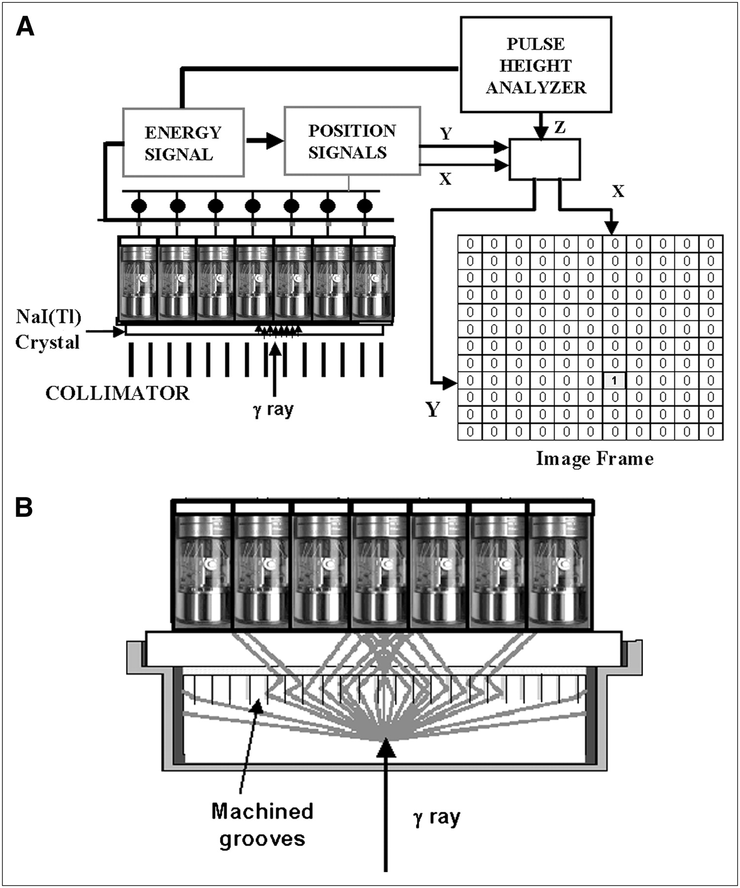
Scintillation camera. (A) In a conventional scintillation camera, light from the scintillation is sampled by an array of photomultipler tubes, which generate an energy signal as well as an x,y-coordinate pair. If the event falls within the pulse–height analyzer energy window, an image matrix element is incremented at the associated x,y-location. (B) With StarBrite technology (Saint-Gobain Crystals and Detectors), grooves are machined into the back surface of the 1-in. thick NaI(Tl) crystal to limit the spread of the scintillation light, resulting in improved intrinsic spatial resolution.
- FIGURE 2.
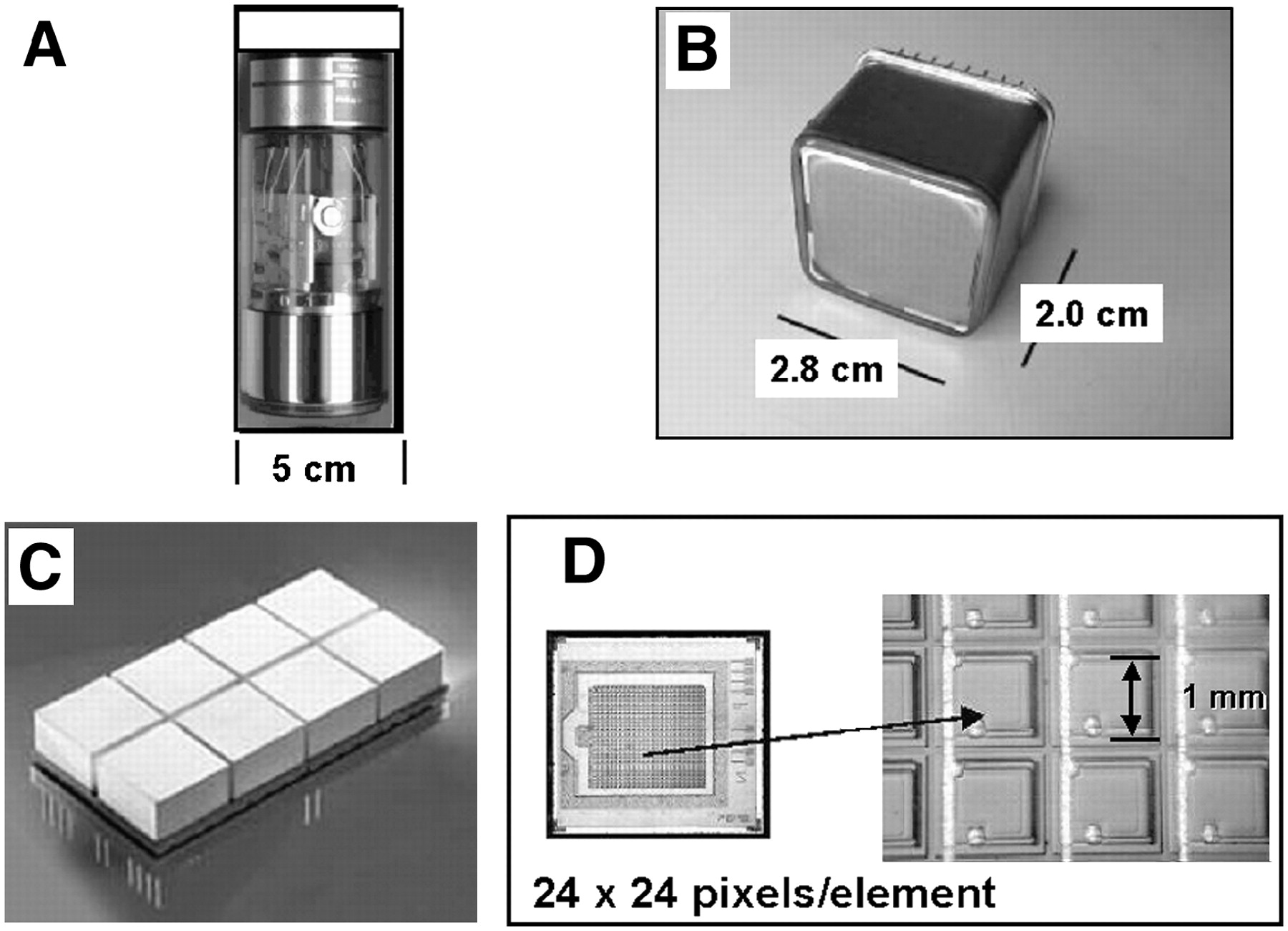
Photon transducers for converting scintillations into an electronic pulse. (A) PMT. (B) Position-sensitive PMT. (C) Avalanche photodiode. (D) Silicon photomultiplier.
- FIGURE 3.
New cardiac SPECT systems. (A) CardiArc uses slit–slat approach to acquire projection information. (B) D-SPECT detector head consists of 10 pixelated CZT detector columns. After a scout scan determines heart location, each detector independently swivels to collect projections.
- FIGURE 4.
SPECT/CT systems. (A) GE Healthcare Infinia Hawkeye. (B) Philips Precedence. (C) Siemens Symbia True Point. (D) Example of clinical 111In-pentetreotide SPECT/CT study of patient with carcinoid.
- FIGURE 5.
Mutual perpendicular cross-sections through submillimeter-resolution 3D myocardial perfusion image volume of living mouse (50). Image data were acquired over 30 min, starting 30 min after administration of 222 MBq (6 mCi) of 99mTc-tetrofosmin. On left a short-axis slice shows myocardial perfusion in right ventricular (RV) and left ventricular (LV) walls. Perfusion in anterior papillary muscle (arrow) can be distinguished from other parts of LV wall. On top right is a vertical long-axis slice; on bottom right is a horizontal long-axis slice. Images at bottom show a hot rod phantom and the reconstructed cross-sectional image, with a slice thickness of 0.5 mm.
- FIGURE 6.
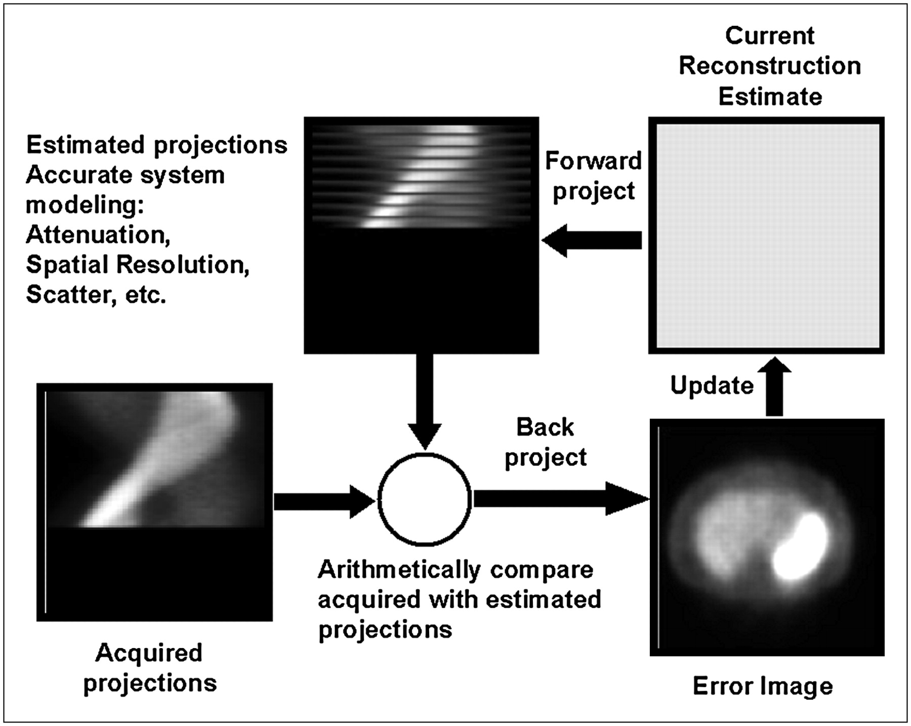
Iterative reconstruction. Projections calculated from the estimated distribution are arithmetically compared with measured data and the result backprojected to update the next estimate. Corrections for attenuation, scatter, and spatial resolution are made by mathematically modeling these factors into the forward and backprojection calculations.
- FIGURE 7.
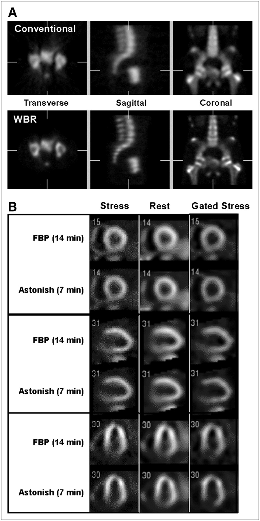
Example images from new image-processing algorithms. (A) Wide Beam Reconstruction (WBR) applied to bone SPECT. The top row shows the conventional SPECT views, whereas the bottom row shows the results when WBR is applied to the same patient data. (Images courtesy of Vanderbilt University Medical Center, Nashville, TN.) (B) Astonish method applied to myocardial perfusion SPECT. The stress, rest, and gated components of a 99mTc-MIBI myocardial SPECT study processed with filtered backprojection are compared with Astonish processed data acquired in half the time. (Images courtesy of Radiologic Associates of Sacramento.)


Tables
Scintillator Atomic number Z effective Density ρ (g/cm3) Decay time τ (ns) Wavelength λ (nm) Relative light output (% of NaI(Tl)) NaI(Tl) 50 3.67 200 415 100 CsI(Tl) 54 4.5 1,000 550 45 (118*) CsI(Na) 54 4.51 630 420 85 LaBr3:Ce 47 5.3 25 360 160 ↵* Represents total light output. Because of the long wavelength of the scintillation, the effective signal for CsI(Tl) is only 45% of NaI(Tl).
No. of detector heads 2 Field of view 40 × 55 cm Energy resolution 9.50% Intrinsic spatial resolution 3.8 mm (FWHM) Planar count sensitivity (LEHR) 190 cps/MBq (95 cps/MBq per head) SPECT spatial resolution (LEHR) 10.5 mm (FWHM) LEHR = low energy, high resolution; FWHM = full width at half maximum; cps = counts per second.
Infinia Hawkeye Precedence True Point Manufacturer GE Healthcare Philips Siemens SPECT System Infinia Skylight Symbia CT system Hawkeye Brilliance T, T2, T6 No. of CT slices 1 or 4 6 or 16 2 or 6 Slice thickness (mm) 10 or 5 0.6–12 0.6–10* Tube rotation (s) 23 0.5 0.6–1.5* Standard HC resolution (lp/cm) (2% MTF) >3 13 15* Room dimensions (cm) 419 × 470 711 × 442 640 × 358 ↵* Values are for Symbia T6 (see text).
HC = high contrast; lp/cm = line pairs per centimeter; MTF = modulation transfer function.
Manufacturer Device Web site URL Bioscan NanoSPECT www.bioscan.com/product.php?p=nanospect Gamma Medica X-SPECT www.gammamedica.com/X-SPECT.php GE Healthcare Explore SPECT-CZT www.gehealthcare.com/usen/fun_img/pcimaging/index.html Molecular Imaging U-SPECT www.milabs.com NeuroPhysics MollyQ www.neurophysics.com/products/products.html Siemens Inveon www.medical.siemens.com/webapp/wcs/stores/servlet/ProductDisplay?storeId=10001&langId=-11&catalogId=-11&catTree=100001,1006503&productId=168892


Jump to section
Related Articles
Cited By...
- Multicenter Study of Quantitative SPECT: Reproducibility of 99mTc Quantitation Using a Conjugated-Gradient Minimization Reconstruction Algorithm
- Approaches to Reducing Radiation Dose from Radionuclide Myocardial Perfusion Imaging
- Breast Imaging Devices for Nuclear Medicine
- Semiconductor Detectors Allow Low-Dose-Low-Dose 1-Day SPECT Myocardial Perfusion Imaging
- Validation of CT Attenuation Correction for High-Speed Myocardial Perfusion Imaging Using a Novel Cadmium-Zinc-Telluride Detector Technique
- Multicenter Trial of High-Speed Versus Conventional Single-Photon Emission Computed Tomography Imaging: Quantitative Results of Myocardial Perfusion and Left Ventricular Function
- Nuclear Myocardial Perfusion Imaging with a Cadmium-Zinc-Telluride Detector Technique: Optimized Protocol for Scan Time Reduction
- SPECT/CT
- Cardiovascular nuclear imaging: from perfusion to molecular function
- High-Speed Myocardial Perfusion Imaging: Initial Clinical Comparison With Conventional Dual Detector Anger Camera Imaging