


Article Figures & Data
Figures
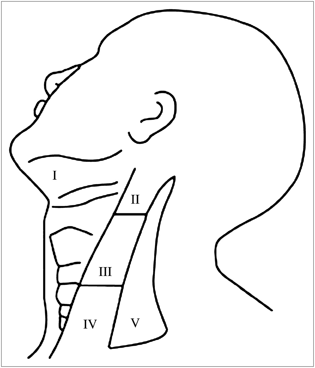
- FIGURE 1.
Schema of neck lymph node levels used for surgical and radiologic assessment.
- FIGURE 2.
Flow chart shows distribution of findings.
- FIGURE 3.
TP PET/CT in 53-y-old man with cancer of left oral tongue. Primary tumor is not well seen on noncontrast CT (A) but is clearly delineated on PET (B) and PET/CT fusion (C) images. (D) CT shows borderline lymph node in right level II neck but no abnormality in left neck. However, PET shows moderate 18F-FDG uptake (SUV, 4.4) in left neck (E), which on fusion images clearly localizes to a small left level II node (F).
- FIGURE 4.
FP PET/CT in 50-y-old man with cancer of right oral tongue. Primary tumor is not well seen on noncontrast CT (A) but is clearly delineated on PET (B) and PET/CT fusion (C) images. (D) CT shows small lymph node in right neck level III, which shows moderate 18F-FDG uptake (SUV, 4.6) on PET image (E). Fusion image shows 18F-FDG uptake clearly within this node (F). Histopathology revealed abundant lymphocytes but no metastatic deposit.


Tables
Characteristic Value Sex Male 21 Female 10 Age (y) Mean 60 ± 12 Range 37–84 No. of primary tumor sites Oral tongue 24 Gum 2 Floor of mouth 4 Base of tongue 1 No. of primary tumors T1 13 T2 14 T3 3 T4 1 No. of neck dissections Unilateral 26 Bilateral 5 Type of neck dissection SOHND (levels I–III) 11 Extended SOHND (levels I–IV) 14 MRND, type III (levels I–V) 10 LND (levels II–IV) 1 SOHND = supraomohyoid neck dissection, levels I–III; extended SOHND = SOHND, levels I–IV; MRND = modified radical neck dissection, type III, levels I–V with preservation of sternocleidomastoid muscle, internal jugular vein, and spinal accessory nerve; LND = lateral neck dissection, levels II–IV.
Neck TP FP TN FN Total Levels 6 6 127 3 142 Sides 6 4 23 3 36 Sensitivity (%) Specificity (%) PPV (%) NPV (%) Accuracy (%) Levels 67 95 50 98 94 Sides 67 85 60 88 80 Levels Level With metastatic nodes (n) Dissected (n) PET TP (n) PET TN (n) PET FP (n) PET FN (n) I 35 2 1 33 0 1 II 36 4 2 27 5 2 III 36 3 3 32 1 0 IV 25 0 0 25 0 0 V 10 0 0 10 0 0 Total 142 9 6 127 6 3 Patient no. Nodal level CT short-axis diameter (mm) Histologic largest diameter of metastasis (mm) ECS (yes/no) PET visual grade SUV TP findings 20 III LN 1: 11 × 8 15 − 5 + 6.2 LN 2: 8 × 6 7 − 3 + 2.8 11 II 8 × 5 3 3 + 3.0 30 III 14 × 10 16 + 5 + 4.4* 8 × 4 11 − − − 31 I (L) 7 × 6 8 + 3 + 4.5 II (R) 14 × 11 10 − 4 + 4.9 26 III 11 × 5 0.3 − 5 + 5.6 FP findings 5 II 12 × 11 5 + 5.7 19 II 10 × 8 4 + 3.9 24 II 10 × 8 3 + 2.7 27 II 11 × 7 5 + 4.6 III 9 × 8 5 + 4.6 26† II 6 × 5 4 + 3.5


Jump to section
Related Articles
Cited By...
- Molecular Imaging to Plan Radiotherapy and Evaluate Its Efficacy
- The Value of PET Compared to MRI in Malignant Head and Neck Tumors
- Contrast-enhanced CT and MRI for detecting neck metastasis of oral cancer: comparison between analyses performed by oral and medical radiologists
- Assessment of a New 18F-FDG PET/CT Protocol in the Staging of Oral Cavity Carcinomas
- PET and PET/CT of the Neck Lymph Nodes Improves Risk Prediction in Patients with Squamous Cell Carcinoma of the Oral Cavity
- 18F-FDG Uptake in Reactive Neck Lymph Nodes of Oral Cancer: Relationship to Lymphoid Follicles
- Improvements in Cancer Staging with PET/CT: Literature-Based Evidence as of September 2006
- Clinical Role of 18F-FDG PET/CT in the Management of Squamous Cell Carcinoma of the Head and Neck and Thyroid Carcinoma