
Main menu

  • Home
  • Content
    • Current
    • Ahead of print
    • Past Issues
    • JNM Supplement
    • SNMMI Annual Meeting Abstracts
    • Continuing Education
    • JNM Podcasts
  • Subscriptions
    • Subscribers
    • Institutional and Non-member
    • Rates
    • Journal Claims
    • Corporate & Special Sales
  • Authors
    • Submit to JNM
    • Information for Authors
    • Assignment of Copyright
    • AQARA requirements
  • Info
    • Reviewers
    • Permissions
    • Advertisers
  • About
    • About Us
    • Editorial Board
    • Contact Information
  • More
    • Alerts
    • Feedback
    • Help
    • SNMMI Journals
  • SNMMI
    • JNM
    • JNMT
    • SNMMI Journals
    • SNMMI

User menu

  • Subscribe
  • My alerts
  • Log in
  • My Cart

Search

  • Advanced search
Journal of Nuclear Medicine
  • SNMMI
    • JNM
    • JNMT
    • SNMMI Journals
    • SNMMI
  • Subscribe
  • My alerts
  • Log in
  • My Cart
Journal of Nuclear Medicine

Advanced Search

  • Home
  • Content
    • Current
    • Ahead of print
    • Past Issues
    • JNM Supplement
    • SNMMI Annual Meeting Abstracts
    • Continuing Education
    • JNM Podcasts
  • Subscriptions
    • Subscribers
    • Institutional and Non-member
    • Rates
    • Journal Claims
    • Corporate & Special Sales
  • Authors
    • Submit to JNM
    • Information for Authors
    • Assignment of Copyright
    • AQARA requirements
  • Info
    • Reviewers
    • Permissions
    • Advertisers
  • About
    • About Us
    • Editorial Board
    • Contact Information
  • More
    • Alerts
    • Feedback
    • Help
    • SNMMI Journals
  • View or Listen to JNM Podcast
  • Visit JNM on Facebook
  • Join JNM on LinkedIn
  • Follow JNM on Twitter
  • Subscribe to our RSS feeds
OtherSPECIAL CONTRIBUTION

The 2006 Henry N. Wagner Lecture: Of Mice and Men (and Positrons)—Advances in PET Imaging Technology

Simon R. Cherry
Journal of Nuclear Medicine November 2006, 47 (11) 1735-1745;
Simon R. Cherry
  • Find this author on Google Scholar
  • Find this author on PubMed
  • Search for this author on this site
  • Article
  • Figures & Data
  • Info & Metrics
  • PDF
Loading

Article Figures & Data

Figures

  • Tables
  • FIGURE 1. 
    • Download figure
    • Open in new tab
    • Download powerpoint
    FIGURE 1. 

    Images and photographs illustrating the major advances in PET instrumentation and methods since 1990 discussed in text: whole-body PET (image courtesy of Dr. Magnus Dahlbom, UCLA) (A), 3D PET (B), LSO and related scintillators (photograph courtesy of Dr. Charles Melcher, Siemens) (C), iterative reconstruction methods (images courtesy of Dr. Richard Leahy, USC) (D), PET/CT (image courtesy of Dr. David Townsend, University of Tennessee) (E), and preclinical PET (mouse image courtesy of Dr. Craig Abbey, UCSB) (F).

  • FIGURE 2. 
    • Download figure
    • Open in new tab
    • Download powerpoint
    FIGURE 2. 

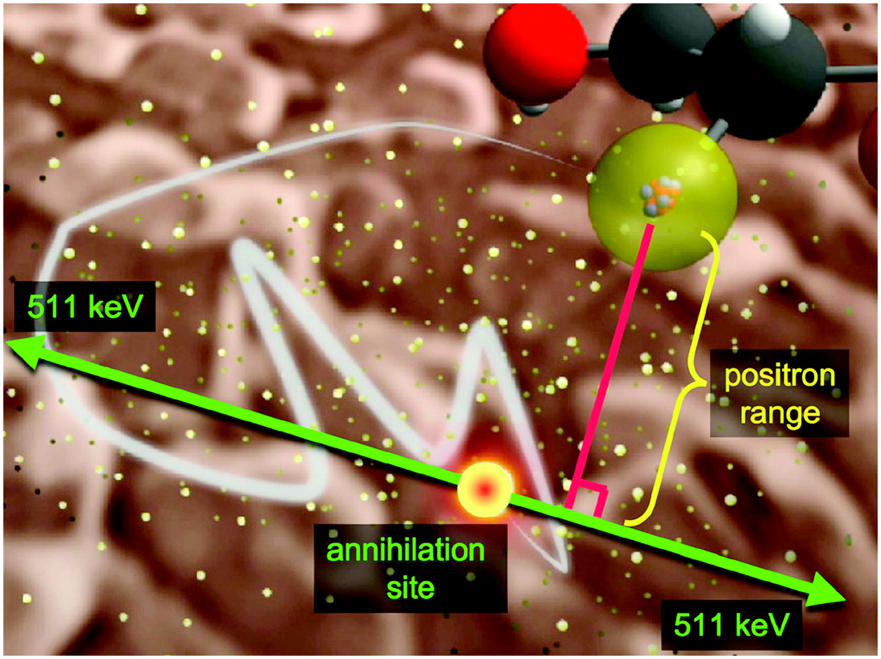
    Basic physics of PET. 18F atom (yellow) on FDG or FDG-6-phosphate molecule decays, emitting positron that scatters in tissue until it loses enough energy to undergo annihilation with an electron, in which mass of positron and electron are converted into 2 back-to-back 511-keV photons.
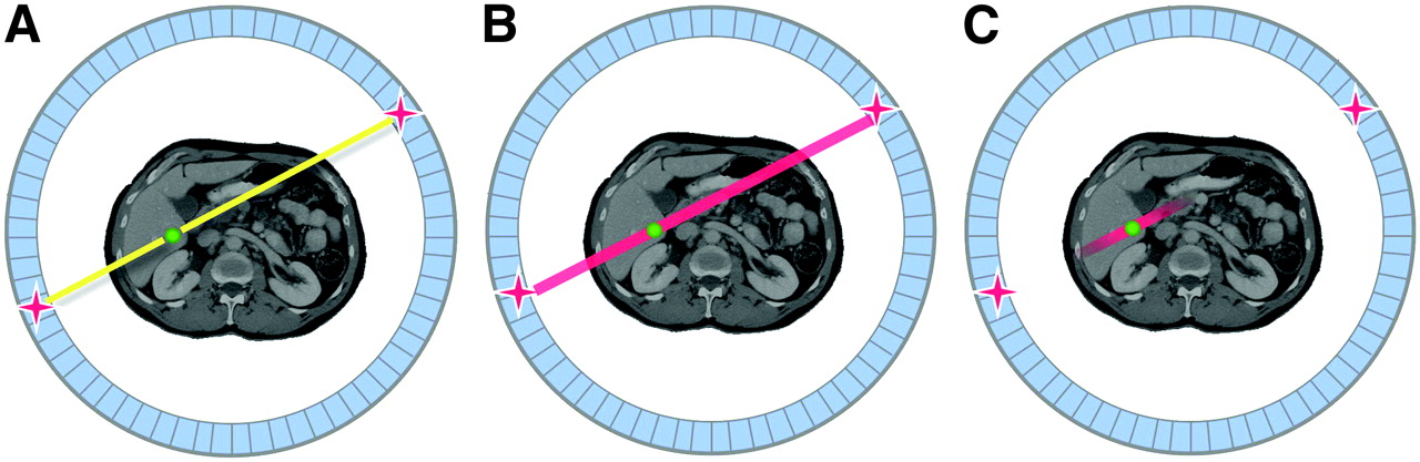
  • FIGURE 3. 
    • Download figure
    • Open in new tab
    • Download powerpoint
    FIGURE 3. 

    Illustration of TOF PET: detection of 2 annihilation photons in PET scanner (A), uniform-probability weighting of annihilation site in standard PET (B), and use of TOF information to constrain location of annihilation site during image reconstruction (C).

  • FIGURE 4. 
    • Download figure
    • Open in new tab
    • Download powerpoint
    FIGURE 4. 

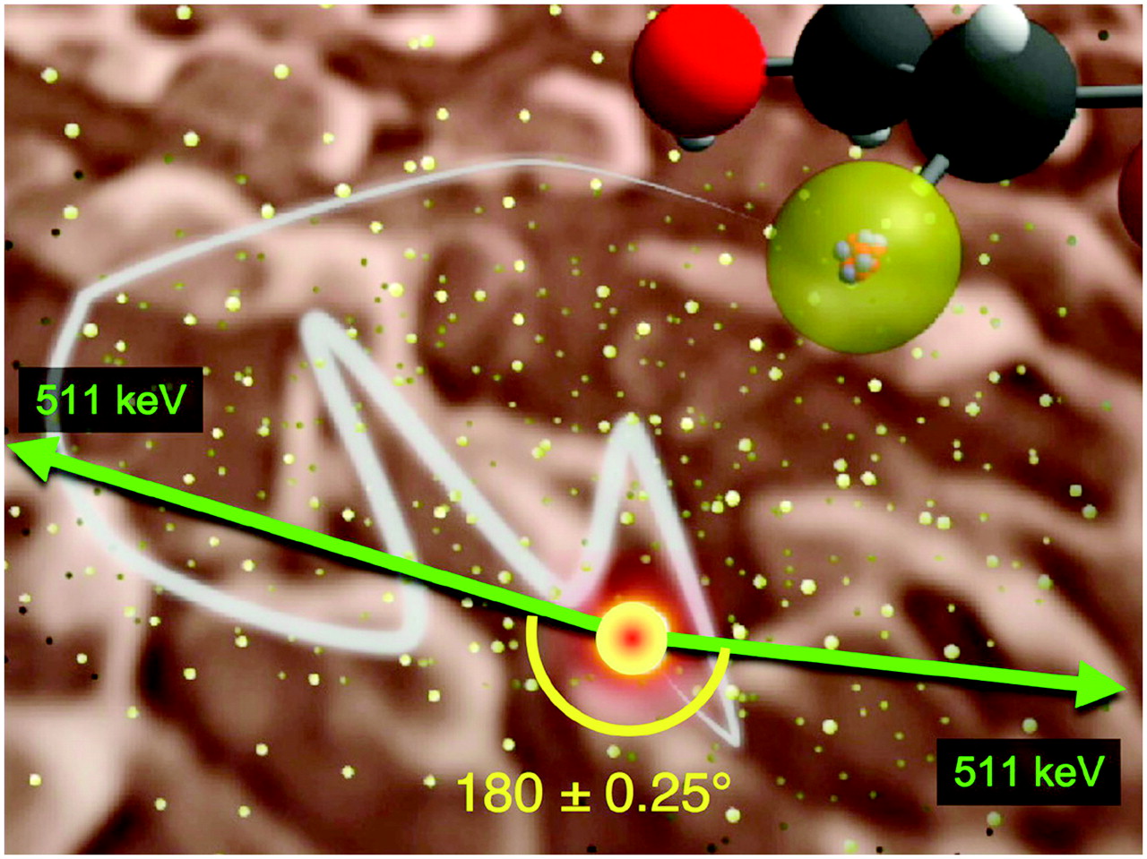
    Illustration of non-colinearity of annihilation photons in PET. Angle is greatly exaggerated; distribution of angles around 180° is gaussian, with SD of only 0.25°.

  • FIGURE 5. 
    • Download figure
    • Open in new tab
    • Download powerpoint
    FIGURE 5. 

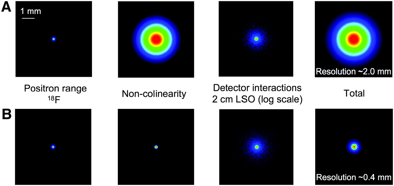
    Contribution of physics factors (positron range, non-colinearity, and detector scatter) to resolution attainable in PET: clinical PET scanner, with detector ring diameter of 80 cm, consisting of 2-cm-thick LSO detectors, and imaging radiotracer labeled with 18F (A); small-animal PET scanner, with detector ring diameter of 8 cm, also consisting of 2-cm-thick LSO detectors and imaging 18F-labeled radiotracer (B). Contribution of positron range, non-colinearity, and detector scatter is shown in each case.

  • FIGURE 6. 
    • Download figure
    • Open in new tab
    • Download powerpoint
    FIGURE 6. 

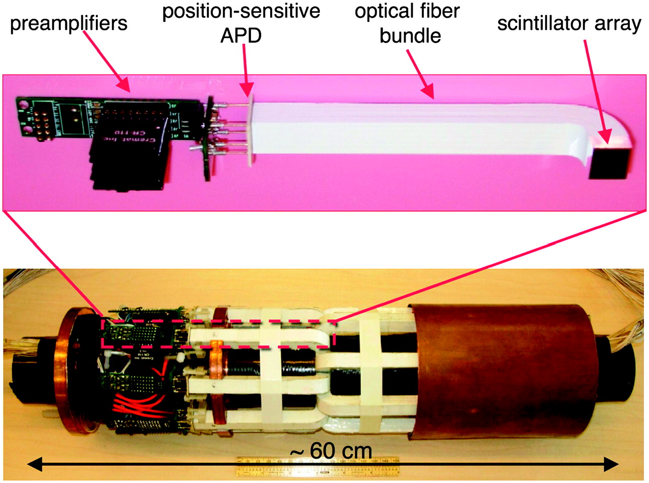
    Photograph of MRI-compatible PET insert based on arrays of LSO scintillator coupled through short lengths of optical fibers to position-sensitive APDs and MRI-compatible electronics. This insert fits inside bore of 7-T animal MRI scanner, permitting simultaneous PET and MRI studies.
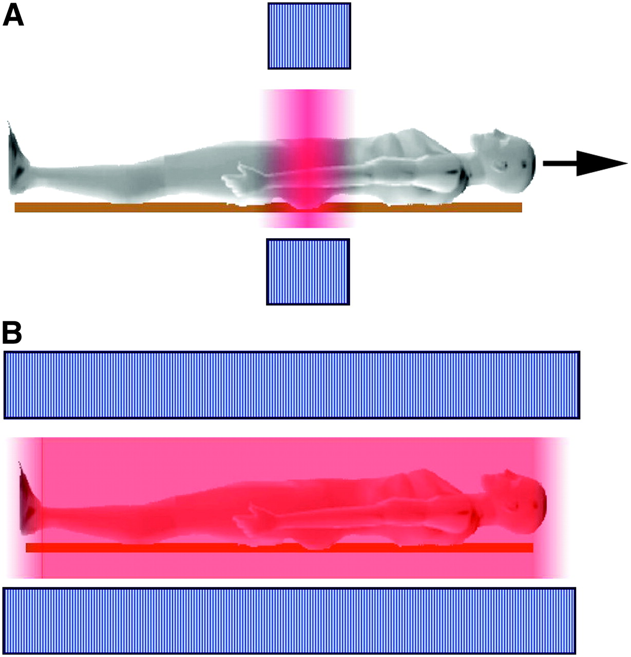
  • FIGURE 7. 
    • Download figure
    • Open in new tab
    • Download powerpoint
    FIGURE 7. 

    Schematic illustration of current configuration and sensitivity for 3D whole-body PET (A) compared with configuration in which whole body is within field of view (B). Axial extent of PET detectors is indicated by blue, and sensitivity is indicated by the intensity of red.

  • FIGURE 8. 
    • Download figure
    • Open in new tab
    • Download powerpoint
    FIGURE 8. 

    Concept of whole-body PET/MRI scanner, and images that such a system might produce (PET image courtesy of Siemens Medical Solutions; whole-body T1-weighted MR image courtesy of Dr. Heinz-Peter Schlemmer, University of Tübingen).

Tables

  • Figures
    • View popup
    TABLE 1

    Properties of PET Scintillators

    ScintillatorRelative light output (NaI(Tl) = 100)Decay time (ns)Thickness for 90% efficiency at 511 keV (cm)
    NaI(Tl)1002306.6
    BGO153002.4
    LSO, LYSO70402.7
    GSO:Ce25603.3
    BaF2280.8 (15%), 640 (85%)5.1
    CsF52.55.4
    LaBr3150354.9
    • GSO = gadolinium oxyorthosilicate.

PreviousNext
Back to top

In this issue

Journal of Nuclear Medicine: 47 (11)
Journal of Nuclear Medicine
Vol. 47, Issue 11
November 2006
  • Table of Contents
  • Table of Contents (PDF)
  • About the Cover
  • Index by author
Print
Download PDF
Article Alerts
Sign In to Email Alerts with your Email Address
Email Article

Thank you for your interest in spreading the word on Journal of Nuclear Medicine.

NOTE: We only request your email address so that the person you are recommending the page to knows that you wanted them to see it, and that it is not junk mail. We do not capture any email address.

Enter multiple addresses on separate lines or separate them with commas.
The 2006 Henry N. Wagner Lecture: Of Mice and Men (and Positrons)—Advances in PET Imaging Technology
(Your Name) has sent you a message from Journal of Nuclear Medicine
(Your Name) thought you would like to see the Journal of Nuclear Medicine web site.
Citation Tools
The 2006 Henry N. Wagner Lecture: Of Mice and Men (and Positrons)—Advances in PET Imaging Technology
Simon R. Cherry
Journal of Nuclear Medicine Nov 2006, 47 (11) 1735-1745;

Citation Manager Formats

  • BibTeX
  • Bookends
  • EasyBib
  • EndNote (tagged)
  • EndNote 8 (xml)
  • Medlars
  • Mendeley
  • Papers
  • RefWorks Tagged
  • Ref Manager
  • RIS
  • Zotero
Share
The 2006 Henry N. Wagner Lecture: Of Mice and Men (and Positrons)—Advances in PET Imaging Technology
Simon R. Cherry
Journal of Nuclear Medicine Nov 2006, 47 (11) 1735-1745;
Twitter logo Facebook logo LinkedIn logo Mendeley logo
  • Tweet Widget
  • Facebook Like
  • Google Plus One
Bookmark this article

Jump to section

  • Article
    • Abstract
    • THE PAST: A REVIEW OF MAJOR ADVANCES IN PET TECHNOLOGY SINCE 1990
    • THE PRESENT AND THE NEAR FUTURE
    • THE FAR FUTURE: WHOLE-BODY PET/MRI?
    • CONCLUSION
    • Acknowledgments
    • Footnotes
    • References
  • Figures & Data
  • Info & Metrics
  • PDF

Related Articles

  • This Month in JNM
  • PubMed
  • Google Scholar

Cited By...

  • Is There a Need for a Pediatric PET/CT Camera?
  • Development and Evaluation of mini-EXPLORER: A Long Axial Field-of-View PET Scanner for Nonhuman Primate Imaging
  • Total-Body PET: Maximizing Sensitivity to Create New Opportunities for Clinical Research and Patient Care
  • A Prototype High-Resolution Small-Animal PET Scanner Dedicated to Mouse Brain Imaging
  • Positron emission tomography/computed tomography surveillance in patients with Hodgkin lymphoma in first remission has a low positive predictive value and high costs
  • Promising New Photon Detection Concepts for High-Resolution Clinical and Preclinical PET
  • Assessing Antibody Pharmacokinetics in Mice with In Vivo Imaging
  • Small-Animal Molecular Imaging Methods
  • The Advantages of Nanoparticles for PET
  • NEMA NU4-2008 Image Quality Performance Report for the microPET Focus 120 and for Various Transmission and Reconstruction Methods
  • New Technologies for Human Cancer Imaging
  • Noninvasive Imaging of Cell-Mediated Therapy for Treatment of Cancer
  • Latest Advances in Molecular Imaging Instrumentation
  • PET Image Denoising Using a Synergistic Multiresolution Analysis of Structural (MRI/CT) and Functional Datasets
  • Use of a Peptide Derived from Foot-and-Mouth Disease Virus for the Noninvasive Imaging of Human Cancer: Generation and Evaluation of 4-[18F]Fluorobenzoyl A20FMDV2 for In vivo Imaging of Integrin {alpha}v{beta}6 Expression with Positron Emission Tomography
  • Google Scholar

More in this TOC Section

  • Computational Nuclear Oncology Toward Precision Radiopharmaceutical Therapies: Ethical, Regulatory, and Socioeconomic Dimensions of Theranostic Digital Twins
  • Consensus Nomenclature for Radionuclide Therapy: Initial Recommendations from Nuclear Medicine Global Initiative
  • Self-Citation Among Highly Cited Nuclear Medicine and Medical Imaging Researchers
Show more SPECIAL CONTRIBUTION

Similar Articles

SNMMI

© 2025 SNMMI

Powered by HighWire