


Article Figures & Data
Figures
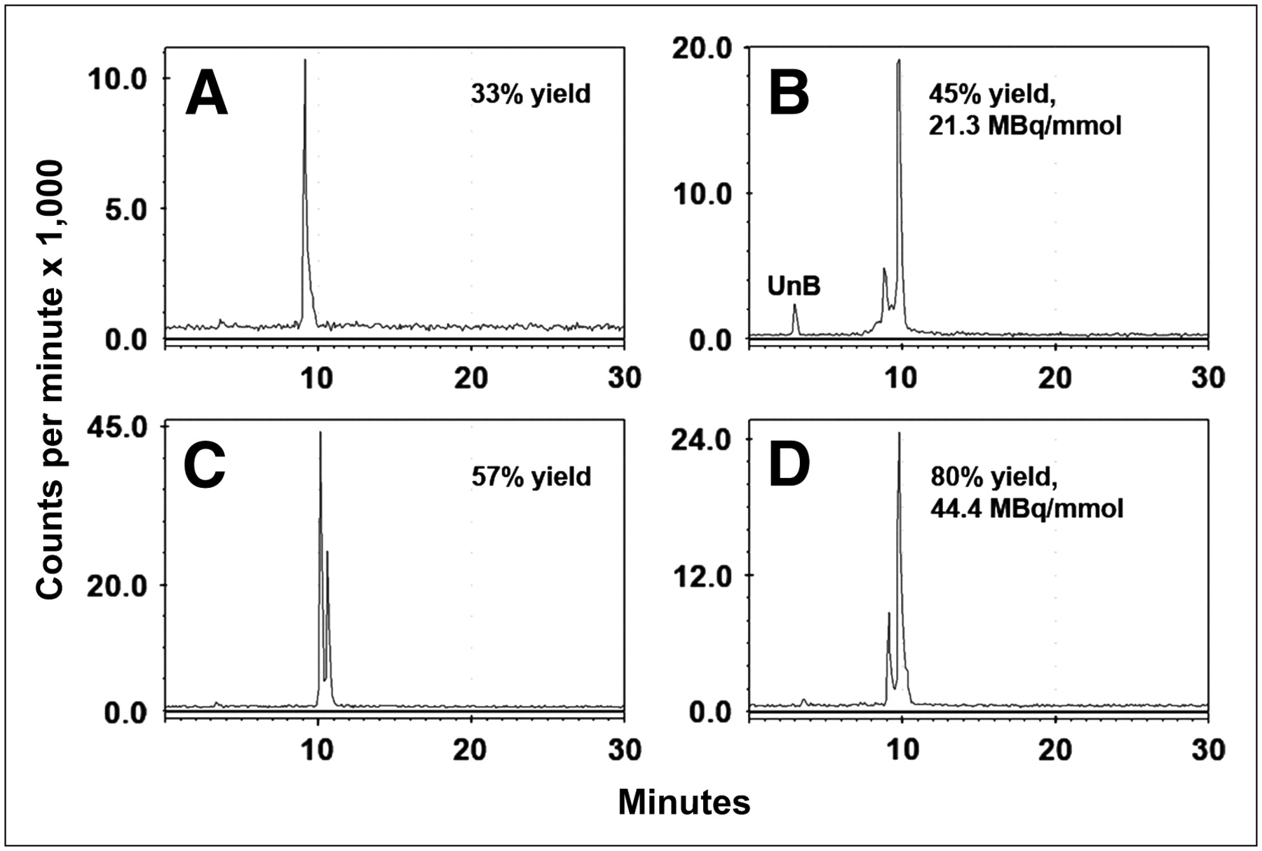
- FIGURE 1.
RP-HPLC illustrates final radiolabeled products obtained under different radioiodination conditions. (A and C) 124I-Labeled products obtained using Na124I with (C) or without (A) addition of carrier-added iodide. Addition of carrier improved yields, with formation of both mono- and diiododinated forms. (B and D) 131I-Labeled products prepared using IODO-GEN–coated vials vs. microparticulate IODO-GEN. UnB = unbound radioiodine.
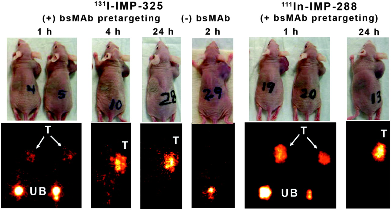
- FIGURE 2.
Whole-body γ-images of 131I-IMP-325 in animals given hBS14 bs-mAb 24 h earlier (+ bs-mAb) or animals given only peptide alone (− bs-mAb). Images on the right show pretargeting under the same conditions using 111In-IMP-288. T = tumor; UB = urinary bladder. Arrows show tumor location on photographs of animals.
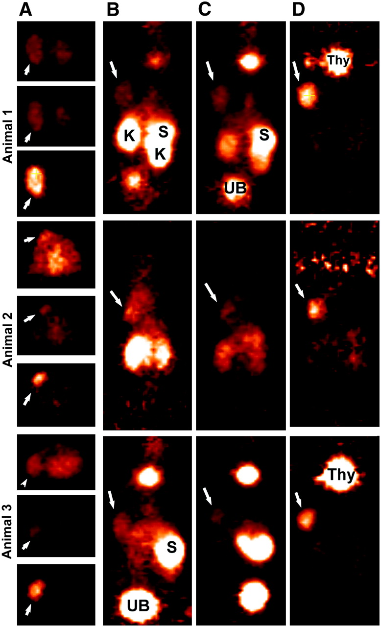
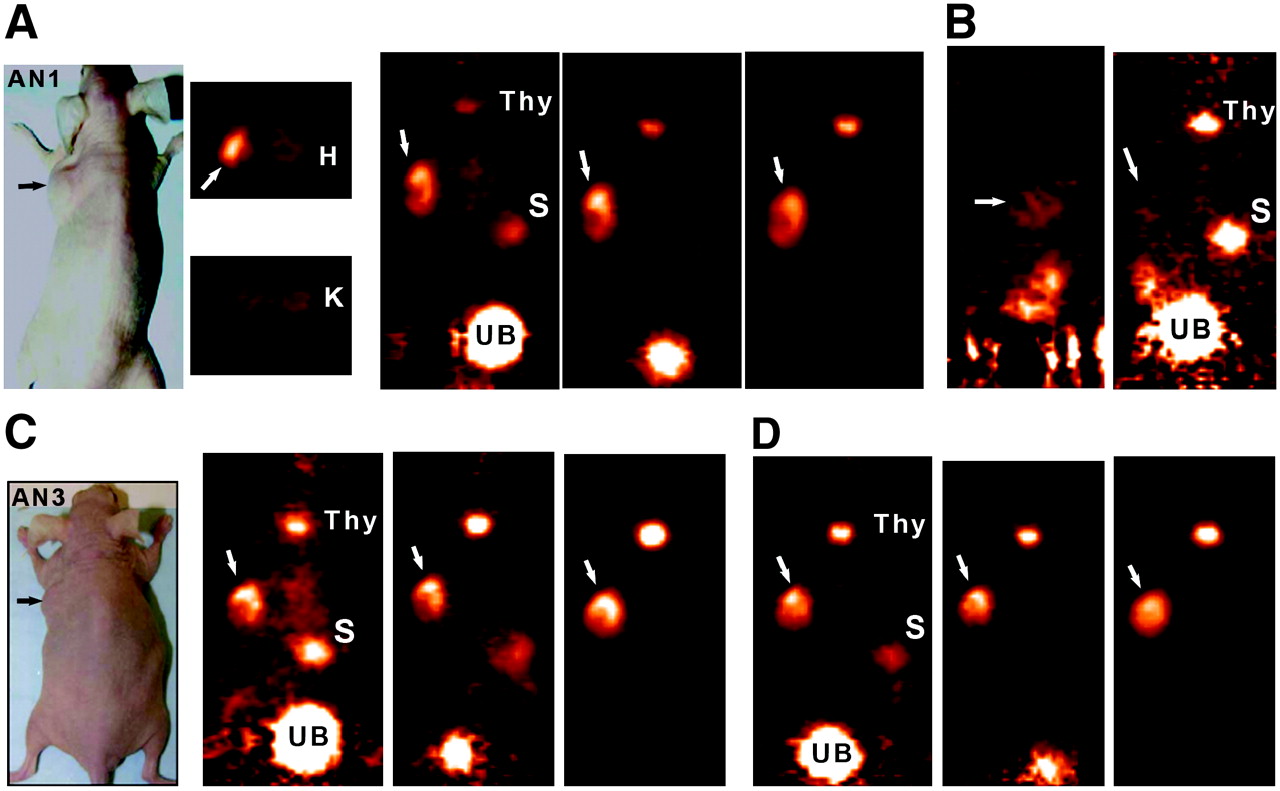
- FIGURE 3.
Focus 120 microPET images of 124I-IMP-325 pretargeted with hBS14 (A, C, and D) or alone (not pretargeted; B). (A) From left to right, photograph of AN1, 2 transverse slices (0.82 mm) at level of tumor (arrows) and kidneys (K), taken ∼2 h after injection, and coronal slices obtained at ∼2, 6, and 24 h after injection. (B) From left to right, sagittal and coronal views obtained 1.9 h after injection. (C) From left to right, photograph of AN3 and coronal slices obtained at ∼2, 6, and 24 h after injection. (D) From left to right, coronal slices obtained at ∼2, 6, and 24 h after injection. All pretargeted images are adjusted to same intensity as 2-h image for each animal to illustrate retention and clearance of 124I-IMP-325 over time. At 24-h necropsy, tumors were 0.448, 0.285, and 0.483 g in pretargeted animals and 0.472 g in animal given 124I-IMP-325 alone. H = heart; S = stomach; Thy = thyroid; UB = urinary bladder.
- FIGURE 4.
Focus 120 microPET images of 3 animals injected with 124I-hMN-14 Fab′. (A) Transverse slices (0.82-mm thick) in same plane to highlight tumor (arrows) localization at ∼2, 6, and 24 h, from top to bottom, respectively, for each animal. Six-hour images are taken at same intensity as 2-h images, but intensity was increased substantially for 24-h images. Coronal sections taken from 2-, 6-, and 24-h intervals (B, C, and D, respectively) illustrate elevated uptake of radiolabeled Fab′ initially in kidneys (K) and stomach (S). Uptake in these tissues was cleared over time, but there was persistent thyroid (Thy) uptake. Tumor weights at necropsy (24 h) were 0.596, 0.173, and 0.280 g, respectively.
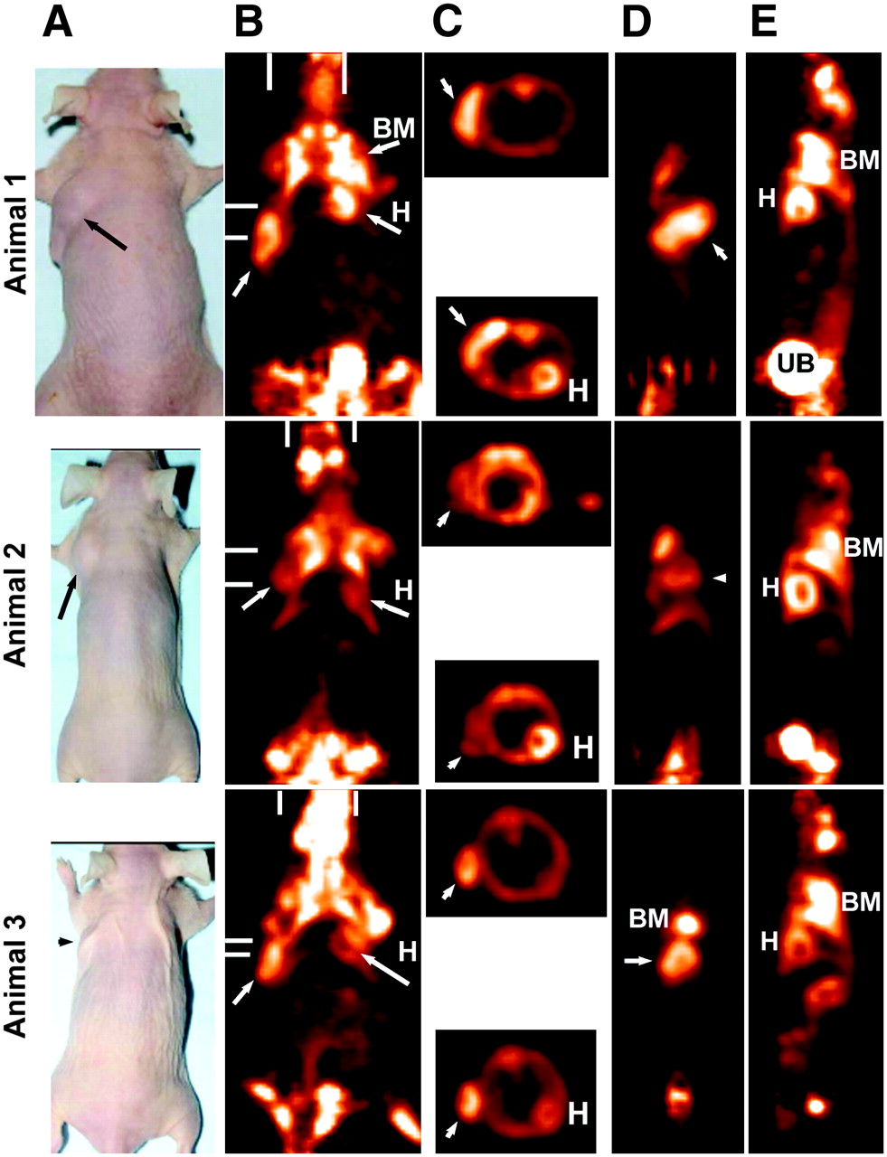
- FIGURE 5.
Focus 120 microPET images of animals bearing LS174T human colorectal cancer xenografts 1.7 h after 18F-FDG. All images were taken at same maximum intensity and background set to 0.0. (A) Photographs of 3 tumor-bearing animals. (B) Coronal slice (0.82-mm thick) taken to highlight uptake in tumor (arrows), as well as in bone marrow (BM) of shoulder and in hind quarters. Heart (H) and brain can also be seen in some images. Bars represent planes used for transverse and sagittal sections. (C) Transverse sections that highlight tumor or heart wall uptake. Sagittal sections illustrate the side view of tumor (D) and a plane that highlights heart, bone marrow, and urinary bladder (UB) activity (E). Tumor weights were 0.425, 0.321, and 0.153 g, respectively.


Tables
- TABLE 1
Tissue Uptake and Targeting Ratios for Pretargeted or Nonpretargeted 131I-IMP-325 and Pretargeted 111In-IMP-288
%ID/organ Pretargeted 131I-IMP-325 131I-Peptide alone, 4 h (n = 2) Pretargeted 111In-IMP-288 Tumor or organ 1 h (n = 5) 4 h (n = 5) 24 h (n = 3) 1 h (n = 5) 24 h (n = 3) Tumor 15.1 ± 4.6* 13.9 ± 3.5 10.5 ± 2.6 0.19, 0.47 18.7 ± 9.6 13.6 ± 2.6 1.10 ± 0.26† 1.15 ± 0.35 1.52 ± 0.65 1.1, 2.7 1.07 ± 0.55 1.40 ± 0.42 Liver 0.48 ± 0.06 0.32 ± 0.04 0.21 ± 0.02 0.15, 0.18 0.36 ± 0.09 0.18 ± 0.02 1.22 ± 0.17 (34.6 ± 5.9)‡ (39.8 ± 2.9) (43.0 ± 9.2) (63.6 ± 24.8) (61.9 ± 9.8) Spleen 0.04 ± 0.00 0.03 ± 0.00 0.01 ± 0.00 0.012, 0.013 0.03 ± 0.01 0.01 ± 0.00 0.10 ± 0.02 (36.4 ± 4.3) (47.2 ± 6.9) (49.4 ± 18.6) (65.5 ± 22.3) (83.9 ± 15.2) Kidney 0.67 ± 0.24 0.37 ± 0.06 0.24 ± 0.02 0.37, 0.35 0.48 ± 0.07 0.21 ± 0.02 0.15 ± 0.02 (3.4 ± 1.3) (4.8 ± 0.2) (4.6 ± 0.9) (6.1 ± 0.9) (6.7 ± 1.0) Lungs 0.13 ± 0.01 0.05 ± 0.01 0.01 ± 0.00 0.02, 0.02 0.11 ± 0.03 0.02 ± 0.00 0.17 ± 0.03 (17.8 ± 3.3) (33.0 ± 1.9) (70.5 ± 20.6) (40.5 ± 3.7) (78.5 ± 9.3) Blood 1.48 ± 0.16§ 0.44 ± 0.06 0.04 ± 0.01 0.30, 0.32 0.96 ± 0.51 0.03 ± 0.01 22.7 ± 1.6‖ (15.5 ± 2.6) (44.3 ± 3.9) (280 ± 10) (40.3 ± 24.0) (517 ± 42) Stomach¶ 1.0 ± 0.1 0.80 ± 0.11 0.05 ± 0.01 0.74, 0.77 0.07 ± 0.03 0.03 ± 0.02 0.4 ± 0.05 (5.7 ± 1.6) (4.5 ± 0.8) (56.8 ± 16.1) (188 ± 200) (179 ± 134) ↵* Values represent mean ± SD, except for peptide alone, where individual animal data are provided.
↵† Italicized values are tissue weights in grams; tumor weights for each group are given, whereas representative tissue weights are provided from group of animals necropsied at 1 h.
↵‡ Tumor-to-nontumor ratios based on %ID/g.
↵§ Total blood volume estimated as 7.4% of body weight.
↵‖ Average body weight of animals necropsied at 1 h.
↵¶ Includes contents.
- TABLE 2
ROIs Derived from microPET Images of Animals Given Pretargeted 124I-IMP-325 or 124I-Humanized Anti-CEA Fab′
Activity/volume (mean ± SD) Tumor/ kidney ratio Animal/time Tumor Kidney Pretargeted 124I-IMP-325 Animal 1 (tumor, 0.448 g) 1.9 h 2.58 ± 1.09 1.11 ± 0.24 2.3 6.3 h 2.44 ± 1.24 0.92 ± 0.23 2.6 24 h 2.44 ± 1.20 0.55 ± 0.24 4.4 Animal 2 (tumor, 0.483 g) 2.7 h 1.82 ± 0.67 0.89 ± 0.38 2.0 6.4 h 1.67 ± 0.68 0.71 ± 0.26 2.4 25 h 1.85 ± 0.64 0.44 ± 0.16 4.2 Animal 3 (tumor, 0.285 g) 2.1 h 2.06 ± 0.75 0.70 ± 0.25 2.9 5.8 h 1.89 ± 0.76 0.44 ± 0.19 4.3 25 h 1.90 ± 0.70 0.29 ± 0.08 6.6 124I-Anti-CEA Fab′ Animal 1 (tumor, 0.596 g) 2.1 h 1.77 ± 0.41 11.28 ± 2.86 0.16 6.5 h 1.76 ± 0.43 3.85 ± 0.53 0.46 26 h 0.49 ± 0.09 0.09 ± 0.05 5.44 Animal 2 (tumor, 0.173 g) 1.8 h 1.07 ± 0.49 8.14 ± 2.73 0.10 6.5 h 0.71 ± 0.33 1.68 ± 0.48 0.42 27 h 0.17 ± 0.09 0.10 ± 0.05 1.7 Animal 3 (tumor, 0.280 g) 2.9 h 1.84 ± 0.58 10.33 ± 3.22 0.18 6.6 h 1.03 ± 0.39 1.42 ± 0.70 0.73 27 h 0.36 ± 0.12 0.09 ± 0.04 4.00 - TABLE 3
Comparative Biodistribution (%ID/g and Tumor-to-Nontumor Ratios) of Pretargeted 124I-Peptide Compared with 124I-Anti-CEA Fab′ and 18F-FDG Based on Necropsy Data
%ID/g (mean ± SD) Pretargeted 124I-peptide 124I-Anti-CEA Fab′ (12 μg) 124I-Anti-CEA Fab′ (100 μg), 24 h (n = 4) 18F-FDG*, 2.0 h (n = 3) Tumor or organ 1 h (n = 5) 3 h (n = 5) 24 h (n = 5) 1 h (n = 5) 3 h (n = 5) 24 h (n = 5) Tumor 15.4 ± 3.1 18.2 ± 4.6 12.5 ± 1.1 5.7 ± 0.4 5.3 ± 0.6 0.8 ± 0.2 0.7 ± 0.2 4.8 ± 1.14 0.15 ± 0.02† 0.19 ± 0.03 0.20 ± 0.04 0.09 ± 0.01 0.10 ± 0.03 0.08 ± 0.02 0.08 ± 0.01 0.41 ± 0.11 Liver 0.6 ± 0.1 0.6 ± 0.2 0.3 ± 0.02 8.2 ± 1.6 3.2 ± 0.5 0.3 ± 0.1 0.1 ± 0.03 0.6 ± 0.1 (26.3 ± 7.8)‡ (31.2 ± 5.5) (43 ± 6) (0.7 ± 0.2) (1.7 ± 0.3) (3.0 ± 1.1) (6.4 ± 1.9) (8.7 ± 1.1) Spleen 0.6 ± 0.1 0.6 ± 0.2 0.2 ± 0.03 17.7 ± 3.9 6.3 ± 1.6 0.2 ± 0.04 0.1 ± 0.02 1.9 ± 0.2 (25.4 ± 6.4) (34.6 ± 8.9) (57 ± 10) (0.3 ± 0.1) (0.9 ± 0.2) (3.2 ± 0.6) (9.3 ± 1.9) (2.6 ± 0.8) Kidney 4.8 ± 0.5 4.3 ± 1.5 2.7 ± 0.3 68.7 ± 7.6 9.2 ± 2.3 0.4 ± 0.2 0.2 ± 0.04 0.7 ± 0.2 (3.3 ± 0.8) (4.5 ± 1.3) (4.6 ± 0.5) (0.1 ± 0.02) (0.6 ± 0.1) (2.6 ± 1.4) (3.0 ± 0.6) (7.1 ± 0.4) Lungs 1.4 ± 0.2 1.1 ± 0.3 0.3 ± 0.03 2.9 ± 0.3 2.9 ± 0.3 0.05 ± 0.01 0.04 ± 0.02 1.7 ± 0.2 (11.6 + 3.6) (17.1 ± 2.9) (44 ± 6) (2.0 ± 0.2) (1.8 ± 0.3) (16 ± 6) (20 ± 5) (2.9 ± 0.4) Blood 1.7 ± 0.2 1.1 ± 0.4 0.05 ± 0.01 4.6 ± 0.3 4.9 ± 0.4 0.07 ± 0.00 0.05 ± 0.03 0.30 ± 0.06 (9.1 ± 2.6) (17.9 ± 3.5) (238 ± 48) (1.3 ± 0.07) (1.1 ± 0.2) (11 ± 3) (15 ± 5) (16.0 ± 0.4) Stomach§ 3.9 ± 0.5 4.0 ± 2.2 0.2 ± 0.1 20.8 ± 4.2 32.4 ± 2.6 0.5 ± 0.2 0.3 ± 0.2 1.8 ± 0.9 (4.0 ± 0.9) (6.0 ± 3.9) (72 ± 24) (0.3 ± 0.05) (0.2 ± 0.02) (1.8 ± 1.0) (3.1 ± 1.0) (2.9 ± 0.4)


Jump to section
Related Articles
Cited By...
- Pretargeting: A Path Forward for Radioimmunotherapy
- {alpha}- Versus {beta}-Emitting Radionuclides for Pretargeted Radioimmunotherapy of Carcinoembryonic Antigen-Expressing Human Colon Cancer Xenografts
- Immuno-PET Using Anticarcinoembryonic Antigen Bispecific Antibody and 68Ga-Labeled Peptide in Metastatic Medullary Thyroid Carcinoma: Clinical Optimization of the Pretargeting Parameters in a First-in-Human Trial
- Diels-Alder Reaction for Tumor Pretargeting: In Vivo Chemistry Can Boost Tumor Radiation Dose Compared with Directly Labeled Antibody
- Quantitative Immuno-SPECT Monitoring of Pretargeted Radioimmunotherapy with a Bispecific Antibody in an Intraperitoneal Nude Mouse Model of Human Colon Cancer
- Phase II Trial of Anticarcinoembryonic Antigen Pretargeted Radioimmunotherapy in Progressive Metastatic Medullary Thyroid Carcinoma: Biomarker Response and Survival Improvement
- Immuno-PET of Cancer: A Revival of Antibody Imaging
- Optimization of Hapten-Peptide Labeling for Pretargeted ImmunoPET of Bispecific Antibody Using Generator-Produced 68Ga
- Pretargeted 177Lu Radioimmunotherapy of Carcinoembryonic Antigen-Expressing Human Colonic Tumors in Mice
- Pretargeted Immuno-Positron Emission Tomography Imaging of Carcinoembryonic Antigen-Expressing Tumors with a Bispecific Antibody and a 68Ga- and 18F-Labeled Hapten Peptide in Mice with Human Tumor Xenografts
- Pretargeted Radioimmunotherapy of Pancreatic Cancer Xenografts: TF10-90Y-IMP-288 Alone and Combined with Gemcitabine
- CD20-targeted tetrameric interferon-{alpha}, a novel and potent immunocytokine for the therapy of B-cell lymphomas
- A Novel Method of 18F Radiolabeling for PET
- Improved Therapeutic Results by Pretargeted Radioimmunotherapy of Non-Hodgkin's Lymphoma with a New Recombinant, Trivalent, Anti-CD20, Bispecific Antibody
- A Novel Bispecific, Trivalent Antibody Construct for Targeting Pancreatic Carcinoma
- Multifunctional Antibodies by the Dock-and-Lock Method for Improved Cancer Imaging and Therapy by Pretargeting
- Bispecific Antibody Pretargeting of Radionuclides for Immuno-Single-Photon Emission Computed Tomography and Immuno-Positron Emission Tomography Molecular Imaging: An Update