


Article Figures & Data
Figures
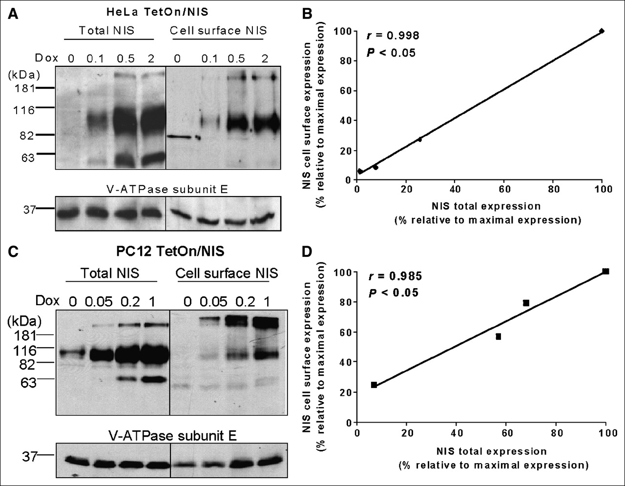
- FIGURE 1.
Doxycycline-inducible NIS expression and radioiodide uptake. (A) Western blot analysis showed that maximal inducible NIS protein level was higher in HeLa TetOn/NIS cells and PC12 TetOn/NIS than in FTC133 TetOn/NIS cells. All TetOn/NIS cells were induced for 48 h with various doxycycline concentrations. Equal loading of proteins was normalized with V-ATPase. Exposure time for detecting NIS protein in FTC133 TetOn/NIS was 30 min, whereas exposure time for HeLa TetOn/NIS and PC12 TetOn/NIS cells was 5 min. Results are representative of 3 independent experiments. (B) Extent of radioiodide uptake generally correlates with level of inducible NIS proteins. Each data point was performed in triplicate, and mean ± SD are shown. Results are representative of 3 independent experiments. RAIU = radioiodide uptake.
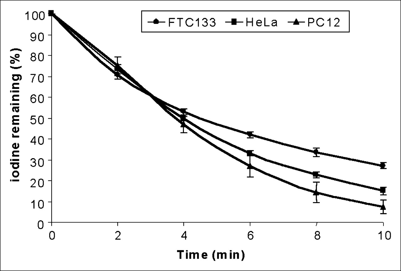
- FIGURE 2.
TetOn/NIS cells have similar rates of iodide efflux. TetOn/NIS cells were induced for 48 h with either 2 μg of doxycycline per milliliter for FTC133 TetOn/NIS and HeLa TetOn/NIS cells or 1 μg of doxycycline per milliliter for PC12 TetOn/NIS cells. Each data point was performed in triplicate, and mean ± SD are shown. Results are representative of 3 independent experiments.
- FIGURE 3.
NIS protein efficiently traffics to cell surface in TetOn/NIS cells. (A and C) Total NIS protein levels correlate with cell-surface NIS levels in both HeLa TetOn/NIS cells (A) and PC12 TetOn/NIS cells (C). Cell-surface biotinylation followed by Western blot analysis using NIS antibodies was performed to detect cell-surface NIS levels, and aliquots of whole-cell lysates were used for Western blot analysis to determine total NIS protein levels. Ratio of 90-kDa NIS vs. ∼181-kDa NIS decreases on surface of TetOn/NIS cells. TetOn/NIS cells were induced for 48 h with various doxycycline concentrations as indicated. Equal loading of proteins was normalized with V-ATPase. Exposure time was 5 min. Results are representative of 3 independent experiments. (B and D) Regression analysis indicates linear relationship between total NIS protein levels and cell-surface NIS levels in both HeLa TetOn/NIS cells (B) and PC12 TetOn/NIS cells (D). Densitometric analysis was performed to determine normalized total NIS protein levels and cell-surface NIS levels. To consolidate values from 3 independent experiments, maximally induced total NIS protein levels and maximally induced cell-surface NIS levels were designated as 100%, and other values were assigned relative to maximal levels. Correlation coefficient was determined. P < 0.05 indicates statistical significance. Dox = doxycycline.
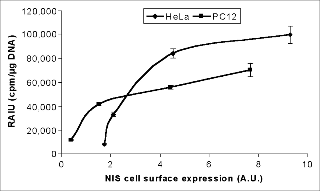
- FIGURE 4.
Extent of radioiodide uptake is not further increased by cell-surface NIS levels beyond a certain range in either HeLa TetOn/NIS or PC12 TetOn/NIS cells. Arbitrary values of cell-surface NIS levels were normalized with cell-surface V-ATPase levels and then correlated with corresponding radioiodide uptake. Results are representative of 3 independent experiments. A.U. = arbitrary units; RAIU = radioiodide uptake.
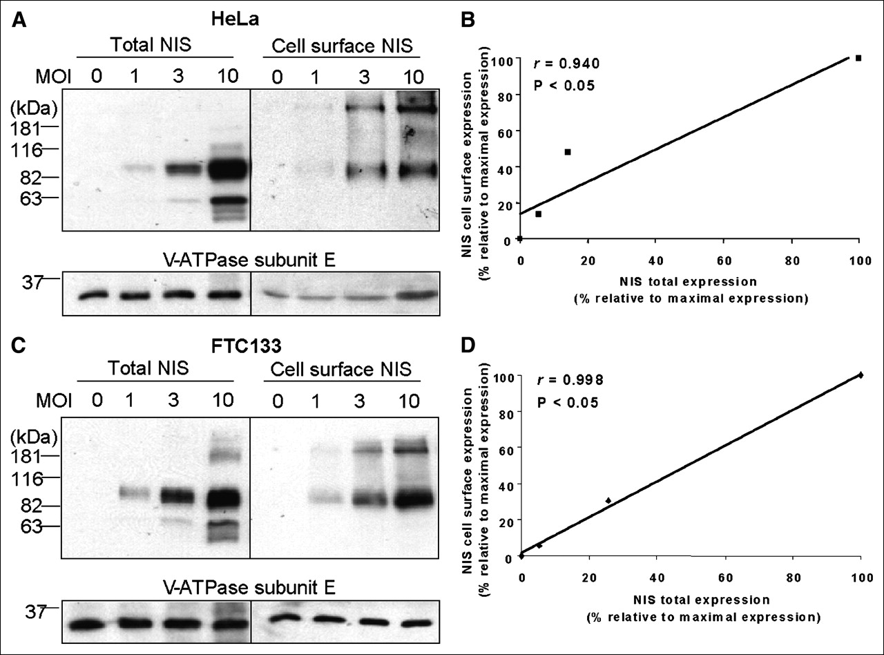
- FIGURE 5.
Recombinant adenovirus confers high NIS protein levels in both parental FTC133 cells and parental HeLa cells. (A and C) Western blot analysis showed that NIS protein levels are comparable between infected HeLa cells (A) and infected FTC133 cells (C). Both cells were infected with recombinant adenovirus for 48 h, with various multiplicity of infection. Equal loading of proteins was normalized with V-ATPase. Exposure time for detecting NIS protein in both cells was 5 s. Results are representative of 2 independent experiments. (B and D) Regression analysis indicates linear relationship between total NIS protein levels and cell-surface NIS levels in both infected HeLa cells (B) and infected FTC133 cells (D). Densitometric analysis was performed to determine normalized total NIS protein levels and cell-surface NIS levels. Maximally induced total NIS protein levels and maximally induced cell-surface NIS levels were designated as 100%, and other values were assigned relative to maximal levels. Correlation coefficient was determined. P < 0.05 indicates statistical significance. MOI = multiplicity of infection.
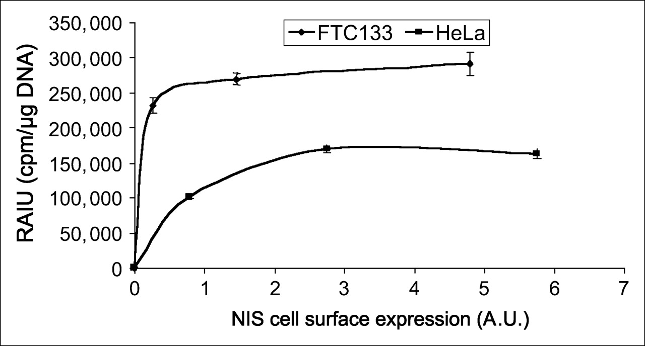
- FIGURE 6.
Radioiodide uptake was higher in infected FTC133 cells than infected HeLa cells at any equivalent NIS cell-surface levels. Arbitrary values of cell-surface NIS levels were normalized with cell-surface V-ATPase levels and then correlated with corresponding radioiodide uptake. Results are representative of 2 independent experiments. Consistent with finding using TetOn/NIS cells, extent of radioiodide uptake is not further increased by cell-surface NIS levels beyond a certain range. A.U. = arbitrary units; RAIU = radioiodide uptake.


Jump to section
Related Articles
Cited By...
- A Nonpump Function of Sodium Iodide Symporter in Thyroid Cancer via Cross-talk with PTEN Signaling
- Specific Activation of Sodium Iodide Symporter Gene in Hepatoma Using Alpha-fetoprotein Promoter Combined with Hepatitis B Virus Enhancer (EIIAPA)
- A "New" Reporter in the Field of Imaging Reporter Genes: Correlating Gene Expression and Function of the Sodium/Iodide Symporter