


Article Figures & Data
Figures
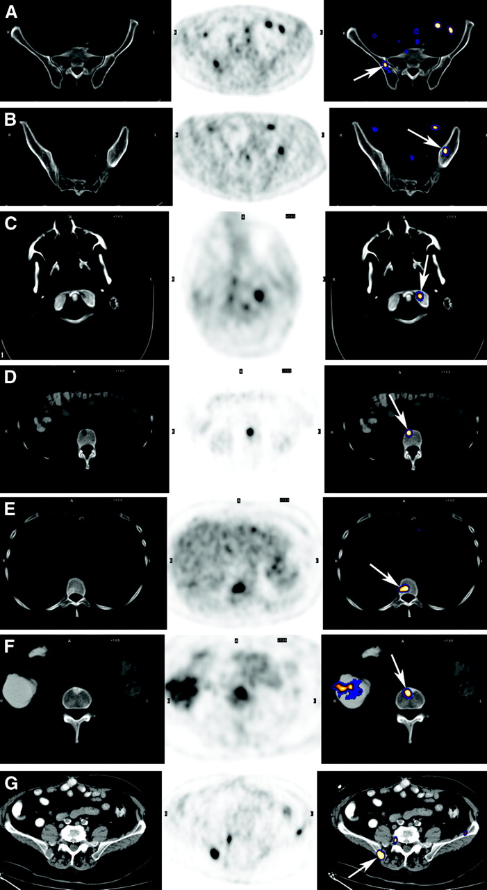
- FIGURE 1.
Different patterns of CT appearance in 18F-FDG–avid bone metastases. Each row represents another metastasis. From left to right: CT image, 18F-FDG PET image, and fused image. Metastases are marked by arrows. (A) CT appearance is normal on early bone marrow metastasis. (B) Early bone marrow metastasis. Minimal asymmetry is detected on CT. Involved marrow is more attenuated than normal marrow. (C) Lytic metastasis. Minor lytic changes are detected on CT at left occipital condyle. This finding was overlooked when CT was interpreted without PET. (D) Lytic lesion. A lytic lesion is clearly detected on CT. (E) Mixed lytic sclerotic metastasis. Location of lesion in posterior part of vertebral body, next to region of pedicle, is characteristic of early vertebral malignant involvement and is caused by its proximity to vertebral venous network. (F) Sclerotic metastasis. (G) Lytic metastasis with soft-tissue component. Margins of bone are disrupted and soft-tissue mass is discernible.
- FIGURE 2.
Detection of accompanying epidural mass by 18F-FDG PET/CT in patient with lymphoma. Top row, transaxial images; bottom row, sagittal images. From left to right: CT image, 18F-FDG PET image, and fused image. Tumor is marked by arrows. Based on fusion with CT, increased 18F-FDG uptake is detected in right aspect of L4 and in accompanying epidural component. Soft-tissue epidural mass appears to displace the thecal sac laterally, on CT.
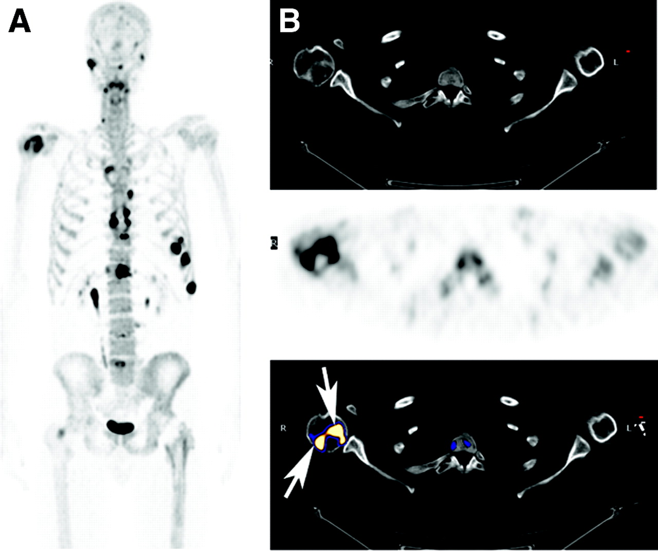
- FIGURE 3.
Multiple myeloma assessment by 18F-fluoride PET/CT. (A) Maximum-intensity-projection image detecting multiple lesions. (B) From left to right: CT image, 18F-fluoride PET image, and fused image. Increased uptake is detected in well-defined margins (arrows) of large lytic lesion detected on CT in head of right humerus.
- FIGURE 4.
Disease progression of multiple myeloma on 18F-FDG PET. (A) Two maximum-intensity-projection images 5 mo apart. Baseline study (left) was performed in patient with monoclonal gammopathy of undetermined significance who presented with vertebral plasmocytoma and spinal compression fracture. In addition to increased uptake at known spinal lesion, increased 18F-FDG uptake was also detected in ribs, which appeared normal on CT data of PET/CT, probably reflecting early malignant involvement in other sites (B). Spinal lesion was treated by radiotherapy. Five months later, numerous new lesions (arrow) were identified on a repeated study, indicative of tumor progression.
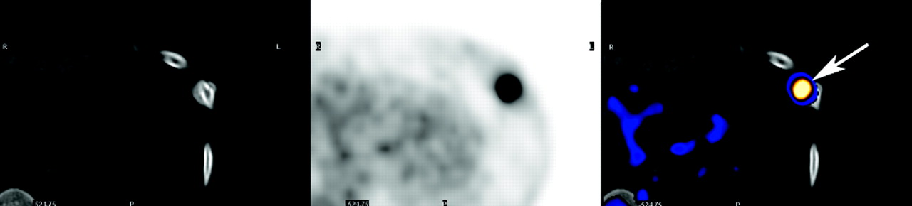
- FIGURE 5.
Benign 18F-FDG uptake (arrow) in recent rib fracture detected on CT data of 18F-FDG PET/CT in patient with lung cancer. From left to right: CT image, 18F-FDG PET image, and fused image.


In this issue


Jump to section
Related Articles
Cited By...
- Bone-Targeted Imaging and Radionuclide Therapy in Prostate Cancer
- Novel Small-Molecule CX3CR1 Antagonist Impairs Metastatic Seeding and Colonization of Breast Cancer Cells
- Evaluation of 18F-Fluoride PET/MR and PET/CT in Patients with Foot Pain of Unclear Cause
- PET/MR in Oncology: Non-18F-FDG Tracers for Routine Applications
- SPECT-CT: applications in musculoskeletal radiology
- MRI for the detection of prostate cancer origin vertebral metastases in the preosteoblastic phase
- Assessment of Patient Exposure to X-Radiation from SPECT/CT Scanners
- Kinetic Analysis of 18F-Fluoride PET Images of Breast Cancer Bone Metastases
- SPECT/CT
- Application of PET/CT in the Development of Novel Anticancer Drugs
- An Introduction to Na18F Bone Scintigraphy: Basic Principles, Advanced Imaging Concepts, and Case Examples
- A prospective comparison of 18F-fluorodeoxyglucose positron emission tomography-computed tomography, magnetic resonance imaging and whole-body planar radiographs in the assessment of bone disease in newly diagnosed multiple myeloma
- Early Detection of Cancer Recurrence: 18F-FDG PET/CT Can Make a Difference in Diagnosis and Patient Care
- SPECT-Guided CT for Evaluating Foci of Increased Bone Metabolism Classified as Indeterminate on SPECT in Cancer Patients
- Use of 99mTc-Sestamibi Scintigraphy in Multiple Myeloma.