
Main menu

  • Home
  • Content
    • Current
    • Ahead of print
    • Past Issues
    • JNM Supplement
    • SNMMI Annual Meeting Abstracts
    • Continuing Education
    • JNM Podcasts
  • Subscriptions
    • Subscribers
    • Institutional and Non-member
    • Rates
    • Journal Claims
    • Corporate & Special Sales
  • Authors
    • Submit to JNM
    • Information for Authors
    • Assignment of Copyright
    • AQARA requirements
  • Info
    • Reviewers
    • Permissions
    • Advertisers
  • About
    • About Us
    • Editorial Board
    • Contact Information
  • More
    • Alerts
    • Feedback
    • Help
    • SNMMI Journals
  • SNMMI
    • JNM
    • JNMT
    • SNMMI Journals
    • SNMMI

User menu

  • Subscribe
  • My alerts
  • Log in
  • My Cart

Search

  • Advanced search
Journal of Nuclear Medicine
  • SNMMI
    • JNM
    • JNMT
    • SNMMI Journals
    • SNMMI
  • Subscribe
  • My alerts
  • Log in
  • My Cart
Journal of Nuclear Medicine

Advanced Search

  • Home
  • Content
    • Current
    • Ahead of print
    • Past Issues
    • JNM Supplement
    • SNMMI Annual Meeting Abstracts
    • Continuing Education
    • JNM Podcasts
  • Subscriptions
    • Subscribers
    • Institutional and Non-member
    • Rates
    • Journal Claims
    • Corporate & Special Sales
  • Authors
    • Submit to JNM
    • Information for Authors
    • Assignment of Copyright
    • AQARA requirements
  • Info
    • Reviewers
    • Permissions
    • Advertisers
  • About
    • About Us
    • Editorial Board
    • Contact Information
  • More
    • Alerts
    • Feedback
    • Help
    • SNMMI Journals
  • View or Listen to JNM Podcast
  • Visit JNM on Facebook
  • Join JNM on LinkedIn
  • Follow JNM on Twitter
  • Subscribe to our RSS feeds
OtherBasic Science Investigations

U-SPECT-I: A Novel System for Submillimeter-Resolution Tomography with Radiolabeled Molecules in Mice

Freek J. Beekman, Frans van der Have, Brendan Vastenhouw, Annemarie J.A. van der Linden, Peter P. van Rijk, J. Peter H. Burbach and Marten P. Smidt
Journal of Nuclear Medicine July 2005, 46 (7) 1194-1200;
Freek J. Beekman
  • Find this author on Google Scholar
  • Find this author on PubMed
  • Search for this author on this site
Frans van der Have
  • Find this author on Google Scholar
  • Find this author on PubMed
  • Search for this author on this site
Brendan Vastenhouw
  • Find this author on Google Scholar
  • Find this author on PubMed
  • Search for this author on this site
Annemarie J.A. van der Linden
  • Find this author on Google Scholar
  • Find this author on PubMed
  • Search for this author on this site
Peter P. van Rijk
  • Find this author on Google Scholar
  • Find this author on PubMed
  • Search for this author on this site
J. Peter H. Burbach
  • Find this author on Google Scholar
  • Find this author on PubMed
  • Search for this author on this site
Marten P. Smidt
  • Find this author on Google Scholar
  • Find this author on PubMed
  • Search for this author on this site
  • Article
  • Figures & Data
  • Info & Metrics
  • PDF
Loading

Article Figures & Data

Figures

  • FIGURE 1.
    • Download figure
    • Open in new tab
    • Download powerpoint
    FIGURE 1.

    (A) Overview of U-SPECT-I system. Triangular lead shielding is placed between camera heads of triple-detector system, tungsten cylinder containing pinholes being centered within the 3 detectors. x,y,z stage with attached bed, placed in front of lower detector, is also visible. (B) Cylinder with 75 gold pinhole apertures that are focused on its center. (C) Cross section of cylinder with tilted pinholes.
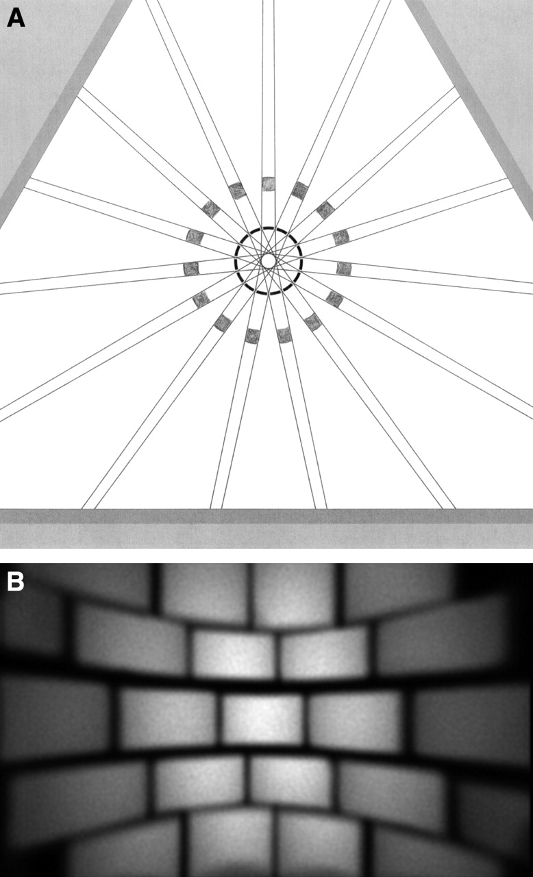
  • FIGURE 2.
    • Download figure
    • Open in new tab
    • Download powerpoint
    FIGURE 2.

    Illustration of highly focusing pinhole geometry of U-SPECT-I. (A) Cross section through 1 of 5 rings with pinholes. Lines emerging from central circle mark triangular cross sections of beams in which emitted γ-quanta can travel from animal toward detector. Pinholes in all rings focus on center to maximize detection yield. Lead cylinder with square holes shown in gray is placed around tungsten cylinder with pinholes to prevent projection overlap. (B) Radiation intensity on 1 of 3 detectors when a bottle with a 99mTc solution is imaged. Image demonstrates how the large detector is divided into a large number of small subcameras.
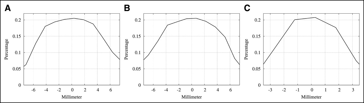
  • FIGURE 3.
    • Download figure
    • Open in new tab
    • Download powerpoint
    FIGURE 3.

    Demonstration of high sensitivity of U-SPECT-I. Shown are sensitivity profiles lying along mutually orthogonal lines that cross the center of field of view, obtained with a scanning point source: along x-axis (A), along y-axis (B), and along transaxial axis (z-axis) (C).

  • FIGURE 4.
    • Download figure
    • Open in new tab
    • Download powerpoint
    FIGURE 4.

    Demonstration of submillimeter resolution of U-SPECT-I images. (A) Photograph of miniature acrylic resolution phantom with capillaries used as test object. (B) Reconstructed cross-sectional image, with a slice thickness of 0.5 mm, of the phantom shown in A. Half-millimeter capillaries are clearly separated on image.

  • FIGURE 5.
    • Download figure
    • Open in new tab
    • Download powerpoint
    FIGURE 5.

    Mutual perpendicular cross sections through submillimeter-resolution 3-dimensional myocardial perfusion image volume of living mouse (named Animal Co-Image of the Year at the annual meeting of the Society of Nuclear Medicine, Philadelphia, 2004). Image data were acquired during 30 min, starting 30 min after administration of 222 MBq (6 mCi) of 99mTc-tetrofosmin. On left is a short-axis slice showing myocardial perfusion in right ventricular (RV) and left ventricular (LV) walls. Perfusion in anterior papillary muscle (arrow) can be distinguished from other parts of left ventricular wall. At top right is a vertical long-axis slice; at bottom right, a horizontal long-axis slice.
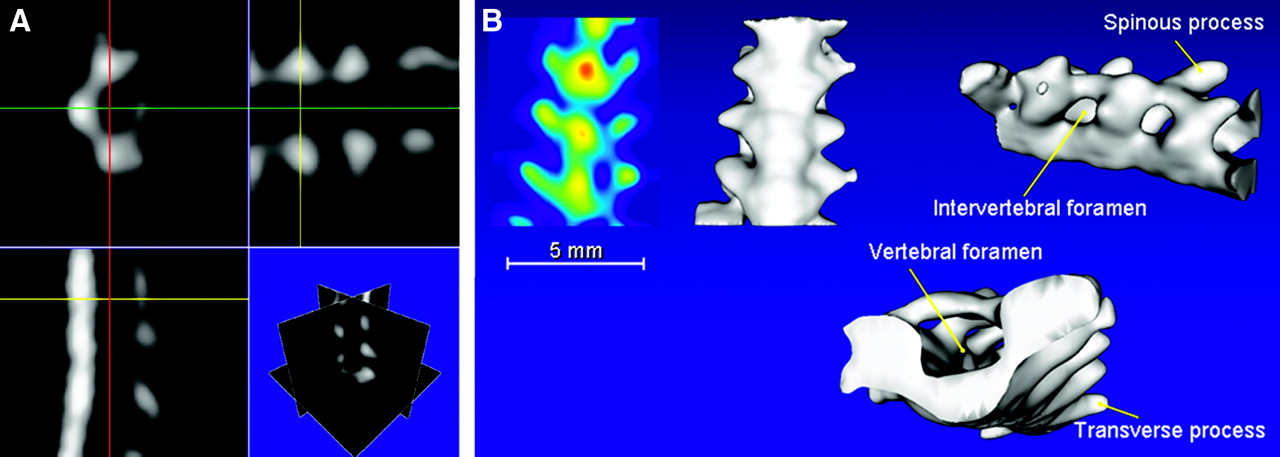
  • FIGURE 6.
    • Download figure
    • Open in new tab
    • Download powerpoint
    FIGURE 6.

    Different representations of reconstructed image volume of lumbar spine, acquired during 22 min, 2 h after injection of 148 MBq (4 mCi) of 99mTc-HDP. (A) Three different orthogonal cross sections. (B) Projection of local image maxima (far left) and 3 isosurface renderings of tracer concentrations in same spinal section (named Animal Co-Image of the Year at the annual meeting of the Society of Nuclear Medicine, Philadelphia, 2004).

PreviousNext
Back to top

In this issue

Journal of Nuclear Medicine: 46 (7)
Journal of Nuclear Medicine
Vol. 46, Issue 7
July 1, 2005
  • Table of Contents
  • About the Cover
  • Index by author
Print
Download PDF
Article Alerts
Sign In to Email Alerts with your Email Address
Email Article

Thank you for your interest in spreading the word on Journal of Nuclear Medicine.

NOTE: We only request your email address so that the person you are recommending the page to knows that you wanted them to see it, and that it is not junk mail. We do not capture any email address.

Enter multiple addresses on separate lines or separate them with commas.
U-SPECT-I: A Novel System for Submillimeter-Resolution Tomography with Radiolabeled Molecules in Mice
(Your Name) has sent you a message from Journal of Nuclear Medicine
(Your Name) thought you would like to see the Journal of Nuclear Medicine web site.
Citation Tools
U-SPECT-I: A Novel System for Submillimeter-Resolution Tomography with Radiolabeled Molecules in Mice
Freek J. Beekman, Frans van der Have, Brendan Vastenhouw, Annemarie J.A. van der Linden, Peter P. van Rijk, J. Peter H. Burbach, Marten P. Smidt
Journal of Nuclear Medicine Jul 2005, 46 (7) 1194-1200;

Citation Manager Formats

  • BibTeX
  • Bookends
  • EasyBib
  • EndNote (tagged)
  • EndNote 8 (xml)
  • Medlars
  • Mendeley
  • Papers
  • RefWorks Tagged
  • Ref Manager
  • RIS
  • Zotero
Share
U-SPECT-I: A Novel System for Submillimeter-Resolution Tomography with Radiolabeled Molecules in Mice
Freek J. Beekman, Frans van der Have, Brendan Vastenhouw, Annemarie J.A. van der Linden, Peter P. van Rijk, J. Peter H. Burbach, Marten P. Smidt
Journal of Nuclear Medicine Jul 2005, 46 (7) 1194-1200;
Twitter logo Facebook logo LinkedIn logo Mendeley logo
  • Tweet Widget
  • Facebook Like
  • Google Plus One
Bookmark this article

Jump to section

  • Article
    • Abstract
    • MATERIALS AND METHODS
    • RESULTS
    • DISCUSSION
    • CONCLUSION
    • Acknowledgments
    • Footnotes
    • REFERENCES
  • Figures & Data
  • Info & Metrics
  • PDF

Related Articles

  • THIS MONTH IN JNM
  • PubMed
  • Google Scholar

Cited By...

  • The aptabot: an inducibly affinity-switching, minimally invasive in vivo contrast agent
  • Ultra-High-Sensitivity Submillimeter Mouse SPECT
  • Imaging Capabilities of the Inveon SPECT System Using Single-and Multipinhole Collimators
  • Fast Spiral SPECT with Stationary {gamma}-Cameras and Focusing Pinholes
  • MIRD Pamphlet No. 23: Quantitative SPECT for Patient-Specific 3-Dimensional Dosimetry in Internal Radionuclide Therapy
  • Using the NEMA NU 4 PET Image Quality Phantom in Multipinhole Small-Animal SPECT
  • Assessing Antibody Pharmacokinetics in Mice with In Vivo Imaging
  • Small-Animal Molecular Imaging Methods
  • Recent Advances in Small-Animal Cardiovascular Imaging
  • U-SPECT-II: An Ultra-High-Resolution Device for Molecular Small-Animal Imaging
  • A Skew-Slit Collimator for Small-Animal SPECT
  • Small-Animal SPECT and SPECT/CT: Important Tools for Preclinical Investigation
  • Virtual-Pinhole PET
  • SPECT Low-Field MRI System for Small-Animal Imaging
  • A Feasibility Study of a Prototype PET Insert Device to Convert a General-Purpose Animal PET Scanner to Higher Resolution
  • Recent Advances in SPECT Imaging
  • Submillimeter Total-Body Murine Imaging with U-SPECT-I
  • Google Scholar

More in this TOC Section

  • Synthesis and Biologic Evaluation of Novel 18F-Labeled Probes Targeting Prostate-Specific Membrane Antigen for PET of Prostate Cancer
  • Tumor-Specific Binding of Radiolabeled PEGylated GIRLRG Peptide: A Novel Agent for Targeting Cancers
  • PET/MRI of Hypoxic Atherosclerosis Using 64Cu-ATSM in a Rabbit Model
Show more Basic Science Investigations

Similar Articles

SNMMI

© 2025 SNMMI

Powered by HighWire