
Main menu

  • Home
  • Content
    • Current
    • Ahead of print
    • Past Issues
    • JNM Supplement
    • SNMMI Annual Meeting Abstracts
  • Subscriptions
    • Subscribers
    • Institutional and Non-member
    • Rates
    • Corporate & Special Sales
    • Journal Claims
  • Authors
    • Submit to JNM
    • Information for Authors
    • Assignment of Copyright
    • AQARA requirements
  • Info
    • Continuing Education
    • Reviewers
    • Permissions
    • Advertisers
  • About
    • About Us
    • Editorial Board
    • Contact Information
  • More
    • Alerts
    • Feedback
    • Help
    • SNMMI Journals
  • SNMMI
    • JNM
    • JNMT
    • SNMMI Journals
    • SNMMI

User menu

  • Subscribe
  • My alerts
  • Log in
  • Log out
  • My Cart

Search

  • Advanced search
Journal of Nuclear Medicine
  • SNMMI
    • JNM
    • JNMT
    • SNMMI Journals
    • SNMMI
  • Subscribe
  • My alerts
  • Log in
  • Log out
  • My Cart
Journal of Nuclear Medicine

Advanced Search

  • Home
  • Content
    • Current
    • Ahead of print
    • Past Issues
    • JNM Supplement
    • SNMMI Annual Meeting Abstracts
  • Subscriptions
    • Subscribers
    • Institutional and Non-member
    • Rates
    • Corporate & Special Sales
    • Journal Claims
  • Authors
    • Submit to JNM
    • Information for Authors
    • Assignment of Copyright
    • AQARA requirements
  • Info
    • Continuing Education
    • Reviewers
    • Permissions
    • Advertisers
  • About
    • About Us
    • Editorial Board
    • Contact Information
  • More
    • Alerts
    • Feedback
    • Help
    • SNMMI Journals
  • Visit JNM on Facebook
  • Join JNM on LinkedIn
  • Follow JNM on Twitter
  • Subscribe to our RSS feeds
OtherClinical Investigations

Optimized Intravenous Contrast Administration for Diagnostic Whole-Body 18F-FDG PET/CT

Thomas Beyer, Gerald Antoch, Andreas Bockisch and Joerg Stattaus
Journal of Nuclear Medicine March 2005, 46 (3) 429-435;
Thomas Beyer
  • Find this author on Google Scholar
  • Find this author on PubMed
  • Search for this author on this site
Gerald Antoch
  • Find this author on Google Scholar
  • Find this author on PubMed
  • Search for this author on this site
Andreas Bockisch
  • Find this author on Google Scholar
  • Find this author on PubMed
  • Search for this author on this site
Joerg Stattaus
  • Find this author on Google Scholar
  • Find this author on PubMed
  • Search for this author on this site
  • Article
  • Figures & Data
  • Info & Metrics
  • PDF
Loading

Article Figures & Data

Figures

  • Tables
  • FIGURE 1.
    • Download figure
    • Open in new tab
    • Download powerpoint
    FIGURE 1.

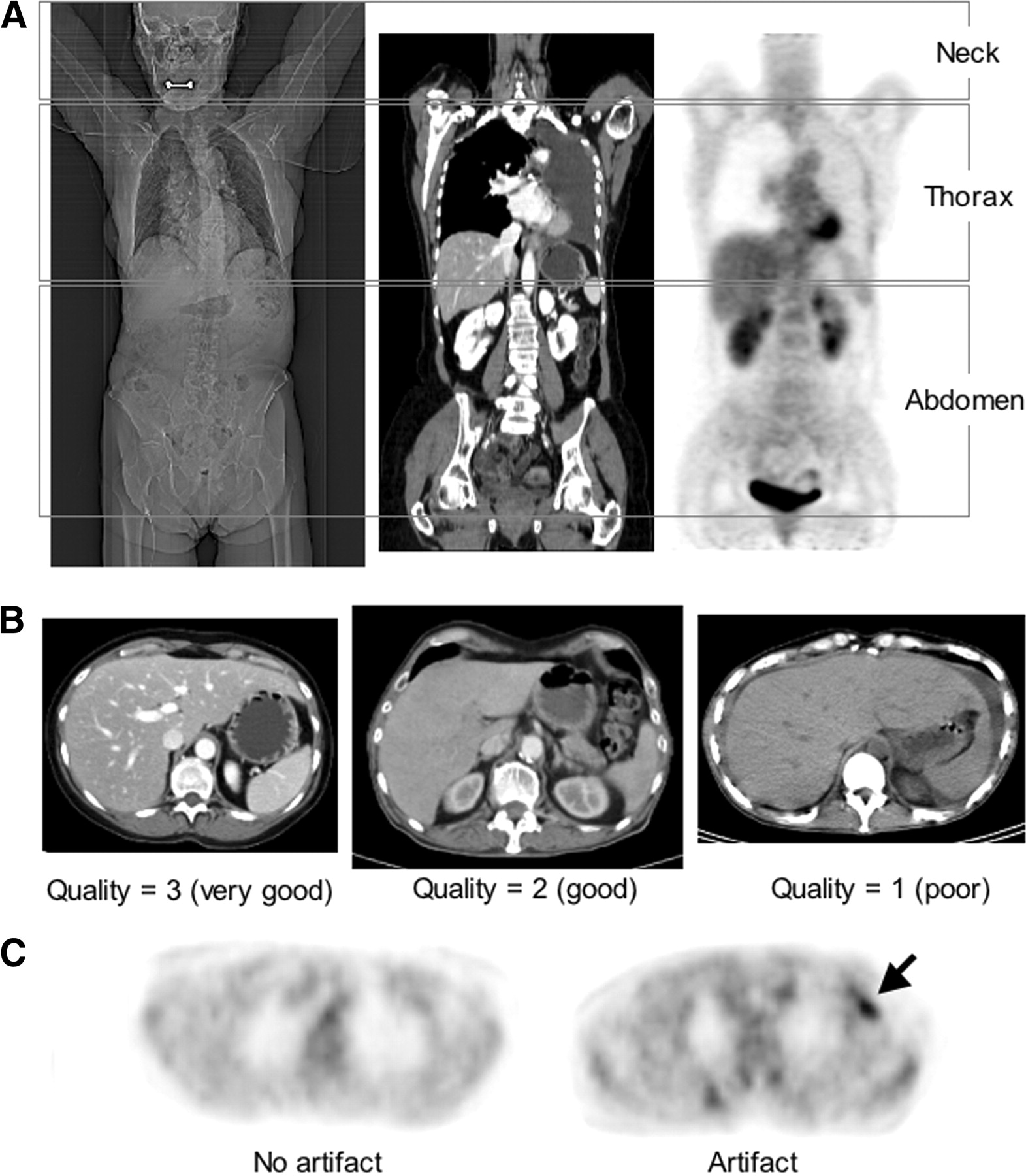
    (A) Diagnostic image quality and contrast-induced abnormalities were assessed independently for 3 axial imaging ranges: neck, thorax, and abdomen. (B) Examples of diagnostic quality of thoracic CT images: 3 (very good), 2 (good), 1 (poor). (C) Examples of PET images after CT-based attenuation correction without and with artifact from intravenous contrast (arrow) are shown.

  • FIGURE 2.
    • Download figure
    • Open in new tab
    • Download powerpoint
    FIGURE 2.

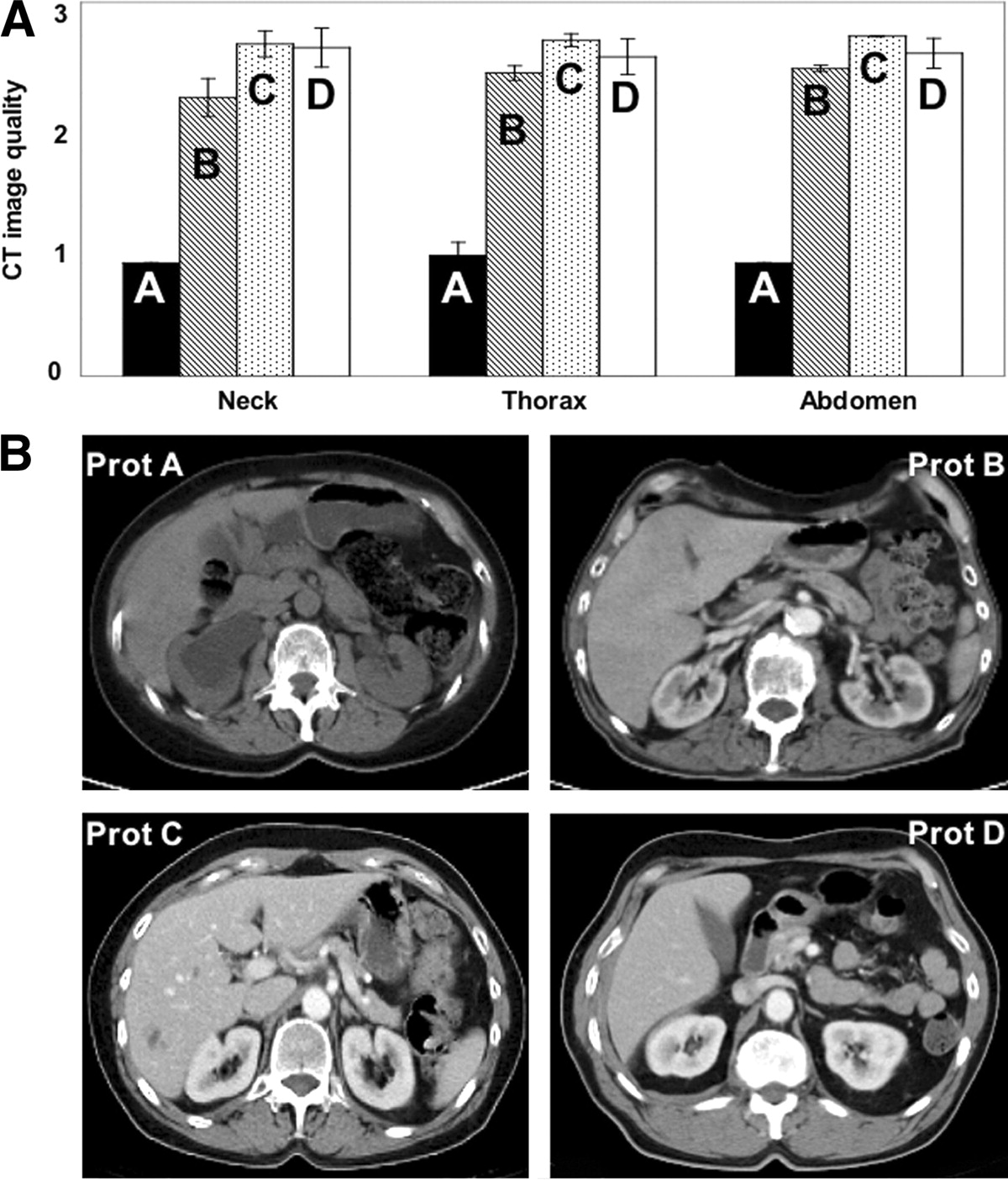
    (A) Diagnostic quality of CT images averaged over ratings by 3 radiology reviewers. CT image quality was lowest for nonenhanced studies (protocol A) but increased throughout whole-body imaging range when intravenous contrast was used (protocols B–D). Examples of CT images (B) of abdomen for subjects enrolled in protocols A (nonenhanced) and B–D (intravenous contrast according to Table 1) are shown. Prot = protocol.

  • FIGURE 3.
    • Download figure
    • Open in new tab
    • Download powerpoint
    FIGURE 3.

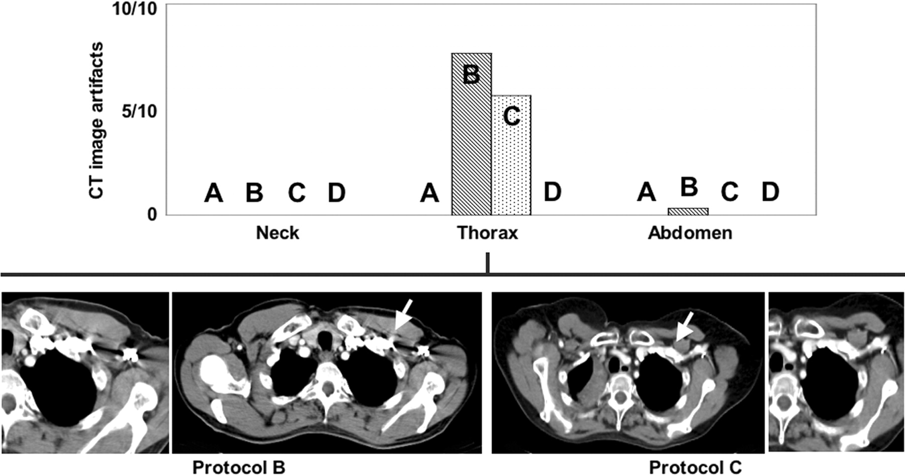
    Average number of CT image artifacts attributed to intravenous contrast in neck, thorax, and abdomen. Examples of CT images are shown to illustrate artifacts (arrows) for protocols B and C. Zoomed inserts are shown to the left and right for B and C, respectively. Protocol B also resulted in 1 case with reported high-density artifacts in abdomen as reported by 1 radiology reader (example not shown).

  • FIGURE 4.
    • Download figure
    • Open in new tab
    • Download powerpoint
    FIGURE 4.

    Average CT attenuation in major vessels of whole-body CT images from PET/CT examinations with contrast administration protocols A–D (Table 1).

  • FIGURE 5.
    • Download figure
    • Open in new tab
    • Download powerpoint
    FIGURE 5.

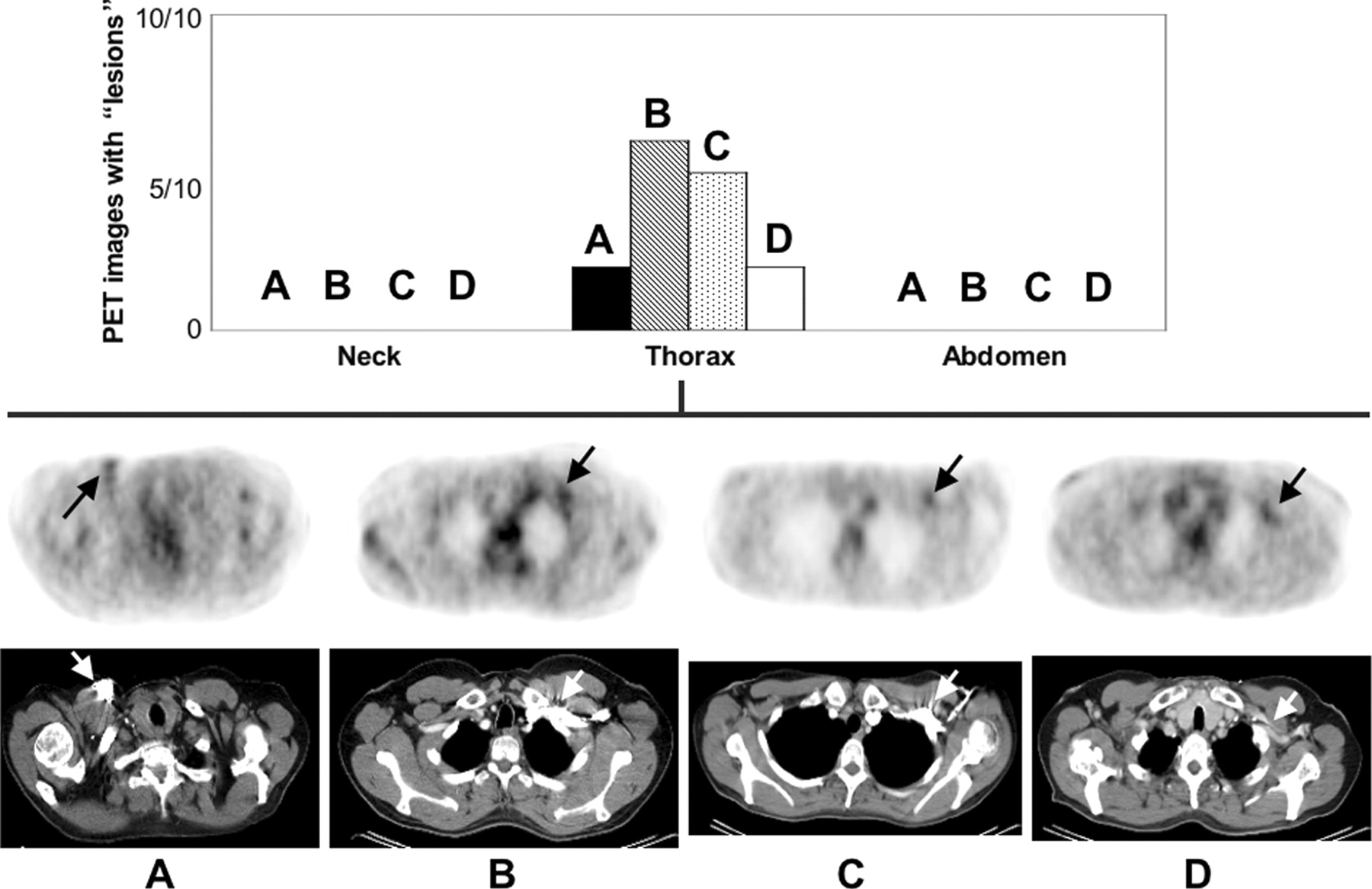
    Number of attenuation-corrected PET studies with abnormal tracer uptake attributed to focal accumulations of intravenous contrast through protocols A–D. Axial PET images (after CT-based attenuation correction) through apex of lungs are shown for protocols A–D with arrows pointing to reported artifactual findings on PET.
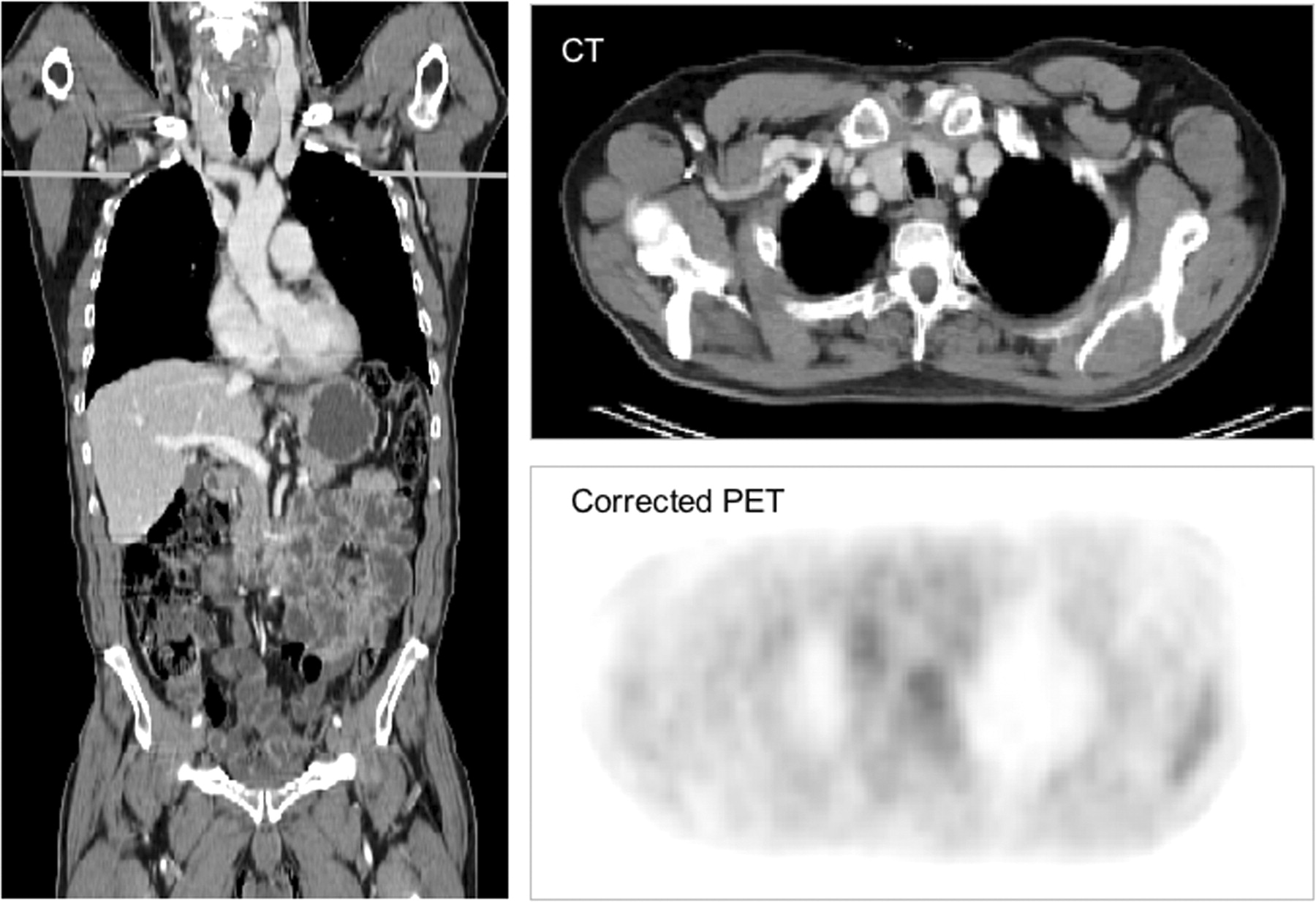
  • FIGURE 6.
    • Download figure
    • Open in new tab
    • Download powerpoint
    FIGURE 6.

    18F-FDG PET/CT study following the modified contrast injection protocol D. Coronal CT view illustrates uniform vessel enhancement throughout whole-body imaging range. Axial CT and corrected PET images are shown through area of subclavian vein to demonstrate absence of contrast-induced abnormalities.

Tables

  • Figures
    • View popup
    TABLE 1

    Patient Groups for Adapted Intravenous Contrast Injection Protocols and Parameters for Intravenous Contrast Injection

    GroupProtocol
    ABCD
    DescriptorNonenhancedStandardExtendedModified
    Patients10101010
        Male/female5/56/46/45/5
        Mean age* (y)61 ± 1461 ± 1260 ± 762 ± 10
        Mean body mass index*24 ± 424 ± 523 ± 425 ± 4
    Intravenous contrast administration
        Intravenous contrastNoYesYesYes
        Contrast materialXenetix 300Xenetix 300Xenetix 300
        PhaseDualTripleDual
        Total contrast volume (mL)140: 90, 50170: 90, 40, 40140: 80, 60
        Flow rate (mL/s)3, 1.53, 2, 1.53, 1.5
        Delay (s)303050
        Scan directionCraniocaudalCraniocaudalCraniocaudalCaudocranial
    • ↵* Mean ± SD.

    • View popup
    TABLE 2

    Mean CT Attenuation (HU) and SD (%) in Major Vessels of Interest in Neck, Thorax, and Abdomen in Whole-Body PET/CT Studies with Intravenous Contrast Injection According to Administration Protocols A–D

    ProtocolNeckThoraxAbdomen, PV
    IJVCCABVAA
    A48 (13)47 (15)46 (17)41 (12)49 (10)
    B140 (43)200 (40)1,300 (31)200 (15)150 (27)
    C220 (27)230 (35)600 (83)170 (18)160 (13)
    D160 (19)160 (19)140 (21)170 (18)180 (22)
PreviousNext
Back to top

In this issue

Journal of Nuclear Medicine: 46 (3)
Journal of Nuclear Medicine
Vol. 46, Issue 3
March 1, 2005
  • Table of Contents
  • About the Cover
  • Index by author
Print
Download PDF
Article Alerts
Sign In to Email Alerts with your Email Address
Email Article

Thank you for your interest in spreading the word on Journal of Nuclear Medicine.

NOTE: We only request your email address so that the person you are recommending the page to knows that you wanted them to see it, and that it is not junk mail. We do not capture any email address.

Enter multiple addresses on separate lines or separate them with commas.
Optimized Intravenous Contrast Administration for Diagnostic Whole-Body 18F-FDG PET/CT
(Your Name) has sent you a message from Journal of Nuclear Medicine
(Your Name) thought you would like to see the Journal of Nuclear Medicine web site.
Citation Tools
Optimized Intravenous Contrast Administration for Diagnostic Whole-Body 18F-FDG PET/CT
Thomas Beyer, Gerald Antoch, Andreas Bockisch, Joerg Stattaus
Journal of Nuclear Medicine Mar 2005, 46 (3) 429-435;

Citation Manager Formats

  • BibTeX
  • Bookends
  • EasyBib
  • EndNote (tagged)
  • EndNote 8 (xml)
  • Medlars
  • Mendeley
  • Papers
  • RefWorks Tagged
  • Ref Manager
  • RIS
  • Zotero
Share
Optimized Intravenous Contrast Administration for Diagnostic Whole-Body 18F-FDG PET/CT
Thomas Beyer, Gerald Antoch, Andreas Bockisch, Joerg Stattaus
Journal of Nuclear Medicine Mar 2005, 46 (3) 429-435;
Twitter logo Facebook logo LinkedIn logo Mendeley logo
  • Tweet Widget
  • Facebook Like
  • Google Plus One
Bookmark this article

Jump to section

  • Article
    • Abstract
    • MATERIALS AND METHODS
    • RESULTS
    • DISCUSSION
    • CONCLUSION
    • Acknowledgments
    • Footnotes
    • REFERENCES
  • Figures & Data
  • Info & Metrics
  • PDF

Related Articles

  • THIS MONTH IN JNM
  • PubMed
  • Google Scholar

Cited By...

  • Quantitative Evaluation of Segmentation- and Atlas-Based Attenuation Correction for PET/MR on Pediatric Patients
  • Standards for PET Image Acquisition and Quantitative Data Analysis
  • Limitations of CT During PET/CT
  • FDG-PET/CT in restaging of patients with recurrent breast cancer: possible impact on staging and therapy
  • Value of contrast-enhanced multiphase CT in combined PET/CT protocols for oncological imaging
  • Can PET/CT Replace Separate Diagnostic CT for Cancer Imaging? Optimizing CT Protocols for Imaging Cancers of the Chest and Abdomen
  • PET/CT in Lymphoma: Prospective Study of Enhanced Full-Dose PET/CT Versus Unenhanced Low-Dose PET/CT
  • Reply: Adequate Evaluation of Image Registration in Hybrid PET/CT.
  • The Role of PET in Lymphoma
  • Optimized Contrast-Enhanced CT Protocols for Diagnostic Whole-Body 18F-FDG PET/CT: Technical Aspects of Single-Phase Versus Multiphase CT Imaging
  • Whole-Body 18F-FDG PET/CT in the Presence of Truncation Artifacts
  • CT in PET/CT: Essential Features of Interpretation
  • Radiation Exposure of Patients Undergoing Whole-Body Dual-Modality 18F-FDG PET/CT Examinations
  • Google Scholar

More in this TOC Section

  • Left Ventricular Strain from Myocardial Perfusion PET Imaging: Method Development and Comparison to 2-Dimensional Echocardiography
  • Prognostic Role of 68Ga-PSMA11 PET–Based Response in Patients with Prostate Cancer Undergoing Taxane-Based Chemotherapy
  • Radioimmunoscintigraphy and Pretreatment Dosimetry of 131I-Omburtamab for Planning Treatment of Leptomeningeal Disease
Show more Clinical Investigations

Similar Articles

SNMMI

© 2023 Journal of Nuclear Medicine

Powered by HighWire