


Article Figures & Data
Figures
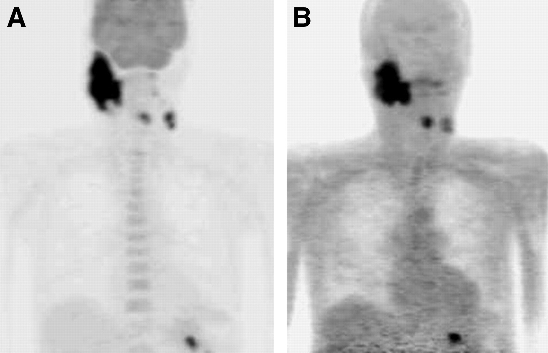
- FIGURE 1.
18F-FDG PET (A) and 18F-FET PET (B) of 81-y-old man (patient 2) with high-grade lymphoma (non-Hodgkin’s lymphoma). 18F-FDG shows high uptake in multiple lymph nodes, whereas 18F-FET uptake is negative in all lymph nodes.
- FIGURE 2.
18F-FDG PET (A) and 18F-FET PET (B) of 63-y-old man (patient 32) with head-neck carcinoma (squamous cell carcinoma). Primary tumor and lymph node metastases are positive for both 18F-FDG and 18F-FET.
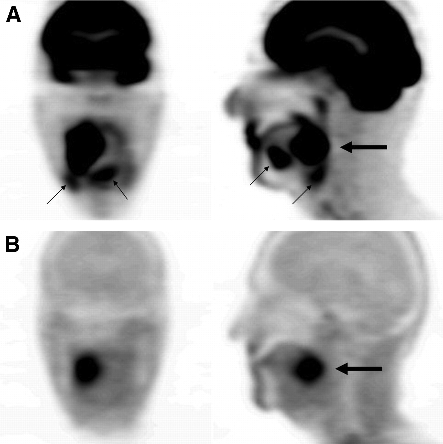
- FIGURE 3.
18F-FDG PET (A) and 18F-FET PET (B) of 61-y-old man with cancer of tongue (squamous cell carcinoma). Increased 18F-FDG uptake was noted in tumor (large arrow), and additional uptake was demonstrated in inflammatory tissues (small arrows). Increased 18F-FET uptake was noted only in tumor (large arrow).


Tables
Patient Age (y) Sex Primary tumor Histologic diagnosis Confirmation Pretreatment 18F-FDG staging* No. of lesions 18F-FET staging* No. of lesions 1 45 F Lymphoma HG NHL Surgery Chemotherapy II 5 (−) 0 2 81 M Lymphoma HG NHL Biopsy Chemotherapy IV >5 (−) 0 3 41 M Lymphoma LG NHL Biopsy Chemotherapy I 1 (−) 0 4 39 F Lymphoma LG NHL Biopsy Chemotherapy IV >5 (−) 0 5 48 F Colorectal Adenoca. Surgery Chemotherapy T(−) N0, M1 >5 T(−) N0, M0 0 6 67 M Colorectal Adenoca. Biopsy None T(+) N0, M0 1 T(−) N0, M0 0 7 59 M Colorectal Adenoca. Surgery Surgery/chemotherapy T(−) N0, M1 2 T(−) N0, M0 0 8 76 M Colorectal Adenoca. Surgery Surgery T(+) N0, M1 >5 T(−) N0, M0 0 9 50 M Colorectal Adenoca. Surgery Surgery/chemotherapy T(−) N0, M1 3 T(−) N0, M0 0 10 77 M Colorectal Adenoca. Surgery Surgery/chemotherapy T(−) N0, M1 1 T(−) N0, M0 0 11 48 M Colorectal Adenoca. Surgery Surgery/chemotherapy T(−) N0, M1 2 T(−) N0, M0 0 12 69 F Pancreatic Adenoca. Surgery Surgery/chemotherapy T(−) N0, M1 2 T(−) N0, M0 0 13 54 M Pancreatic Adenoca. Surgery None T(+) N0, M0 1 T(−) N0, M0 0 14 56 F Pancreatic Adenoca. Biopsy Chemotherapy T(+) N0, M0 1 T(−) N0, M0 0 15 62 M Pancreatic Adenoca. Biopsy None T(+) N1, M1 >5 T(−) N0, M0 0 16 57 M Pancreatic Adenoca. Biopsy None T(+) N1, M1 >5 T(−) N0, M0 0 17 76 M Pancreatic Adenoca. Surgery None T(+) N0, M0 1 T(−) N0, M0 0 18 69 M Lung SCC Biopsy None T(+) N1, M1 >5 T(+) N1, M1 >5 19 72 F Lung SCC Biopsy None T(+) N1, M1 >5 T(+) N1, M1 >5 20 69 M Lung Mixed CC Biopsy None T(+) N0, M1 3 T(−) N0, M0 0 21 52 F Ovarian Adenoca. Biopsy None T(−) N1, M1 >5 T(−) N0, M0 0 22 46 F Ovarian Adenoca. Surgery Surgery T(−) N0, M1 4 T(−) N0, M0 0 23 63 F Ovarian Adenoca. Surgery Surgery/chemotherapy T(−) N1, M1 >5 T(−) N0, M0 0 24 81 M Prostatic Adenoca. Biopsy Antiandrogen T(+) N1, M1 >5 T(−) N0, M0 0 25 76 M Prostatic Adenoca. Biopsy Antiandrogen T(−) N0, M1 1 T(−) N0, M0 0 26 76 F Breast IDC Surgery None T(+) N3, M1 >5 T(−) N0, M0 0 27 30 F Breast IDC Biopsy None T(+) N3, M0 3 T(+) N3, M0 3 28 65 F Breast IDC Biopsy None T(+) N1, M0 2 T(+) N1, M0 2 29 65 F Breast IDC Biopsy None T(+) N1, M0 2 T(+) N1, M0 2 30 47 M Head-neck SCC Surgery None T(+) N2c, M0 >5 T(+) N2c, M0 >5 31 41 M Head-neck SCC Surgery None T(+) N0, M1 3 T(+) N0, M0 1 32 63 M Head-neck SCC Surgery None T(+) N2c, M0 >5 T(+) N2c, M0 3 33 68 M Head-neck SCC Surgery Surgery T(+) N0, M0 1 T(+) N0, M0 1 34 73 M Head-neck SCC Surgery Surgery T(+) N0, M1 2 T(+) N0, M0 1 35 73 M Head-neck Adenoca. Surgery None T(−) N0, M0 0 T(−) N0, M0 0 36 59 M Head-neck SCC Surgery None T(+) N1, M0 2 T(+) M0, M0 1 37 61 M Head-neck SCC Biopsy None T(+) N1, M1 3 T(+) N0, M0 1 38 73 F Head-neck SCC Biopsy None T(+) N1, M1 4 T(+) N0, M0 1 ↵* Clinical staging was done according to International Union Against Cancer with modification of T status as follows: T(+) = tumor present; T(−) = tumor absent. HG NHL = high-grade non-Hodgkin’s lymphoma; LG NHL = low-grade non-Hodgkin’s lymphoma; adenoca. = adenocarcinoma; SCC = squamous cell carcinoma; mixed CC = mixed-cell carcinoma; IDC = infiltrating duct carcinoma of breast.
In this issue


Jump to section
Related Articles
Cited By...
- Amino Acid PET in Neurooncology
- Amino Acid PET in Neurooncology
- Combined PET Imaging of the Inflammatory Tumor Microenvironment Identifies Margins of Unique Radiotracer Uptake
- Pituitary Incidentaloma Found on O-(2-18F-Fluoroethyl)-L-Tyrosine PET
- Comparison of O-(2-18F-Fluoroethyl)-L-Tyrosine and L-3H-Methionine Uptake in Cerebral Hematomas
- Innovations in Radiotherapy Planning of Head and Neck Cancers: Role of PET
- Radiopharmaceuticals in Preclinical and Clinical Development for Monitoring of Therapy with PET
- Tumor Cell Metabolism Imaging
- Differential Uptake of O-(2-18F-Fluoroethyl)-L-Tyrosine, L-3H-Methionine, and 3H-Deoxyglucose in Brain Abscesses
- Fluorine-18-{alpha}-Methyltyrosine Positron Emission Tomography for Diagnosis and Staging of Lung Cancer: A Clinicopathologic Study
- Molecular Imaging with 123I-FIAU, 18F-FUdR, 18F-FET, and 18F-FDG for Monitoring Herpes Simplex Virus Type 1 Thymidine Kinase and Ganciclovir Prodrug Activation Gene Therapy of Cancer
- Dynamic Imaging of Transient Metabolic Processes by Small-Animal PET for the Evaluation of Photosensitizers in Photodynamic Therapy of Cancer
- 18F-FET PET Differentiation of Ring-Enhancing Brain Lesions
- 18F-FET PET Compared with 18F-FDG PET and CT in Patients with Head and Neck Cancer