
Main menu

  • Home
  • Content
    • Current
    • Ahead of print
    • Past Issues
    • JNM Supplement
    • SNMMI Annual Meeting Abstracts
    • Continuing Education
    • JNM Podcasts
  • Subscriptions
    • Subscribers
    • Institutional and Non-member
    • Rates
    • Journal Claims
    • Corporate & Special Sales
  • Authors
    • Submit to JNM
    • Information for Authors
    • Assignment of Copyright
    • AQARA requirements
  • Info
    • Reviewers
    • Permissions
    • Advertisers
  • About
    • About Us
    • Editorial Board
    • Contact Information
  • More
    • Alerts
    • Feedback
    • Help
    • SNMMI Journals
  • SNMMI
    • JNM
    • JNMT
    • SNMMI Journals
    • SNMMI

User menu

  • Subscribe
  • My alerts
  • Log in
  • My Cart

Search

  • Advanced search
Journal of Nuclear Medicine
  • SNMMI
    • JNM
    • JNMT
    • SNMMI Journals
    • SNMMI
  • Subscribe
  • My alerts
  • Log in
  • My Cart
Journal of Nuclear Medicine

Advanced Search

  • Home
  • Content
    • Current
    • Ahead of print
    • Past Issues
    • JNM Supplement
    • SNMMI Annual Meeting Abstracts
    • Continuing Education
    • JNM Podcasts
  • Subscriptions
    • Subscribers
    • Institutional and Non-member
    • Rates
    • Journal Claims
    • Corporate & Special Sales
  • Authors
    • Submit to JNM
    • Information for Authors
    • Assignment of Copyright
    • AQARA requirements
  • Info
    • Reviewers
    • Permissions
    • Advertisers
  • About
    • About Us
    • Editorial Board
    • Contact Information
  • More
    • Alerts
    • Feedback
    • Help
    • SNMMI Journals
  • View or Listen to JNM Podcast
  • Visit JNM on Facebook
  • Join JNM on LinkedIn
  • Follow JNM on Twitter
  • Subscribe to our RSS feeds
OtherClinical Investigations

Superimposed Display of Coronary Artery on Gated Myocardial Perfusion Scintigraphy

Yoshihiro Nishimura, Kazuki Fukuchi, Tetsuro Katafuchi, Masayoshi Sagou, Hisashi Oka, Yoshio Ishida and Kenya Murase
Journal of Nuclear Medicine September 2004, 45 (9) 1444-1449;
Yoshihiro Nishimura
  • Find this author on Google Scholar
  • Find this author on PubMed
  • Search for this author on this site
Kazuki Fukuchi
  • Find this author on Google Scholar
  • Find this author on PubMed
  • Search for this author on this site
Tetsuro Katafuchi
  • Find this author on Google Scholar
  • Find this author on PubMed
  • Search for this author on this site
Masayoshi Sagou
  • Find this author on Google Scholar
  • Find this author on PubMed
  • Search for this author on this site
Hisashi Oka
  • Find this author on Google Scholar
  • Find this author on PubMed
  • Search for this author on this site
Yoshio Ishida
  • Find this author on Google Scholar
  • Find this author on PubMed
  • Search for this author on this site
Kenya Murase
  • Find this author on Google Scholar
  • Find this author on PubMed
  • Search for this author on this site
  • Article
  • Figures & Data
  • Info & Metrics
  • PDF
Loading

Article Figures & Data

Figures

  • FIGURE 1.
    • Download figure
    • Open in new tab
    • Download powerpoint
    FIGURE 1.

    Basic flow chart of image fusion between coronary angiography and gated MPI.

  • FIGURE 2.
    • Download figure
    • Open in new tab
    • Download powerpoint
    FIGURE 2.

    Principles of MPI reorientation. To obtain the same oblique angle as in biplane coronary angiography (A), we changed the conventional setting of the long axis of the left ventricle for reorientation (B) to a new setting for the long axis that was consistent with both LAO-60° and RAO-30° views (C).
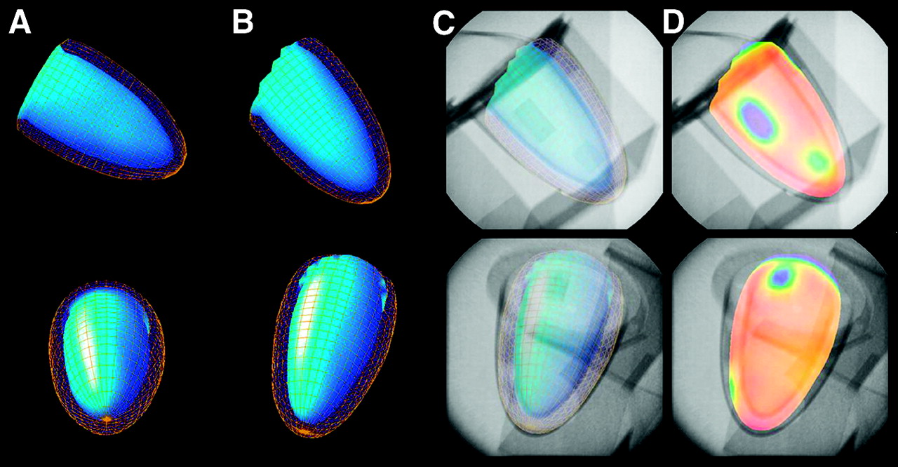
  • FIGURE 3.
    • Download figure
    • Open in new tab
    • Download powerpoint
    FIGURE 3.

    Results of phantom studies confirming the accuracy of reorienting the long axis of the left ventricle for image registration (top: RAO-30°; bottom: LAO-60°). (A and B) QGS 3D surface maps with inner and outer cardiac walls generated by the conventional method (A) and after reorientation (B). (C) Reoriented 3D surface maps were superimposed on radiographic images at RAO-30° and LAO-60° views. (D) Changing the surface display allowed the colored 3D map to reflect the relative count distribution with the fused radiographic image.
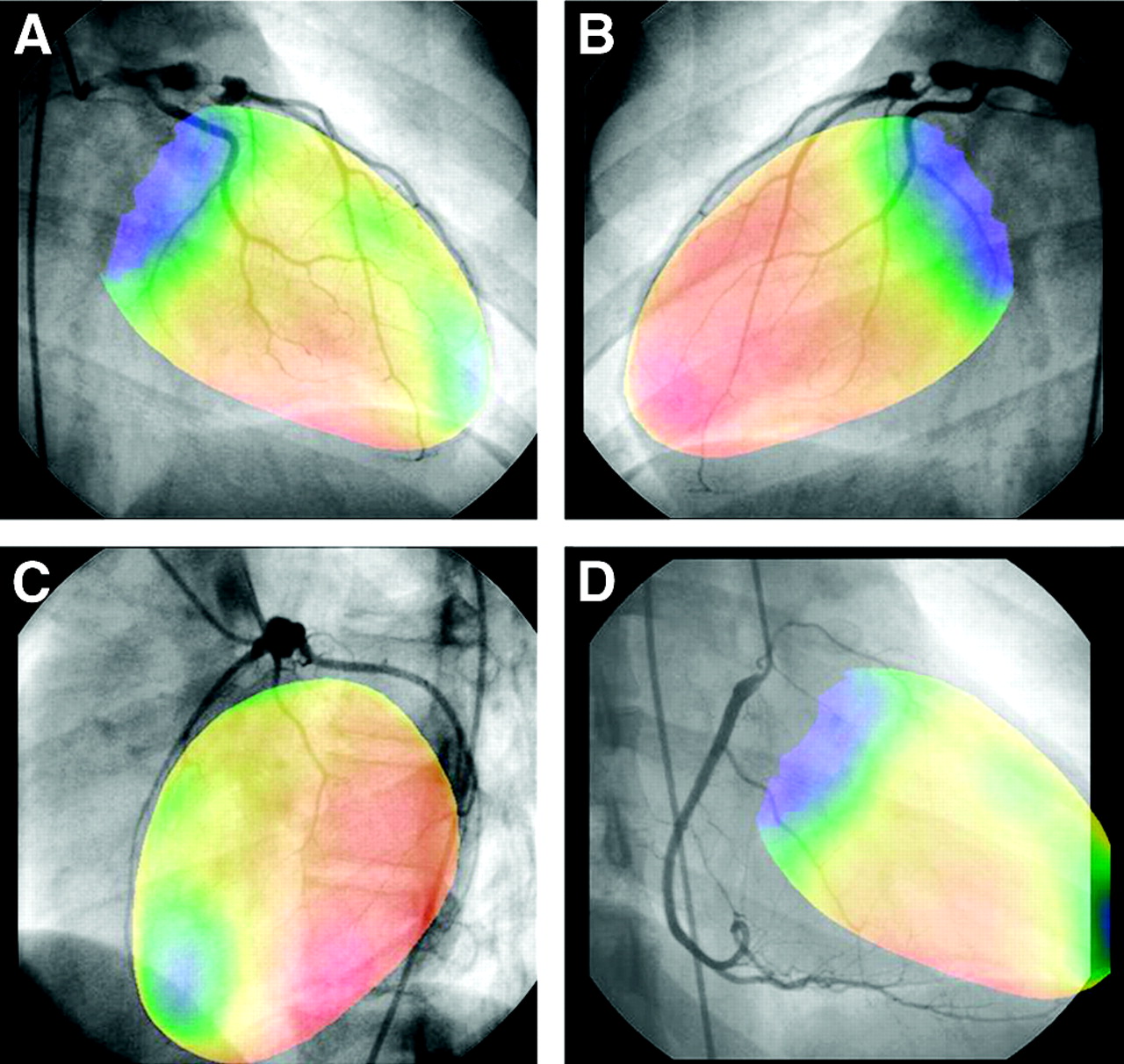
  • FIGURE 4.
    • Download figure
    • Open in new tab
    • Download powerpoint
    FIGURE 4.

    Superimposed displays of a coronary angiogram and an exercise 3D surface map obtained from a 15-y-old girl with Kawasaki disease. An aneurysmal dilatation with distal stenosis (90% narrowing) was located in the proximal portion of the left anterior descending artery on coronary angiography. MPI showed anteroapical hypoperfusion during exercise. The perfusion abnormality was obviously distributed in the distal territory of the left descending coronary artery (A) and not in the diagonal branch territory (B). Meanwhile, the anterior LAO-60° view (C) and superimposition of the right coronary artery on the septal view of MPI (D) are also available with our technique.
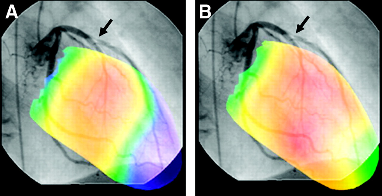
  • FIGURE 5.
    • Download figure
    • Open in new tab
    • Download powerpoint
    FIGURE 5.

    Superimposed displays of images from left coronary angiography and MPI at exercise (A) and rest (B) from a 57-y-old man with chest pain. The unified images show exercise-induced ischemia in the anteroapical myocardium in geometric correspondence with the diseased left anterior descending artery (arrow).

  • FIGURE 6.
    • Download figure
    • Open in new tab
    • Download powerpoint
    FIGURE 6.

    Superimposed displays of a coronary angiogram and an exercise 3D surface map obtained from an 11-y-old girl with Kawasaki disease. (A) The unified image depicts apical and inferior hypoperfusion and a coronary aneurysm (arrow) with distal stenosis in the left anterior descending artery. The right coronary artery (arrowheads) was filled by collateral vessels through the septal branch because the proximal portion of the right coronary artery was occluded. (B) After bypass surgery, the nongated unified image shows homogeneous left ventricular perfusion with patency of the left internal mammary artery bypass graft at exercise. (C and D) ECG-gated unified images at end-diastole (C) and end-systole (D) show normal regional wall motion and count increase in the left ventricular wall.

PreviousNext
Back to top

In this issue

Journal of Nuclear Medicine: 45 (9)
Journal of Nuclear Medicine
Vol. 45, Issue 9
September 1, 2004
  • Table of Contents
  • About the Cover
  • Index by author
Print
Download PDF
Article Alerts
Sign In to Email Alerts with your Email Address
Email Article

Thank you for your interest in spreading the word on Journal of Nuclear Medicine.

NOTE: We only request your email address so that the person you are recommending the page to knows that you wanted them to see it, and that it is not junk mail. We do not capture any email address.

Enter multiple addresses on separate lines or separate them with commas.
Superimposed Display of Coronary Artery on Gated Myocardial Perfusion Scintigraphy
(Your Name) has sent you a message from Journal of Nuclear Medicine
(Your Name) thought you would like to see the Journal of Nuclear Medicine web site.
Citation Tools
Superimposed Display of Coronary Artery on Gated Myocardial Perfusion Scintigraphy
Yoshihiro Nishimura, Kazuki Fukuchi, Tetsuro Katafuchi, Masayoshi Sagou, Hisashi Oka, Yoshio Ishida, Kenya Murase
Journal of Nuclear Medicine Sep 2004, 45 (9) 1444-1449;

Citation Manager Formats

  • BibTeX
  • Bookends
  • EasyBib
  • EndNote (tagged)
  • EndNote 8 (xml)
  • Medlars
  • Mendeley
  • Papers
  • RefWorks Tagged
  • Ref Manager
  • RIS
  • Zotero
Share
Superimposed Display of Coronary Artery on Gated Myocardial Perfusion Scintigraphy
Yoshihiro Nishimura, Kazuki Fukuchi, Tetsuro Katafuchi, Masayoshi Sagou, Hisashi Oka, Yoshio Ishida, Kenya Murase
Journal of Nuclear Medicine Sep 2004, 45 (9) 1444-1449;
Twitter logo Facebook logo LinkedIn logo Mendeley logo
  • Tweet Widget
  • Facebook Like
  • Google Plus One
Bookmark this article

Jump to section

  • Article
    • Abstract
    • MATERIALS AND METHODS
    • RESULTS
    • DISCUSSION
    • CONCLUSION
    • Acknowledgments
    • Footnotes
    • REFERENCES
  • Figures & Data
  • Info & Metrics
  • PDF

Related Articles

  • THIS MONTH IN JNM
  • PubMed
  • Google Scholar

Cited By...

  • Cardiac Image Fusion from Stand-Alone SPECT and CT: Clinical Experience
  • Google Scholar

More in this TOC Section

  • Feasibility of Ultra-Low-Activity 18F-FDG PET/CT Imaging Using a Long–Axial-Field-of-View PET/CT System
  • Cardiac Presynaptic Sympathetic Nervous Function Evaluated by Cardiac PET in Patients with Chronotropic Incompetence Without Heart Failure
  • Validation and Evaluation of a Vendor-Provided Head Motion Correction Algorithm on the uMI Panorama PET/CT System
Show more Clinical Investigations

Similar Articles

SNMMI

© 2025 SNMMI

Powered by HighWire