
Main menu

  • Home
  • Content
    • Current
    • Ahead of print
    • Past Issues
    • JNM Supplement
    • SNMMI Annual Meeting Abstracts
    • Continuing Education
    • JNM Podcasts
  • Subscriptions
    • Subscribers
    • Institutional and Non-member
    • Rates
    • Journal Claims
    • Corporate & Special Sales
  • Authors
    • Submit to JNM
    • Information for Authors
    • Assignment of Copyright
    • AQARA requirements
  • Info
    • Reviewers
    • Permissions
    • Advertisers
  • About
    • About Us
    • Editorial Board
    • Contact Information
  • More
    • Alerts
    • Feedback
    • Help
    • SNMMI Journals
  • SNMMI
    • JNM
    • JNMT
    • SNMMI Journals
    • SNMMI

User menu

  • Subscribe
  • My alerts
  • Log in
  • My Cart

Search

  • Advanced search
Journal of Nuclear Medicine
  • SNMMI
    • JNM
    • JNMT
    • SNMMI Journals
    • SNMMI
  • Subscribe
  • My alerts
  • Log in
  • My Cart
Journal of Nuclear Medicine

Advanced Search

  • Home
  • Content
    • Current
    • Ahead of print
    • Past Issues
    • JNM Supplement
    • SNMMI Annual Meeting Abstracts
    • Continuing Education
    • JNM Podcasts
  • Subscriptions
    • Subscribers
    • Institutional and Non-member
    • Rates
    • Journal Claims
    • Corporate & Special Sales
  • Authors
    • Submit to JNM
    • Information for Authors
    • Assignment of Copyright
    • AQARA requirements
  • Info
    • Reviewers
    • Permissions
    • Advertisers
  • About
    • About Us
    • Editorial Board
    • Contact Information
  • More
    • Alerts
    • Feedback
    • Help
    • SNMMI Journals
  • View or Listen to JNM Podcast
  • Visit JNM on Facebook
  • Join JNM on LinkedIn
  • Follow JNM on Twitter
  • Subscribe to our RSS feeds
OtherClinical Investigations

Preoperative Assessment of Postoperative Remnant Liver Function Using Hepatobiliary Scintigraphy

Roelof J. Bennink, Sander Dinant, Deha Erdogan, Bob H. Heijnen, Irene H. Straatsburg, Arlene K. van Vliet and Thomas M. van Gulik
Journal of Nuclear Medicine June 2004, 45 (6) 965-971;
Roelof J. Bennink
  • Find this author on Google Scholar
  • Find this author on PubMed
  • Search for this author on this site
Sander Dinant
  • Find this author on Google Scholar
  • Find this author on PubMed
  • Search for this author on this site
Deha Erdogan
  • Find this author on Google Scholar
  • Find this author on PubMed
  • Search for this author on this site
Bob H. Heijnen
  • Find this author on Google Scholar
  • Find this author on PubMed
  • Search for this author on this site
Irene H. Straatsburg
  • Find this author on Google Scholar
  • Find this author on PubMed
  • Search for this author on this site
Arlene K. van Vliet
  • Find this author on Google Scholar
  • Find this author on PubMed
  • Search for this author on this site
Thomas M. van Gulik
  • Find this author on Google Scholar
  • Find this author on PubMed
  • Search for this author on this site
  • Article
  • Figures & Data
  • Info & Metrics
  • PDF
Loading

Article Figures & Data

Figures

  • Tables
  • FIGURE 1.
    • Download figure
    • Open in new tab
    • Download powerpoint
    FIGURE 1.

    Preoperative HBS in patient 13, with a proximal cholangiocarcinoma. (A) Reframed images of the dynamic acquisition. Homogeneous liver uptake with moderate cholestasis is seen in the left side, without functional repercussion. (B) A summed image from 150 to 350 s after intravenous injection of 80 MBq of 99mTc-mebrofenin, with an ROI drawn semiautomatically (threshold, 20%) around the entire liver and a second ROI drawn in the mediastinum (blood pool). (C) A blood-pool–corrected liver-uptake time–activity curve. Liver uptake (d) is calculated as the increase in specific (corrected for blood pool) 99mTc-mebrofenin uptake (y-axis) per minute over a period of 200 s (x-axis).

  • FIGURE 2.
    • Download figure
    • Open in new tab
    • Download powerpoint
    FIGURE 2.

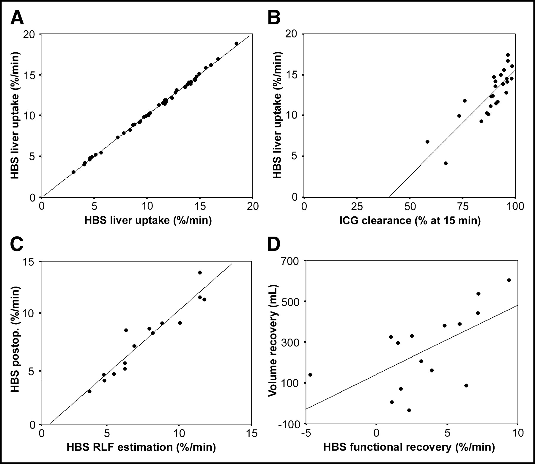
    Scatter plots with linear regression line of HBS liver function calculation reproducibility (A), HBS and ICG clearance LFR assessment (B), HBS preoperative and postoperative (postop.) RLF measurement (C), and liver volume and function recovery (D).

  • FIGURE 3.
    • Download figure
    • Open in new tab
    • Download powerpoint
    FIGURE 3.

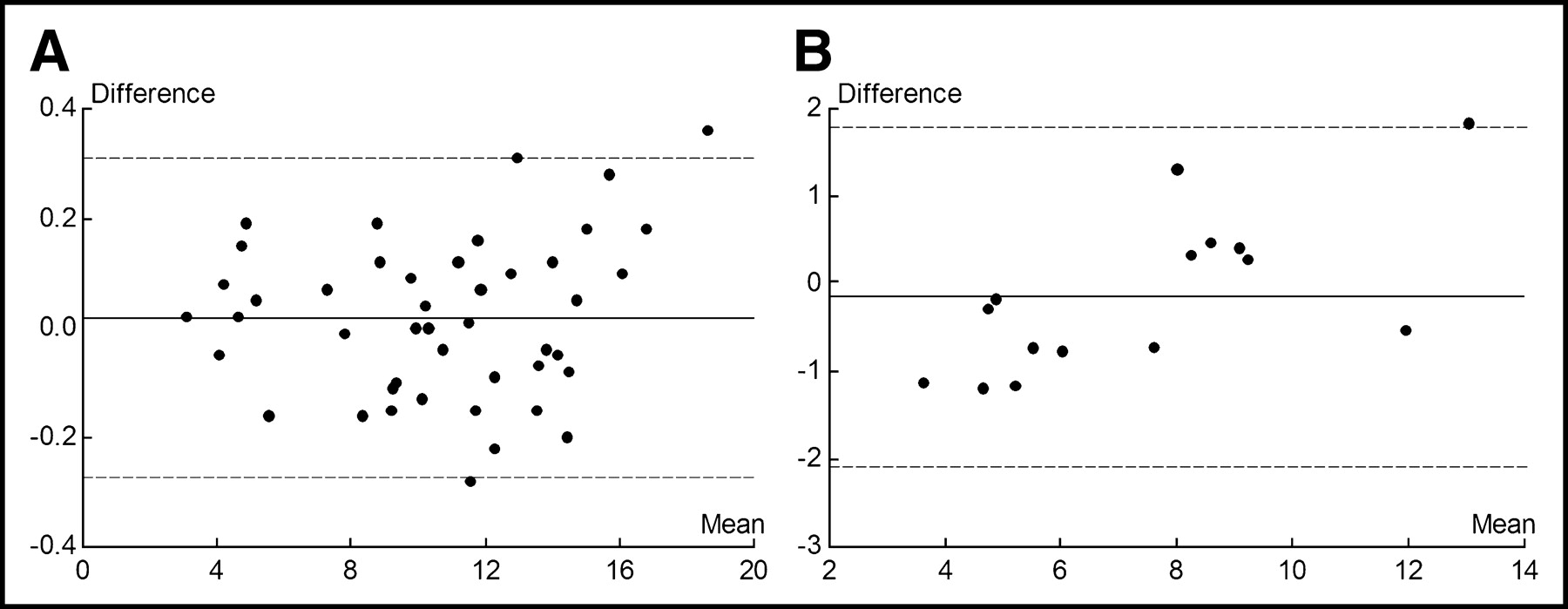
    Bland–Altman plots of hepatic 99mTc-mebrofenin uptake, expressed as percentage uptake per minute. (A) A plot of the mean of the repeated liver uptake function calculation on HBS in 45 studies (horizontal axis) vs. the differences in the repeated calculations (vertical axis). (B) A plot of the mean of FLR determination on HBS before and after surgery in 15 studies (horizontal axis) vs. the differences in the repeated measurements (vertical axis). The horizontal solid lines indicate the mean difference between the 2 calculations. The horizontal dashed lines indicate the 95% limits of agreement (mean ± 1.96 SD).

  • FIGURE 4.
    • Download figure
    • Open in new tab
    • Download powerpoint
    FIGURE 4.

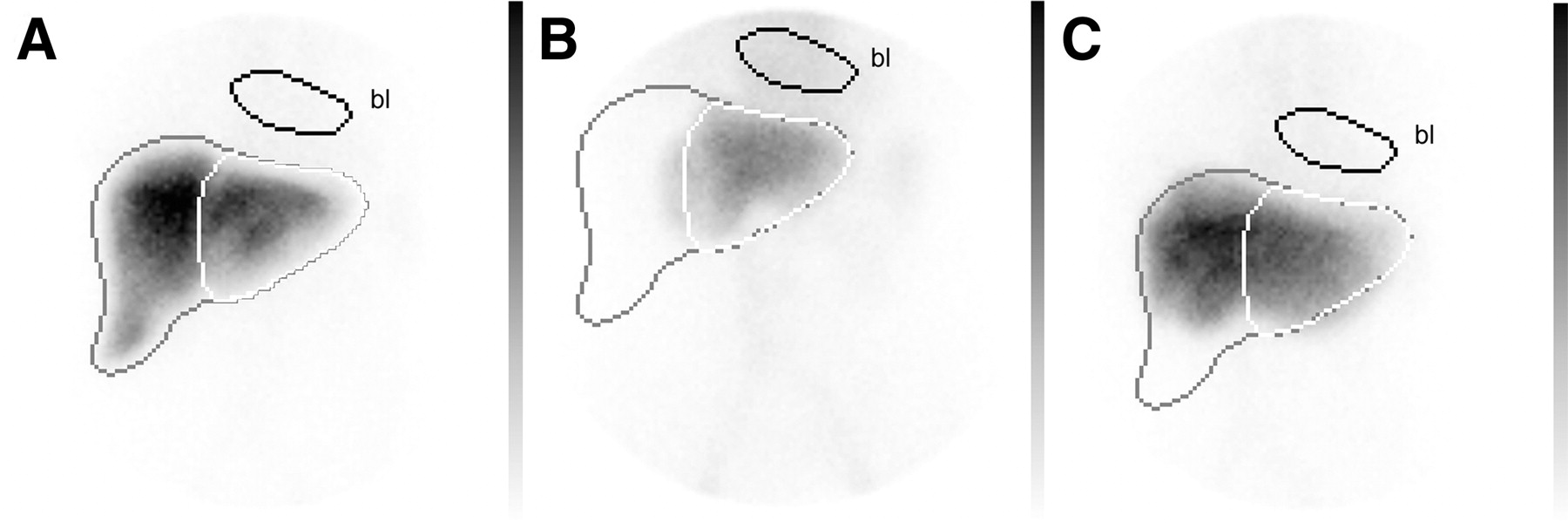
    Summed images of patient 3 from 150 to 350 s after intravenous injection of 80 MBq of 99mTc-mebrofenin preoperatively (A), 1 d postoperatively after right-sided hemihepatectomy (B), and 3 mo postoperatively (C). Images are normalized to the preoperative HBS. In A, an ROI is drawn over the entire liver (black), the future remnant liver (white), and the mediastinal blood pool (bl). The ROIs were copied on the HBS performed 1 d and 3 mo postoperatively. The total liver function was 16.05%/min, and the RLF was estimated at 5.88%/min on preoperative HBS. The measured RLF 1 d postoperatively was 5.15%/min. After 3 mo, liver function recovered to 12.80%/min, with hypertrophy visible on HBS.

  • FIGURE 5.
    • Download figure
    • Open in new tab
    • Download powerpoint
    FIGURE 5.

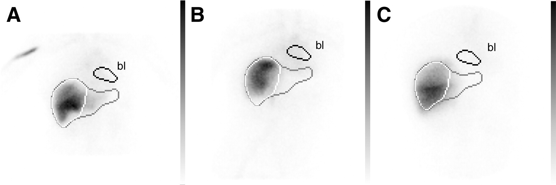
    Summed images of patient 15 from 150 to 350 s after intravenous injection of 80 MBq of 99mTc-mebrofenin preoperatively (A), 1 d postoperatively after left-sided hemihepatectomy (B), and 3 mo postoperatively (C). Images are normalized to the preoperative HBS. In A, an ROI is drawn over the entire liver (black), the future remnant liver (white), and the mediastinal blood pool (bl). The ROIs were copied on the HBS performed 1 d and 3 mo postoperatively. The total liver function was 10.30%/min, and the RLF was estimated at 8.99%/min on preoperative HBS. The measured RLF 1 d postoperatively was 9.30%/min. After 3 mo, liver function recovered to 11.79%/min, with hypertrophy visible on HBS.

Tables

  • Figures
    • View popup
    TABLE 1

    Patient Volumetric and Functional Data

    Patient no.PreoperativePerioperativePostoperative
    ICG (%)CT (mL)HBS (%/min)RLFHBS (%/min)VolRes (mL)VolRem (mL)HBS (%/min)
    195.801,31814.556.418005185.64
    291.631,73911.704.891,0007394.61
    398.571,28516.055.888004855.15
    496.312,34618.458.109501,3968.43
    590.811,51811.517.379006188.67
    694.541,35315.5512.2350085311.7
    789.691,88714.7112.147001,18713.97
    887.201,61610.188.352001,4168.82
    996.371,75916.727.978009597.25
    1090.612,82513.624.191,3001,5253.06
    1194.263,31213.859.101,1002,2129.38
    1293.231,66814.965.277009664.08
    1389.241,94212.395.811,2007424.65
    1487.231,28210.774.964008824.79
    1586.4498210.308.894005829.30
    • RLFHBS = RLF determined on preoperative HBS.

    • VolRes = Surgically resected volume.

    • VolRem = Volume of remnant liver.

    • View popup
    TABLE 2

    Liver Recovery Data

    Patient no.HBSPost (%/min)HBS3M (%/min)VolPost (mL)Vol3M (mL)HBSRec (%/min)VolRec (mL)
    15.6412.805189607.16442
    24.6111.847391,2777.23537
    35.1514.524851,0869.37601
    48.4312.331,3961,5583.90162
    58.6710.186189141.51296
    611.712.718531,1771.01324
    713.979.301,1871,327−4.67140
    88.8211.141,4161,3812.32−35
    97.2513.609591,0476.3587
    103.064.151,5251,5301.095
    119.3814.202,2122,5924.82380
    124.089.949661,3565.86389
    134.657.837429473.18206
    144.799.768829554.9772
    159.3011.795829132.49331
    • Post = Assessment 1 d after surgery.

    • 3M = Assessment 3 mo after surgery.

    • HBSRec = Functional recovery determined with HBS.

    • VolRec = Volume recovery determined with CT.

PreviousNext
Back to top

In this issue

Journal of Nuclear Medicine: 45 (6)
Journal of Nuclear Medicine
Vol. 45, Issue 6
June 1, 2004
  • Table of Contents
  • About the Cover
  • Index by author
Print
Download PDF
Article Alerts
Sign In to Email Alerts with your Email Address
Email Article

Thank you for your interest in spreading the word on Journal of Nuclear Medicine.

NOTE: We only request your email address so that the person you are recommending the page to knows that you wanted them to see it, and that it is not junk mail. We do not capture any email address.

Enter multiple addresses on separate lines or separate them with commas.
Preoperative Assessment of Postoperative Remnant Liver Function Using Hepatobiliary Scintigraphy
(Your Name) has sent you a message from Journal of Nuclear Medicine
(Your Name) thought you would like to see the Journal of Nuclear Medicine web site.
Citation Tools
Preoperative Assessment of Postoperative Remnant Liver Function Using Hepatobiliary Scintigraphy
Roelof J. Bennink, Sander Dinant, Deha Erdogan, Bob H. Heijnen, Irene H. Straatsburg, Arlene K. van Vliet, Thomas M. van Gulik
Journal of Nuclear Medicine Jun 2004, 45 (6) 965-971;

Citation Manager Formats

  • BibTeX
  • Bookends
  • EasyBib
  • EndNote (tagged)
  • EndNote 8 (xml)
  • Medlars
  • Mendeley
  • Papers
  • RefWorks Tagged
  • Ref Manager
  • RIS
  • Zotero
Share
Preoperative Assessment of Postoperative Remnant Liver Function Using Hepatobiliary Scintigraphy
Roelof J. Bennink, Sander Dinant, Deha Erdogan, Bob H. Heijnen, Irene H. Straatsburg, Arlene K. van Vliet, Thomas M. van Gulik
Journal of Nuclear Medicine Jun 2004, 45 (6) 965-971;
Twitter logo Facebook logo LinkedIn logo Mendeley logo
  • Tweet Widget
  • Facebook Like
  • Google Plus One
Bookmark this article

Jump to section

  • Article
    • Abstract
    • MATERIALS AND METHODS
    • RESULTS
    • DISCUSSION
    • CONCLUSION
    • Footnotes
    • REFERENCES
  • Figures & Data
  • Info & Metrics
  • PDF

Related Articles

  • No related articles found.
  • PubMed
  • Google Scholar

Cited By...

  • Liver Intrinsic Function Evaluation (LIFE): Multi-parametric Liver Function Profiles of Patients Undergoing Hepatectomy
  • A Pilot Study on Hepatobiliary Scintigraphy to Monitor Regional Liver Function in 90Y Radioembolization
  • Evaluation of liver regeneration and post-hepatectomy liver failure after hemihepatectomy in patients with hepatocellular carcinoma
  • The Role of Organic Anion Transporters in Diagnosing Liver Diseases by Magnetic Resonance Imaging
  • Quantitative Assessment of Hepatic Function During Liver Regeneration in a Standardized Rat Model
  • Nuclear Imaging Techniques for the Assessment of Hepatic Function in Liver Surgery and Transplantation
  • Comparison Between the Values of the Hepatic Uptake Rate Obtained by 2 Methods, Using Hepatobiliary Scintigraphy in Patients with Nonalcoholic Steatohepatitis
  • 99mTc-Mebrofenin Hepatobiliary Scintigraphy with SPECT for the Assessment of Hepatic Function and Liver Functional Volume Before Partial Hepatectomy
  • 99mTc-GSA Scintigraphy with SPECT for Assessment of Hepatic Function and Functional Volume During Liver Regeneration in a Rat Model of Partial Hepatectomy
  • Risk Assessment of Posthepatectomy Liver Failure Using Hepatobiliary Scintigraphy and CT Volumetry
  • Google Scholar

More in this TOC Section

  • Cardiac Presynaptic Sympathetic Nervous Function Evaluated by Cardiac PET in Patients with Chronotropic Incompetence Without Heart Failure
  • Validation and Evaluation of a Vendor-Provided Head Motion Correction Algorithm on the uMI Panorama PET/CT System
  • Left Ventricular Strain from Myocardial Perfusion PET Imaging: Method Development and Comparison to 2-Dimensional Echocardiography
Show more Clinical Investigations

Similar Articles

SNMMI

© 2025 SNMMI

Powered by HighWire