


Article Figures & Data
Figures
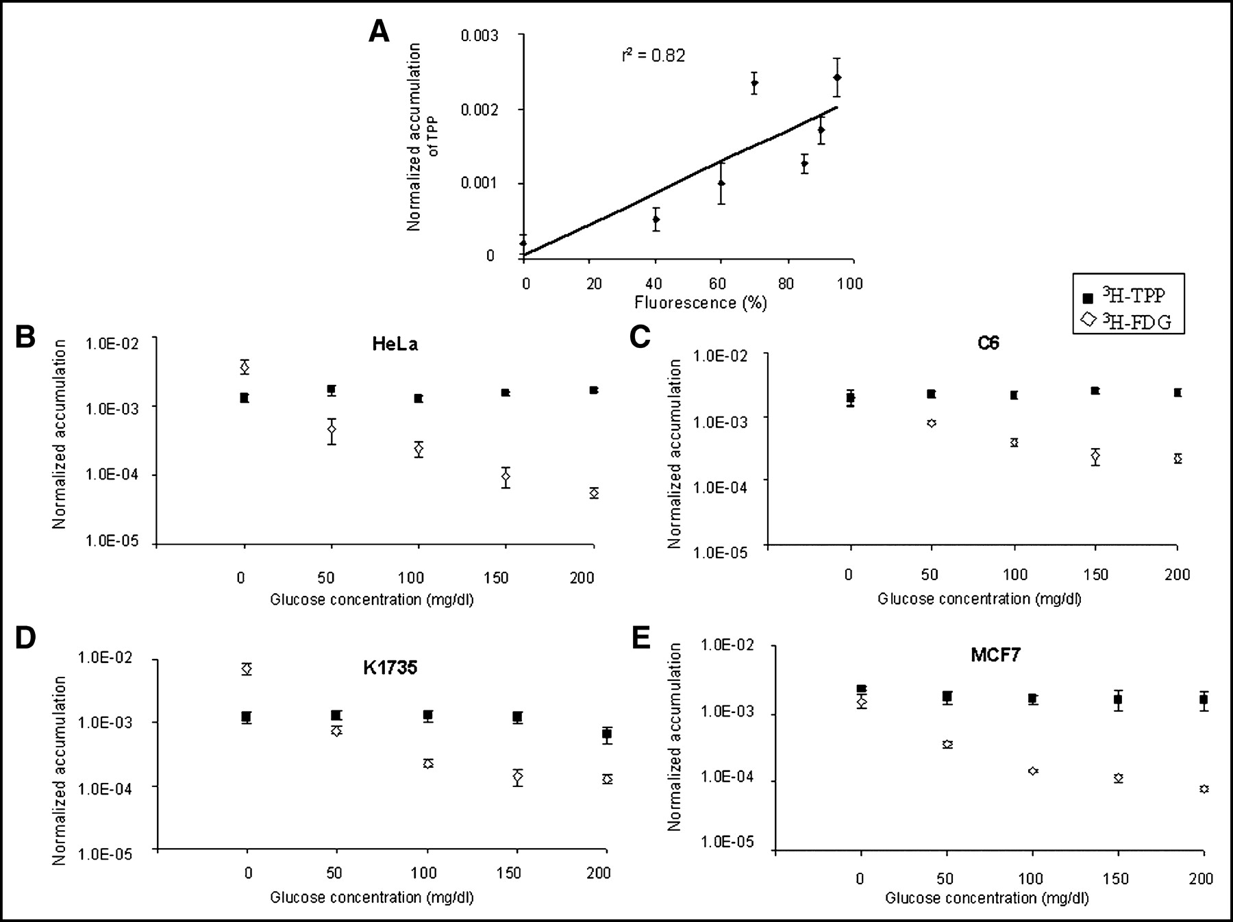
- FIGURE 1.
(A) ΔΨm data (2) and current 3H-TPP uptake data showed good correlation (r2 = 0.82) in HeLa, LnCap, MCF7, A549, N2A, SCC-15, and K1735 cell lines. 3H-TPP accumulation data are expressed as normalized accumulation of probe in (dpm cells/dpm medium/μg total protein) ± SE. (B–E) 3H-TPP significantly accumulates in HeLa (B), C6 (C), K1735 (D), and MCF7 (E) cell lines in cell culture. Cellular uptake studies with 3H-TPP and 3H-FDG were performed under 5 different glucose concentrations of culture media (0, 50, 100, 150, 200 mg/dL). Data are expressed as normalized accumulation of probe in (dpm cells/dpm medium/μg total protein) ± SE. Though 3H-FDG accumulation in cell culture is dependent on medium glucose concentration, 3H-TPP accumulation is constant for all glucose concentrations. 3H-TPP accumulation is significantly (P < 0.05) greater than that of 3H-FDG for glucose ≥50 mg/dL in C6, HeLa, and MCF7 cells. 3H-TPP accumulation is significantly (P < 0.05) greater than that of 3H-FDG for glucose ≥100 mg/dL in K1735 cells.
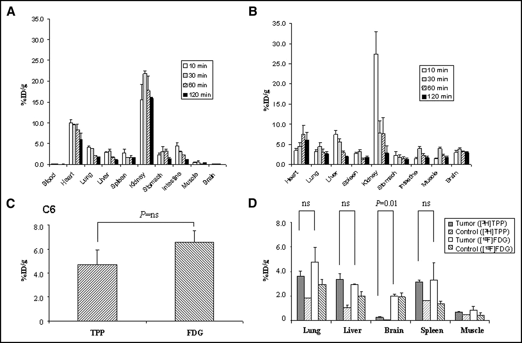
- FIGURE 2.
Biodistribution of 3H-TPP and 18F-FDG in normal nude mice and mouse models with tumor xenografts or metastases. (A) Biodistribution of 3H-TPP in normal nude mice (n = 12) showed relatively high accumulation in heart (8.3 ± 1.4 %ID/g at 1 h) and kidneys (17.8 ± 3.5 %ID/g at 1 h) but low uptake in other tissues (0.04–3.2 %ID/g at 1 h). 3H-TPP exhibits very low values in blood as early as 10 min and in brain. (B) Biodistribution of 18F-FDG in different organs of nude mice (n = 12) shows high accumulation in heart (7.6 ± 2.2 %ID/g at 1 h) and kidneys (7.7 ± 4.0 %ID/g at 1 h). Radioactivity of 18F-FDG in other organs is relatively low (1.4–3.1 %ID/g at 1 h). (C) Comparison of 3H-TPP and 18F-FDG accumulation in nude mice carrying xenograft tumor (n = 8). 3H-TPP accumulation in C6 xenograft tumor (4.7 ± 1.2 %ID/g) is slightly lower than that of 18F-FDG (6.5 ± 1.0 %ID/g; P = not significant [ns]). (D) Comparison of 3H-TPP and 18F-FDG accumulation in metastatic lesions of malignant melanoma in SCID mice (n = 6). 3H-TPP vs. 18F-FDG accumulation in metastatic lung (3.6 ± 0.4 %ID/g vs. 4.8 ± 0.7 %ID/g), liver (3.4 ± 0.4 %ID/g vs. 2.9 ± 0.1 %ID/g), and spleen (3.2 ± 0.1 %ID/g vs. 3.3 ± 0.7 %ID/g) is not significantly different. 18F-FDG accumulation in metastatic brain is significantly higher than that of 3H-TPP (2.0 ± 0.2 %ID/g vs. 0.3 ± 0.1 %ID/g; P = 0.01). All values are mean ± SE.
- FIGURE 3.
Autoradiographic images of 3H-TPP and 18F-FDG in C6 xenograft tumor. C6 cells were subcutaneously implanted in left shoulders of mice to develop xenograft tumor model. When tumors reached 8 mm of size, DWBA was performed with tail-vein injection of 3H-TPP (370 kBq [10 μCi]) and 18F-FDG (7.4 MBq [200 μCi)]) using the same animal. (Left) 3H-TPP accumulation in C6 xenograft tumor (arrow). This image exhibits relatively low background radioactivity except for strong renal activity. (Right) 18F-FDG accumulation in C6 xenograft tumor (arrow). 18F-FDG uptake is observed in kidneys, blood vessels, and dorsal upper back, indicating brown adipose tissue (arrowhead). (Middle) Tissue section (45-μm thickness) corresponding to DWBA images.
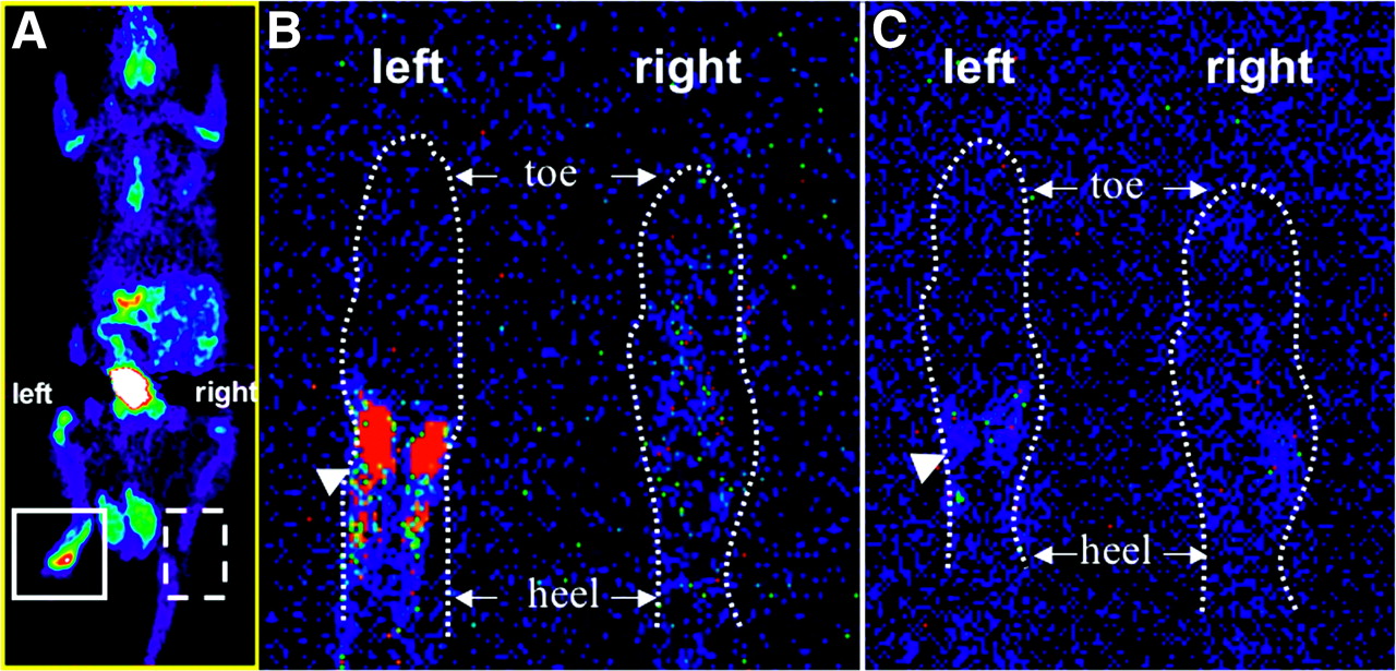
- FIGURE 4.
Comparison of 18F-FDG and 3H-TPP accumulation in inflammatory tissue. Inflammatory reaction was induced by subcutaneous injection of 50 μL of CFA (10 mg/mL) in left hind paw of Sprague–Dawley rat. Right hind paw was injected with saline as negative control. 3H-TPP (3.7 MBq [100 μCi]) and 18F-FDG (74 MBq [2 mCi]) were injected via tail vein to perform autoradiography and microPET studies, respectively. (A) Whole-body microPET image of Sprague–Dawley rat shows strong accumulation of 18F-FDG in inflammatory tissue of left hind paw. After killing the rat, autoradiographic studies for 18F-FDG and 3H-TPP were performed using the same animal. (B) 18F-FDG autoradiographic image of coronal section of inflammatory (left) and control (right) hind paw. CFA pretreatment in left hind paw as compared with control right hind paw (saline injected) of rat caused significant increase in 18F-FDG accumulation (arrowhead) corresponding to microPET image. (C) 3H-TPP autoradiographic image of coronal section of inflammatory (left) and control (right) hind paw. This slice was taken right after slice for 18F-FDG (45 μm). 3H-TPP shows very mild possible accumulation in inflammatory tissue of left hind paw (arrowhead).


In this issue


Jump to section
Related Articles
Cited By...
- Perspectives on Brown Adipose Tissue Imaging: Insights from Preclinical and Clinical Observations from the Last and Current Century
- Comparison of 18F-Labeled Fluoroalkylphosphonium Cations with 13N-NH3 for PET Myocardial Perfusion Imaging
- Evaluation of a Mitochondrial Voltage Sensor, (18F-Fluoropentyl)Triphenylphosphonium Cation, in a Rat Myocardial Infarction Model
- 4-[18F]-Tetraphenylphosphonium as a PET Tracer for Myocardial Mitochondrial Membrane Potential
- Imaging of Apoptosis
- Impact of Animal Handling on the Results of 18F-FDG PET Studies in Mice
- A New Strategy to Screen Molecular Imaging Probe Uptake in Cell Culture Without Radiolabeling Using Matrix-Assisted Laser Desorption/Ionization Time-of-Flight Mass Spectrometry
- Phosphonium compounds as new and specific inhibitors of bovine serum amine oxidase
- Re: Tetraphenylphosphonium as a Novel Molecular Probe for Imaging Tumors