


Abstract
Despite substantial advances in the diagnosis of cardiovascular disease, there is a need for 18F-labeled myocardial perfusion agents for the diagnosis of ischemic heart disease because current PET tracers for myocardial perfusion imaging have a short half-life that limits their widespread clinical use in PET. Thus, 18F-labeled fluoroalkylphosphonium derivatives (18F-FATPs), including (5-18F-fluoropentyl)triphenylphosphonium cation (18F-FPTP), (6-18F-fluorohexyl)triphenylphosphonium cation (18F-FHTP), and (2-(2-18F-fluoroethoxy)ethyl)triphenylphosphonium cation (18F-FETP), were synthesized. The myocardial extraction and image quality of the 18F-FATPs were compared with those of 13N-NH3 in rat models. Methods: The first-pass extraction fraction (EF) values of the 18F-FATPs (18F-FPTP, 18F-FHTP, 18F-FETP) and 13N-NH3 were measured in isolated rat hearts perfused with the Langendorff method (flow velocities, 0.5, 4.0, 8.0, and 16.0 mL/min). Normal and myocardial infarction rats were imaged with small-animal PET after intravenous injection of 37 MBq of 18F-FATPs and 13N-NH3. To determine pharmacokinetics, a region of interest was drawn around the heart, and time–activity curves of the 18F-FATPs and 13N-NH3 were generated to obtain the counts per pixel per second. Defect size was analyzed on the basis of polar map images of 18F-FATPs and 13N-NH3. Results: The EF values of 18F-FATPs and 13N-NH3 were comparable at low flow velocity (0.5 mL/min), whereas at higher flows EF values of 18F-FATPs were significantly higher than those of 13N-NH3 (4.0, 8.0, and 16.0 mL/min, P < 0.05). Myocardium-to-liver ratios of 18F-FPTP, 18F-FHTP, 18F-FETP, and 13N-NH3 were 2.10 ± 0.30, 4.36 ± 0.20, 3.88 ± 1.03, and 0.70 ± 0.09, respectively, 10 min after injection, whereas myocardium-to-lung ratios were 5.00 ± 0.25, 4.33 ± 0.20, 7.98 ± 1.23, and 2.26 ± 0.14, respectively. Although 18F-FATPs and 13N-NH3 sharply delineated myocardial perfusion defects, defect size on the 13N-NH3 images was significantly smaller than on the 18F-FATP images soon after tracer injection (0–10 min, P = 0.027). Conclusion: 18F-FATPs exhibit higher EF values and more rapid clearance from the liver and lung than 13N-NH3 in normal rats, which led to excellent image quality in a rat model of coronary occlusion. Therefore, 18F-FATPs are promising new PET radiopharmaceuticals for myocardial perfusion imaging.
- 18F-fluoroalkylphosphonium cations
- perfusion
- first-pass extraction fraction
- cardiac PET
- myocardial infarction
Since the 1980s, SPECT has been the most widely applied method to assess myocardial blood flow and diagnose coronary artery disease (1). Notably, 99mTc-sestamibi, 99mTc-tetrofosmin, and 201Tl are attractive surrogates for myocardial perfusion imaging using SPECT (2,3). However, the technical limitations of SPECT imaging, such as the absence of a standardized method for correction of photon attenuation and significant uptake of SPECT tracers in organs adjacent to the heart, may compromise the delineation of small lesions and limit the diagnostic accuracy of SPECT (4). PET has several technical advantages over SPECT including higher spatial resolution and a standardized method for the correction of photon attenuation. PET imaging also enables quantitative measurements of myocardial blood flow because it allows accurate determinations of rapidly changing tissue counts over time. Because of these advantages, it allows quantitative measures of myocardial tracer uptake and regional myocardial blood flow (5). However, the PET tracers currently used for myocardial perfusion imaging have a short half-life (13N-NH3, 9.97 min; 82Rb, 1.27 min; 15O-H2O, 2.04 min), limiting widespread clinical use of PET because of the need for an on-site cyclotron or generator (6,7). The use of 18F-labeled myocardial perfusion agents could overcome these limitations and simplify clinical protocols as a result of the longer half-life (109.8 min) and better spatial resolution and quantification (5,8).
To address this unmet clinical need, several publications have proposed 18F-labeled phosphonium cations as myocardial imaging agents (9–16). Similar to SPECT tracers such as 99mTc-sestamibi and 99mTc-tetrofosmin, phosphonium cations accumulate in cardiomyocytes as a result of the higher mitochondrial membrane potential (11,17–20). Previously, we reported the synthesis and characterization of novel 18F-labeled fluoroalkylphosphonium derivatives (18F-FATPs), including (5-18F-fluoropentyl)triphenylphosphonium cation (18F-FPTP), (6-18F-fluorohexyl)triphenylphosphonium cation (18F-FHTP), and (2-(2-18F-fluoroethoxy)ethyl)triphenylphosphonium cation (18F-FETP), as a voltage sensor for myocardial imaging (9,10,16). In vivo biodistribution and imaging studies of 18F-FATPs demonstrated intense initial myocardial uptake and retention of these tracers, with rapid clearance from liver and lung. Sharp delineation of areas of myocardial ischemia was achieved, suggesting the potential for use of these moieties as tracers of myocardial perfusion for clinical imaging. Therefore, further characterization of 18F-FATPs to evaluate first-pass myocardial extraction and a comparison with the standard PET tracer 13N-NH3 are required. To determine whether 18F-FATPs are comparable to 13N-NH3 as PET perfusion tracers and to provide better image quality than that currently provided by 13N-NH3 for the detection of myocardial infarction in rats, we compared first-pass extraction fraction (EF) values and image characteristics of the 18F-FATPs with those of 13N-NH3 in isolated rat hearts or in a rat myocardial infarction (MI) model.
MATERIALS AND METHODS
Full details of tracer preparation and animal model are presented in the supplemental materials (available at http://jnm.snmjournals.org).
Perfused Isolated Rat Heart Study
The first-pass EF values of radiotracers were measured in isolated rat hearts perfused by the Langendorff method. Hearts were quickly excised from anesthetized rats (intraperitoneal sodium pentobarbital [0.25 mL]; Merial GmbH), placed in ice-chilled Krebs-Henseleit bicarbonate buffer (glucose at 10 mmol/L), and cannulated via the aorta (n = 8, each tracer, total rat hearts = 32). Krebs-Henseleit buffer, oxygenated with a mixture of 95% oxygen and 5% carbon dioxide, was used to perfuse the heart (flow velocities, 0.5, 4.0, 8.0, and 16.0 mL/min). The heart was stabilized for a period of 15 min, after which a bolus injection of approximately 0.925 MBq each of 18F-FATP or 13N-NH3 (0.037 MBq/μL) was administered. Whole-heart radioactivity was measured over 10 min with a pair of bismuth germanate detectors interfaced to coincidence detection circuitry. Care was taken to keep the counting rate within the linear response range of the system, to prevent dead-time–induced counting rate losses during tracer injection. The total coincidence counting rate was measured as a function of time and corrected for decay. The curve was analyzed by fitting an exponentially decaying function to the data representing the tissue washout component (from 100 to 600 s). The fitted curve was extrapolated to the time of the maximum counting rate, and the value obtained was divided by the measured maximum counting rate. This ratio is a measure of the EF. As the curve was essentially linear in the fit region, possible differences between an exponential fit and a linear fit were studied. No differences were found for the determination of the EF (5).
Small-Animal PET Study and Image Analysis
A dedicated small-animal PET/CT scanner (Inveon; Siemens Medical Solutions) was used for in vivo imaging of the 18F-FATPs and 13N-NH3. Normal (n = 3, each radiotracer) or MI (n = 3, each radiotracer) rats were anesthetized with isoflurane, placed in a cradle, and equipped with masks for anesthetic gas supply and warm water pads at the tail veins for injection. MI rats underwent imaging studies 24 h after left coronary artery occlusion (21,22). Dynamic small-animal PET images were acquired for 70 min after injection of 37 MBq of 18F-FATP and for 30 min after injection of 37 MBq of 13N-NH3.
Acquired images were reconstructed using a 3-dimensional ordered-subset expectation maximization algorithm with 4 iterations. Reconstructed pixel sizes were 0.78 mm in the transverse and axial directions. The dimensions of the reconstructed images were 128 × 128 pixels in each of the 159 transverse slices. Data were normalized and corrected for randoms, dead time, and decay.
Analysis of the small-animal PET images was performed with PMOD software (PMOD Technologies Ltd.) (6). To determine pharmacokinetics, a region of interest was drawn around the heart. Time–activity curves of the 18F-FATPs and 13N-NH3 were generated to obtain the counts per pixel per second. Image contrast was calculated on the basis of the Michelson contrast formula as follows (Eq. 1) (23): Eq. 1where maximum and minimum represent the highest and lowest pixel values, respectively, in the myocardial perfusion polar map of the 18F-FATPs and 13N-NH3 image.
To measure the infarction size in the small-animal PET images, reconstructed PET data were reoriented into 17 segments of polar map images. The perfusion value of each segment was measured as the average value of all pixels in the segment. The infarct area, which was defined as the fraction of the polar map segment with a perfusion value of 60% or less relative to that of the segment with the highest value, was expressed as a percentage of the left ventricular myocardium (16).
Statistical Analysis
Values are reported as the mean ± SD. The first-pass EF values and myocardium-to-liver and myocardium-to-lung ratios of 18F-FATPs or 13N-NH3 were compared by Mann–Whitney U test. Differences in infarction size between 0–10 and 20–30 min or 0–3 and 0–6 min were compared by Wilcoxon signed-rank test. Statistical analysis was performed with SPSS software (version 21.0; IBM). P values of less than 0.05 were considered statistically significant.
RESULTS
Tracer Preparation
The chemical structures of 18F-FATPs are shown in Supplemental Figure 1. The total reaction time of the 18F-FATPs was within 60 min, and the overall decay-corrected radiochemical yield was approximately 15%–30%. Radiochemical purity was greater than 98% as measured by high-performance liquid chromatography. The specific activity of 18F-FATPs was greater than 6.1 TBq/μmol. The non–decay-corrected radiochemical yield of 13N-NH3 was approximately 60%–70%.
Perfused Isolated Rat Heart Study
EF values for all flow rates are shown in Table 1 and Figure 1. Lower EFs were found at higher flows, which was consistent with the 18F-FATPs and 13N-NH3. The EF values of 18F-FPTP, 18F-FHTP, and 18F-FETP were significantly higher than those of 13N-NH3 at flow rates of 4.0, 8.0, and 16 mL/min (P < 0.05). At the lower flow rate of 0.5 mL/min, there were no significant differences among 18F-FPTP, 18F-FHTP, and 13N-NH3. Only 18F-FETP demonstrated a significantly lower EF value than 13N-NH3 at a flow velocity of 0.5 mL/min.
First-Pass EF Values (%) of 18F-FATPs and 13N-NH3 at Different Flow Rates
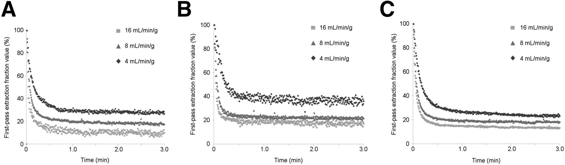
First-pass EFs as function of blood flow. Smoothed time–activity curves obtained from sequential measurements of single rat heart are shown. Flow velocities and EFs are indicated (n = 6, each flow). (A) 18F-FPTP. (B) 18F-FHTP. (C) 18F-FETP.
Small-Animal PET Image Characteristics
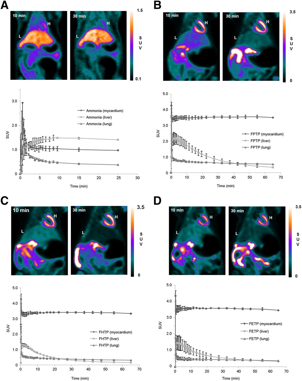
Static small-animal PET images of normal rats 10 and 30 min after intravenous injection of the 18F-FATPs or 13N-NH3 are shown in Figure 2 and Supplemental Videos 1–4. The 18F-FATP images demonstrated good visualization of the heart, with excellent heart-to-background contrast at each time point. High contrast between the myocardium and liver or lung was seen, whereas 13N-NH3 images showed higher liver uptake than heart uptake until 30 min after tracer injection. Time–activity curves and ratios between myocardium and liver or lung after tracer injection are shown in, respectively, Figure 2 and Tables 2 and 3. The 18F-FATP time–activity curves demonstrated rapid accumulation in the myocardium (1–2 min), with stable retention for at least 60 min. The myocardium-to-liver ratios of 18F-FPTP, 18F-FHTP, and 18F-FETP were 1.67 ± 0.21, 2.49 ± 0.08, and 2.60 ± 0.85, respectively, whereas myocardium-to-lung ratios were 3.65 ± 0.20, 5.67 ± 0.52, and 6.31 ± 0.80, respectively, 1 min after intravenous injection. These ratios increased until 30 min. By contrast, the 13N-NH3 time–activity curve revealed that the myocardium-to-liver ratio and the myocardium-to-lung ratio were 0.80 ± 0.14 and 0.93 ± 0.07, respectively, at 5 min after injection. Thereafter, liver and lung uptake of 13N-NH3 was higher than that of the myocardium. The myocardium-to-liver ratios of 18F-FPTP, 18F-FHTP, and 18F-FETP were over 7-, 17-, and 10-fold higher, whereas myocardium-to-lung ratios were 2-, 3-, and 3-fold higher, respectively, than those of 13N-NH3 at 30 min after injection.

Coronal small-animal PET images and time–activity curves of normal rats after intravenous injection of 37 MBq of 13N-NH3 (A), 18F-FPTP (B), 18F-FHTP (C), or 18F-FETP (D) (n = 3, each tracer). Heart was visible, with excellent heart-to-background contrast at each time point after 18F-FATP injection. H = heart; L = liver; SUV = standardized uptake value.
Myocardium-to-Liver Ratios of 18F-FATPs and 13N-NH3 in Normal Rats
Myocardium-to-Lung Ratios of 18F-FATPs and 13N-NH3 in Normal Rats
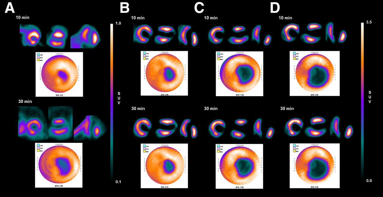
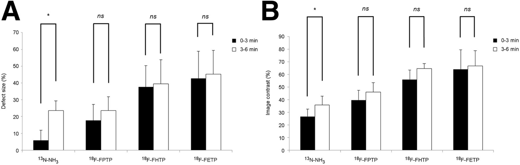
Representative images of MI rats acquired after acute ligation of the left coronary artery in the short-, vertical long-, and horizontal long-axes and a polar map collected 10 and 30 min after injection of 18F-FATPs or 13N-NH3 are shown in Figure 3. Sharply defined myocardial defects were present with 18F-FATPs or 13N-NH3 (10 min). However, defect size on the early (0–10 min) 13N-NH3 images was significantly smaller than that on 18F-FATPs images (0–10 min, P = 0.027), which might be due to spillover of radioactivity into the left ventricular or right ventricular myocardium (Supplemental Fig. 2). To confirm the spillover effect of 13N-NH3 early after intravenous injection, we compared polar map images acquired for 0–3 min and 3–6 min (Supplemental Fig. 3). The defect size and contrast ratio of 13N-NH3 images at 0–3 min were significantly lower than those at 3–6 min (P = 0.043), whereas 18F-FATPs showed similar values for defect size and contrast ratio at 0–3 and 3–6 min (Fig. 4).

Short-, vertical long-, and horizontal long-axis and polar map images of 13N-NH3 (A), 18F-FPTP (B), 18F-FHTP (C), or 18F-FETP (D) in each representative animal. Data were collected between 0–10 and 20–30 min after radiotracer injection (37 MBq). SUV = standardized uptake value.

Defect size (%) (A) and image contrast (%) (B) of 13N-NH3, 18F-FPTP, 18F-FHTP, and 18F-FETP between 0–3 and 3–6 min (n = 3, each tracer). *P < 0.05. ns = not significant.
DISCUSSION
The mitochondrial membrane potential is higher in cardiomyocytes than in normal epithelial cells, and loss of mitochondrial membrane potential is an early event in cell death caused by myocardial ischemia (24). Similar to SPECT agents such as 99mTc-sestamibi and 99mTc-tetrofosmin, phosphonium cations accumulate in cardiomyocytes. This accumulation occurs through the higher density of mitochondria and higher electrochemical transmembrane (inside-negative) potential of cardiomyocytes, which facilitate mitochondrial uptake of phosphonium cations (17). Therefore, radiolabeled phosphonium cations are promising candidates for myocardial imaging (25).
The first radiolabeled phosphonium cation studied for use in PET was 11C-triphenylmethylphosphonium (11C-TPMP) (26). PET studies with 11C-TPMP were performed to evaluate the membrane potential of heart tissue in a canine model, which was determined to be 148.1 ± 6.0 mV (inside-negative). In addition, PET studies showed that 11C-TPMP accumulated in the heart immediately after intravenous injection, with a high heart-to-blood ratio (>46:1) and heart-to-lung ratio (14:1) (27,28). However, the use of 11C-TPMP was limited because of the short half-life of 11C (20 min). To provide a tracer similar to 11C-TPMP but with a longer half-life, the synthesis and evaluation of 18F-labeled phosphonium cations were investigated (28). Among them, 4-18F-fluorobenzyltriphenylphosphonium (18F-FBnTP) showed excellent results as a myocardial perfusion agent. 18F-FBnTP was synthesized using a 4-step procedure. The 4-trimethylammoniumbenzaldehyde was used as a precursor and a third intermediate, 4-18F-fluorobenzyl bromide, was reacted with triphenylphosphine to yield 18F-FBnTP. 18F-FBnTP was metabolically stable and demonstrated excellent characteristics as a cardiac imaging agent in healthy animals and coronary artery disease models (11,13,18). In a separate study, direct fluorination of tetraphenylphosphonium was investigated (14). Synthesis of (4-18F-fluorophenyl)triphenylphosphonium (18F-TPP) was performed via a direct nucleophilic substitution reaction of 18F-fluoride using 4-nitrophenyltriphenylphosphonium as a precursor.
Myocardial selectivity and hepatic clearance depends on the lipophilicity and functional groups of the radiotracer. Although there is little information available with regard to the optimal lipophilicity needed for high myocardial selectivity, cationic radiotracers with log P values in the range of 0.5–1.3 are particularly useful for imaging organs with high mitochondrial density as a result of their fast membrane-penetration kinetics (29). Furthermore, the lipophilic interaction between the triphenylphosphonium cation and the lipid layer is attractive because the alkyl group increases lipophilicity (30,31). Thus, we synthesized several radiolabeled phosphonium cations and assessed lipophilicity at different carbon chain lengths and with different functional groups to derive the uptake kinetics (9,10,12,15,16). The three 18F-FATP derivatives in this study (18F-FPTP, 18F-FHTP, and 18F-FETP) were easily synthesized from the reaction of triphenylphosphine with a precursor and exhibited appropriate cationic activity and lipophilicity for penetration of the mitochondrial membrane and accumulation within (log P = 1.31 ± 0.02, 1.78 ± 0.05, and 0.89 ± 0.02, respectively).
Measurements of first-pass EFs of 18F-FATPs or 13N-NH3 in isolated rat hearts showed that the EF values of 18F-FATPs were higher than those of 13N-NH3 (4.0–16.0 mL/min). Despite some limitations of the Langendorff method, including the absence of normal humoral influences and neuronal regulation, a variety of cardiovascular researchers still use this technique because it has many advantages, such as simplicity, measurement accuracy, high reproducibility, and relatively low cost (32). The flow rate tested in this study (0.5–16.0 mL/min) would be relevant because the expected flow rate of the Langendorff method is 7–9 mL/min for rats, and the myocardial blood flow of normal rats under isoflurane anesthesia is 4.2 ± 0.9 mL/min (33). Generally, flow rates in isolated rat hearts are considerably higher than those under physiologic conditions and lower EFs have been found at higher flow rates. Thus, perfusion tracers showed considerably lower values in the isolated heart model (5). The SPECT agent 99mTc-sestamibi demonstrated an average myocardial EF of 38% (SD = 9%) for a flow range of 0.52–3.19 mL/min/g in isolated rabbit hearts. In another study, 82Rb exhibited a value of 42% (SD = 6%), which was found after injection into the femoral vein of mongrel dogs at normal resting flow rates (0.75–1.5 mL/min/g) (34,35). The 18F-FATPs demonstrated EF values similar to 99mTc-sestamibi and 82Rb. Future studies will use labeled microspheres as the gold standard in a pig model to define the relationship between 18F-FATP PET flow measurements and microsphere-derived blood flow in an animal model more closely related to the human heart.
PET imaging studies of 18F-FATPs in normal rats indicated rapid accumulation in the heart (1–2 min), with stable retention for at least 60 min. Hepatic clearance was fast and produced high heart-to-liver ratios. On the basis of the time–activity curve from small-animal PET images, 18F-FATPs demonstrated high heart-to-liver and heart-to-lung ratios 10 min after injection. These results are consistent with high-quality myocardial images obtained with 18F-FATPs. Intense liver uptake, caused by prominent hepatobiliary excretion, is frequently observed on 99mTc-based myocardial imaging (36,37). High liver uptake is the cause of photon scatter that may mask the detection of flow abnormalities, particularly in the inferior and inferoapical left ventricular wall (37,38).
The defect size and contrast ratio of 13N-NH3 images at 0–3 min were significantly lower than those at 3–6 min, whereas 18F-FATPs showed similar values for defect size and contrast ratio at 0–3 and 3–6 min, which might be due to spillover effect of 13N-NH3. The spillover effect describes blurring of activity from one region of interest to another. Spillover from the left ventricle to the adjacent myocardium accounts for, on average, 10%–40% of the activity derived in the initial part of the tissue time–activity curve (39,40). Without spillover correction, the bidirectional cross-contamination of activities in 13N-NH3 PET images may affect the accuracy of the infarct size and image contrast estimates. However, the 18F-FATPs showed little difference in perfusion defect size at 0–3 and 3–6 min, possibly because myocardial activity reaches a plateau within 1 min of injection.
On the basis of the EF values and parameters of the images, most measurements failed to show a statistically significant difference between 18F-FATP derivatives. Furthermore, the values were obtained in an experimental study using rodent models. Further preclinical evaluation in pig models, whose hearts more closely resemble those of humans, is now under way. Further studies are needed to find the best 18F-FATP derivative.
CONCLUSION
The present study was performed to evaluate the imaging characteristics of 18F-FATPs and compare the results with those of 13N-NH3. The 18F-FATPs exhibited higher EF values in isolated heart, higher uptake in the myocardium, and a more rapid clearance from the liver and lung than 13N-NH3 in normal rats. All these characteristics enabled excellent image quality in a rat model of coronary occlusion. Moreover, 18F-FATPs might accurately evaluate MI size early after tracer injection because they showed little difference in perfusion defect size at 0–3 and 3–6 min. Thus, 18F-FATPs are promising 18F-labeled radiopharmaceuticals for the evaluation of myocardial perfusion by PET. The use of 18F-FATP will support high-throughput clinical protocols and the widespread application of PET myocardial perfusion imaging in hospitals that do not have a cyclotron.
DISCLOSURE
The costs of publication of this article were defrayed in part by the payment of page charges. Therefore, and solely to indicate this fact, this article is hereby marked “advertisement” in accordance with 18 USC section 1734. This study was supported by a grant from the Korean Health Technology R&D Project, Ministry of Health and Welfare, Republic of Korea (HI13C0163). No other potential conflict of interest relevant to this article was reported.
Acknowledgments
We gratefully acknowledge the technical assistance of Hwa Youn Jang at the Department of Nuclear Medicine of Chonnam National University Hwasun Hospital.
Footnotes
Published online Jun. 11, 2015.
- © 2015 by the Society of Nuclear Medicine and Molecular Imaging, Inc.
REFERENCES
- Received for publication March 2, 2015.
- Accepted for publication May 29, 2015.


