


Abstract
Numerous new molecular targets for diseases are rapidly being identified and validated in the postgenomic era, urging scientists to explore novel techniques for accelerating molecular probe development. In this study, matrix-assisted laser desorption/ionization time-of-flight mass spectrometry (MALDI-TOF-MS) was investigated as a potential tool for high-throughput screening and characterization of molecular imaging probes. Specifically, MALDI-TOF-MS was used to screen a small library of phosphonium cations for their ability to accumulate in cells. Methods: C6 cells incubated with phosphonium cations at room temperature were collected and lysed for experiments. Calibration curves for the internal standard, methyltriphenyl phosphonium, and for tetraphenylphosphonium bromide (TPP) and other phosphonium cations were first established. The time course of TPP uptake by C6 cells was then quantified using both MALDI-TOF-MS and liquid scintillation counting with 3H-TPP. In addition, MALDI-TOF-MS was used to screen a library of 8 phosphonium cations and subsequently rank their ability to penetrate membranes and accumulate in cells. Finally, the accumulation of 4-fluorophenyltriphenyl phosphonium (FTPP) in the membrane potential-modulated cells was also measured by MALDI-TOF-MS. Results: MALDI-TOF-MS spectra clearly revealed that TPP was easily identified from cell lysates even as early as 10 min after incubation and that levels as low as 0.11 fmol of TPP per cell could be detected, suggesting the high sensitivity of this technique. The time course of TPP influx determined by both MALDI-TOF-MS and radioactivity counting showed no statistically significant difference (P > 0.05 for all time points). These data validated MALDI-TOF-MS as an alternative approach for accurately measuring uptake of phosphonium cations by cells. TPP and FTPP demonstrated greater accumulation in cells than did the other cations evaluated in this study. Furthermore, uptake profiles suggested that FTPP preserves the membrane potential-dependent uptake property of TPP in cell cultures. Taken together, these data justify further synthesis and evaluation of 18F-FTPP as a molecular probe for imaging mitochondrial dysfunction. Conclusion: These results demonstrate that MALDI-TOF-MS is a powerful analytic tool for rapid screening and characterization of phosphonium cations as molecular probes. This technique can potentially be applied to the evaluation of other imaging probes or drugs and thus may facilitate their rational design and development.
Molecular imaging is a fast-growing research discipline able to study diseases noninvasively and at the molecular level in living subjects (1–3). Advances in molecular and cellular biology have generated many powerful tools for rapid identification and validation of new targets. Currently, molecular targets number approximately 500, and it is estimated that 5,000–10,000 drug targets soon will be discovered and explored (2). Therefore, to fully realize the power of molecular imaging, we need to be able to develop numerous probes in a relatively short time and use them to image specific molecular targets.
Technologies such as combinatorial techniques, computer-aided drug design, and high-throughput drug screening have been used for therapeutic drug development, and these have pushed forward drug discovery dramatically. However, the strategies for developing imaging probes, especially radiolabeled tracers, remain essentially the same. The procedure typically starts with the identification of lead biologic compounds or structure components, incorporation of radionuclides into the lead compound or its derivatives, and evaluation of radiolabeled agents in cell culture, animals, and humans. Before any evaluation of biologic activity, this strategy requires radioactive-probe preparation, which can be time consuming and expensive. Moreover, the method of incorporating the radioisotope into the lead compound while preserving the biologic properties of the probe is not an exact science.
Many criteria exist for a good molecular imaging probe. The ability of a probe to overcome biologic delivery barriers such as the cell membrane is a key issue that may ultimately determine its utility in practice. Therefore, the earlier this information can be obtained, the sooner the probe development strategy and any conclusions about its potential utility may be reached. To this end, this study explored the feasibility of using matrix-assisted laser desorption/ionization time-of-flight mass spectrometry (MALDI-TOF-MS) to screen uptake of molecular imaging probes by cell culture without radiolabeling. In MALDI-TOF-MS, a matrix is introduced into sample preparation to enable soft and efficient ionization of different samples ranging from small molecules to peptides and proteins (4). Then, a time-of-flight system is usually incorporated for coupling with matrix-assisted laser desorption/ionization sources, rendering this technique capable of examining a broad range of masses. MALDI-TOF-MS is an analytic tool with advantages including high-speed analysis and sample throughput, high detection sensitivity, and relatively low cost. In this study, we applied this technique for high-throughput screening and characterization of the mitochondria-targeting property of a library of phosphonium cations (Fig. 1) to facilitate the development of radiolabeled agents for imaging mitochondrial dysfunction.

Chemical structure and molecular weight (M.W.) of a library of phosphonium cations: BrTPP (1), BuTPP (2), CoTPP (3), MeTPP (4), TPP (5), PyTPP (6), TBuP (7), and FTPP (8).
Mitochondria are vital cellular organelles that play a central role in the energy metabolism of the cell. They are also involved in cell apoptosis and cardioprotection. Consequently, mitochondrial dysfunction contributes to a variety of human diseases such as cancer, diabetes, obesity, neurodegenerative disorders, and ischemia-reperfusion injury (5–8). It has long been recognized that lipophilic cations such as tetraphenylphosphonium bromide (TPP) and the fluorescent dye rhodamine-123 have an affinity to, and accumulate selectively in, the mitochondrial matrix because of the combination of elevated plasma and mitochondrial membrane potentials (5,9–11). Several laboratories, including ours, have shown that 11C-triphenylmethylphosphonium (12) and 3H-TPP (13–15) can function as molecular imaging probes for monitoring diseases that involve mitochondrial damage. Considering the rapid increase in research and application of this class of agent, a technique that can quickly identify and characterize useful lipophilic cations for mitochondrial imaging will definitely move forward the field of mitochondrial medicine significantly. Furthermore, many of these delocalized organic cations display antineoplastic effects both in vitro and in vivo (16–21) and have actively been explored as a drug delivery system that targets mitochondria. TPP analogs, cationic triarylalkylphosphonium salts, are particularly interesting because of their high synthetic accessibility for conjugation with a variety of biologic motifs (9,21–22). Therefore, the MALDI-TOF-MS approach toward fast screening of phosphonium cations for cell-targeting capability should help the development of not only molecular imaging probes but also drug carriers and anticancer agents.
In this study, MALDI-TOF-MS was used to compare the amount of phosphonium cation accumulated in cultured glioma C6 cells and thus determine the ability of these cations to penetrate biologic membranes. MALDI-TOF-MS allowed the quick identification of candidate probes worthy of further development for in vivo imaging applications. This result demonstrated that MALDI-TOF-MS should prove to be a powerful analytic tool for rapid screening and characterization of phosphonium cations as molecular imaging probes.
MATERIALS AND METHODS
Materials
α-Cyano-4-hydroxycinnamic acid (α-CHCA), (4-bromobutyl) triphenylphosphine bromide (compound 1, BrTPP), butyltriphenylphosphonium chloride (compound 2, BuTPP), (4-carboxybutyl)triphenylphosphonium bromide (compound 3, CoTPP), methyltriphenylphosphonium bromide (compound 4, MeTPP), TPP (compound 5), triphenyl(2-pyridylmethyl)phosphonium chloride hydrochloride (compound 6, PyTPP), tetrabutylphosphonium bromide (compound 7, TBuP), carbonylcyanide-m-chlorophenylhydrazone (CCCP), and valinomycin were purchased from Sigma-Aldrich Chemical Co. and Fluka. 4-Fluorophenyltriphenylphosphonium (compound 8, FTPP) was synthesized in our laboratory, using the method reported previously (23). Stock solutions of the phosphonium cations for uptake studies were prepared by dissolution in a 1 mol/L concentration of 4-(2-hydroxyethyl)piperazine-1-ethanesulfonic acid (HEPES) buffer and dilution to the desired concentration. 3H-TPP (1.11 TBq/mmol [30 Ci/mmol], 37 kBq/μL [1 μCi/μL]) was obtained from Moravek Biochemicals, Inc.
MALDI-TOF-MS
MALDI-TOF-MS experiments were performed using a Voyager-DE RP Biospectrometry instrument (PerSeptive Biosystems Inc.). On the MALDI-TOF-MS target plate, 1 μL of sample solution and 1 μL of matrix were mixed. α-CHCA (prepared as 10 g/L in 33.3% acetonitrile:33.3% ethyl alcohol:33.3% H2O:0.1% trifluoroacetic acid) was used as solid matrix. All samples were analyzed under the following conditions: reflector positive ion mode with an accelerating voltage of 20 kV, a pulse delay time of 100 ns, a grid voltage of 74.5%, a guidewire voltage adjusted to 0.05%, a laser intensity of 2,710, and 120 shots per spectrum. For each analysis, the laser spot randomly hit the sample to generate an averaged MALDI-TOF-MS spectrum. Three spectra were produced for each sample, and an analyte-to-internal standard response ratio was averaged.
Cell Culture and Uptake Studies of Phosphonium Cations
A rat glioma C6 cell line was grown in Dulbecco’s modified Eagle medium (Invitrogen Life Technologies), high glucose (containing KCl, 5.3 mmol/L, and NaCl, 110.34 mmol/L), plus 10% fetal bovine serum and 1% penicillin-streptomycin at 37°C in 5% CO2 and 95% air. After trypsinization, the cells were washed twice with phosphate-buffered saline (0.01 mol/L; pH, 7.4) and suspended at 10 × 106 cells/mL in low-K+ HEPES buffer (NaCl, 135 mmol; KCl, 5 mmol/L; CaCl2, 1.8 mmol/L; MgSO4, 0.8 mmol/L; HEPES, 50 mmol/L; dextrose, 5.5 mmol/L; pH, 7.4). C6 cells (0.5 × 10⁶/50 μL) were incubated with 10 μL of a 100 μmol/L stock solution of phosphonium salts (final concentration, 5 μmol/L) and 140 μL of HEPES buffer from 10 to 90 min at room temperature. The cells were centrifuged (250g) for 3 min and washed twice with cold phosphate-buffered saline at specified times. Cell pellets were then lysed with 150 μL of cold spectral-grade water, following by freezing in dry ice. Right before the MALDI-TOF-MS analysis, the frozen cell solution was thawed and centrifuged at 12,000g for 5 min. An aliquot of 90 μL of cell lysate and 10 μL of a 10 μmol/L concentration of internal standard MeTPP were mixed together, and 1 μL of solution was subjected to MALDI-TOF-MS analysis. Samples collected from 3 independent cell lysates were prepared for each study and subjected to MALDI-TOF-MS analysis.
Calibration Curves
Phosphonium cations BrTPP, BuTPP, CoTPP, MeTPP, TPP, PyTPP, TBuP, and FTPP were dissolved in methanol. They then were diluted with C6 cell lysate (0.5 × 10⁶ cells/150 μL; these cells were not incubated with any phosphonium compound before lysis) to make various concentrations of compound and stored at 0°C. MeTPP, 1 μmol/L in a 10-μL volume, which was used as an internal standard, was mixed together thoroughly with 10 μL of phosphonium cations with concentrations ranging from 0.1 to 1.5 μmol/L. Subsequently, a 1-μL aliquot and 1 μL of matrix α-CHCA were deposited on the target plate and dried for MALDI-TOF-MS analysis. Triplicate samples were prepared and analyzed.
Uptake of 3H-TPP
Uptake of 3H-TPP by C6 cells was also studied for direct comparison with uptake of TPP and FTPP determined through MALDI-TOF-MS. Briefly, 0.5 × 10⁶ cells were incubated with a 5 μmol/L concentration of 3H-TPP (3H-TPP was spiked with TPP to make a final concentration of 12.21 GBq/mmol [0.33 Ci/mmol]), from 10 to 90 min in 200 μL of HEPES buffer at room temperature. Cell lysate was prepared using the same procedure as described for the uptake studies of phosphonium cations, and the radioactivity of the cell lysate was counted. Triplicate samples were obtained for all uptake studies. Data were expressed as the accumulation of the amount (in fmol) of probe in each cell or the percentage of uptake by cells. 3H analyses were performed with an LS-6500 liquid scintillation counter (Beckman) with Biosafe II scintillation fluid (Research Products International). The attenuation and quenching effects of the cell lysate were found to be negligible, and the number of disintegrations per minute was obtained by correcting for background activity and efficiency (69.3% for 3H).
Mitochondrial Membrane Potential-Dependent Uptake of FTPP
The extent of mitochondrial membrane potential and the cell membrane potential-dependent cellular uptake of FTPP were further assessed using the C6 cells treated with CCCP, valinomycin, and high-K+ HEPES buffer (NaCl, 5 mmol/L; KCl, 135 mmol/L; CaCl2, 1.8 mmol/L; MgSO4, 0.8 mmol/L; HEPES, 50 mmol/L; dextrose, 5.5 mmol/L; pH, 7.4). CCCP, a known protonphore that selectively abolishes the mitochondrial membrane potential (24), was dissolved in dimethyl sulfoxide and diluted to the desired concentration with low-K+ HEPES buffer. The K+-ionophore valinomycin was dissolved in ethanol and also diluted with low-K+ buffer. The final concentrations of ethanol and dimethyl sulfoxide were less than 0.1%. Varying concentrations of CCCP and valinomycin were added to the 0.5 × 10⁶ cells in low-K+ HEPES buffer 30 min before the start of the experiment. After 1 h of incubation with FTPP, 5 μmol/L, the Eppendorf vials were centrifuged, cell lysate were prepared as already described, and the uptake of FTPP was determined using MALDI-TOF-MS. Five sets of 0.5 × 10⁶ cells were also suspended in high-K+ HEPES buffer for 60 min, then exposed to FTPP, 5 μmol/L, for 10–90 min in high-K+ HEPES buffer. Uptake of FTPP was measured by MALDI-TOF-MS. The viability and integrity of the cells after treatment with inhibitors and high-K+ buffer were examined using trypan blue. It showed that the cells were not affected by the inhibitors and high-K+ buffer used within the time when uptake of the phosphonium analogs was measured.
Statistical Analysis
Statistical analysis was performed using the Student t test for unpaired data. A 95% confidence level was chosen to determine the significance of differences between 2 groups, with P < 0.05 indicating a significant difference. Linear regression analysis was performed to assess the relationship between the MALDI-TOF-MS ion-intensity ratios (phosphonium cations/MeTPP) and the molar ratios (phosphonium cations/MeTPP), with MeTPP (1.0 μmol/L) as an internal standard. The lines are based on linear least-squares fitting of each set of data points. The degree of correlation between them was quantified in terms of the square of the Pearson product moment correlation coefficient (r2).
RESULTS
Quantitative Analysis Using MALDI-TOF-MS
α-CHCA is a popular matrix and useful for analysis of low-molecular-weight compounds (4,25). Our preliminary results demonstrated, for all the phosphonium cations shown in Figure 1, that no fragment peaks or sodium or potassium ion adducts were observed, and only the M+ ions showed a good signal without interference from α-CHCA matrix ion peaks. This made quantitative analysis readily possible and reproducible. We therefore used the α-CHCA matrix to study uptake of different phosphonium cations by C6 cells. Furthermore, to maximize intraexperimental (sample-to-sample) and interexperimental (point-to-point and shot-to-shot signal) reproducibility, an internal standard is usually applied for quantitative analysis in the low-molecular-weight range (4). In this study, MeTPP was used as an internal standard for measurement of the amount of phosphonium cations in the cell using MALDI-TOF-MS because of their chemical similarity, although TPP can be used as an internal standard to determine the uptake of MeTPP itself. Figure 2 shows the representative MALDI-TOF-MS spectra for the time course of TPP uptake. MeTPP and TPP could easily be assigned and labeled separately on the spectra. The unlabeled peaks in Figure 2 were generated by the matrix. The ion intensity of the internal standard, MeTPP, was set at 100. The spectra show that the ion intensity of analyte, TPP, obviously increases over time and reaches a maximum uptake at 60 min of incubation. The peak area of the M+ ion (mass-to-charge ratio [m/z], 277.1 for MeTPP and 339.1 for TPP) was then determined, and the ion-intensity ratio (TPP/MeTPP) was calculated. The amount of TPP in the cells at each time point was quantified using a calibration curve. A representative MALDI-TOF-MS spectrum of the uptake of TBuP at 90 min is presented in Figure 3 for comparison with Figure 2. Although the TBuP peak (M+ m/z, 259.2) was small, identifying the peak and following the change in peak intensity were still easy, since the signal-to-noise ratio was more than 5. The lower TBuP/MeTPP than TPP/MeTPP suggests lower uptake for TBuP than for TPP.
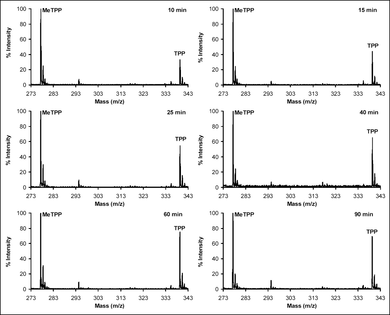
Representative MALDI-TOF-MS spectra for time course of uptake of TPP. Ion intensity of internal standard (MeTPP+; m/z, 277.1) was set as 100 in each spectrum. Ion intensity of analyte (TPP+; m/z, 339.1) increased over time and reached maximum uptake at 60 min of incubation. Peak area of M+ ion was determined, and ion-intensity ratio (TPP/MeTPP) was calculated. Therefore, amount of TPP in cells at each time point could be quantified in conjunction with calibration curve (unlabeled peaks are generated from matrix).
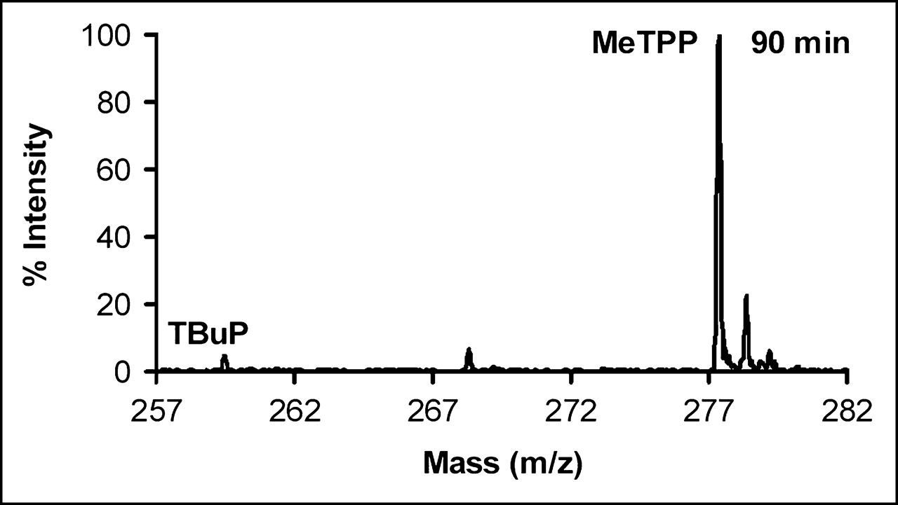
Representative MALDI-TOF-MS spectrum for uptake of TBuP at 90 min of incubation. TBuP peak (M+; m/z, 259.2) is relatively low but can still be easily identified because signal-to-noise ratio is more than 5.
The calibration curves for quantitative analysis were obtained by mixing together a series of phosphonium cations containing 0.1, 0.2, 0.3, 0.5, 0.7, 1.0, or 1.5 pmol of analytes in 1 μL of solution and 1.0 pmol of internal standard (MeTPP) in 1 μL of solution and subjecting them to MALDI-TOF-MS analysis. Triplicate samples were prepared, and 3 averaged spectra were collected for each sample. The IPCs/MeTPP (where PC stands for phosphonium cation) was measured as the ratio of the M+ ion peak area. The plotted data are shown in Figure 4. A very good correlation between MALDI-TOF-MS ion-intensity ratios (phosphonium cations/MeTPP) and molar ratios (phosphonium cations/MeTPP) was observed. The linear correlation coefficients (r2) were all greater than 0.98. Furthermore, the slopes of the calibration curves were different for each cation, indicating a different detection sensitivity for each cation.
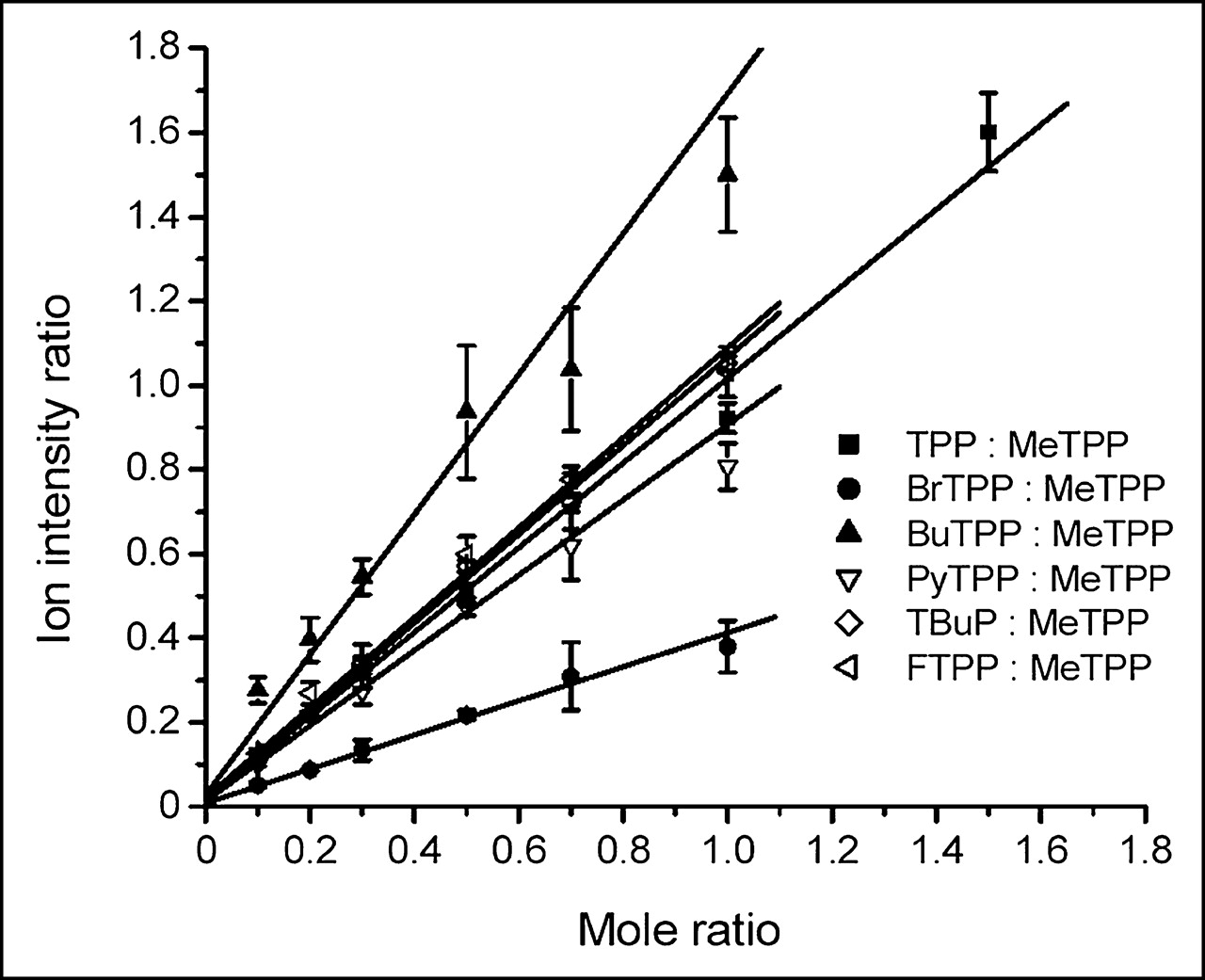
Linear correlation of MALDI-TOF-MS ion-intensity ratios (phosphonium cations/MeTPP) versus molar ratios (phosphonium cations/MeTPP) with 1.0 μmol of MeTPP per liter as internal standard. One microliter of solution or suspension containing 0.1–1.5 pmol of phosphonium cations was used for each analysis. Averages of results were from triplicate analyses. Lines are based on linear least-squares fitting of each of these sets of data points: TPP:MeTPP, y = 0.012225 + 1.00453x, r2 = 0.99915; BrTPP:MeTPP, y = 0.00815 + 0.40528x, r2 = 0.99713; BuTPP:MeTPP, y = 0.02725 + 1.66651x, r2 = 0.98602; PyTPP:MeTPP, y = 0.01386 + 0.89319x, r2 = 0.99525; TBuP:MeTPP, y = 0.01565 + 1.05212x, r2 = 0.99872; FTPP:MeTPP, y = 0.0215 + 1.06846x, r2 = 0.99824.
Comparison of Uptake of TPP Measured by MALDI-TOF-MS and Scintillation Counting
C6 cells at a concentration of 0.5 × 10⁶ were exposed to a 5 μmol/L concentration of TPP and 3H-TPP separately by incubation at 25°C for 90 min. The drug-influx kinetics were followed either by MALDI-TOF-MS, for TPP, or by scintillation counting, for 3H-TPP. The time course of TPP uptake, as determined with the 2 techniques, was almost identical in pattern and in level of uptake (P < 0.05) (Fig. 5).

Uptake of TPP by C6 cells over time at room temperature. Unlabeled TPP uptake was determined with MALDI-TOF-MS, and tritiated TPP uptake was measured with scintillation counting. Values are expressed as mean percentage of uptake ± SD of 3 independent determinations. Time course of TPP influx, as determined with the 2 techniques, exhibited no significant difference in patterns or levels of uptake (P < 0.05).
Uptake of Phosphonium Cations
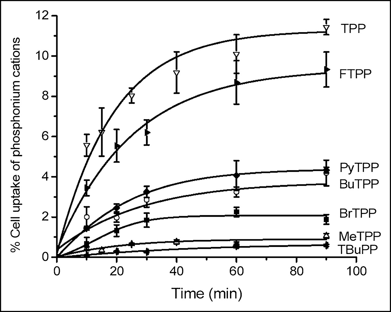
Figure 6 shows the uptake of phosphonium cations (5 μmol/L was used for incubation in all experiments) by C6 cells at 25°C during a 90-min incubation. The phosphonium cations exhibited similar uptake kinetics. The cations rapidly increased in cells on introduction into the buffer and reached a steady-state concentration at 60 min. The steady-state levels of uptake for various phosphonium cations ranged from 0.6% to 11.4%. Interestingly, the molecular peak, fragments, and molecular adduct for the compound CoTPP could not be identified from MALDI-TOF-MS for the cell lysate resulting from incubation with CoTPP. This result can be explained by the fact that CoTPP is a neutral compound under physiologic conditions and either could not enter cells or accumulated at levels lower than could be detected.

Uptake of various phosphonium cations by C6 cells over time at room temperature as determined by MALDI-TOF-MS. Values are expressed as mean percentage of uptake ± SD of 3 independent determinations.
Mitochondrial Membrane Potential-Dependent Uptake of FTPP
The effects of manipulating mitochondrial membrane potential on cellular accumulation of FTPP (determined by MALDI-TOF-MS) were assessed through uptake studies on C6 cells using 4 different concentrations of CCCP (0.1, 1, 10, and 50 μmol/L) and 3 different concentrations of valinomycin (0.1, 1, and 10 μmol/L) in low-K+ HEPES buffer. For control experiments, in which the mitochondrial membrane potentials were unaltered, uptake was determined in a near-physiologic buffer (low-K+ HEPES buffer) without addition of inhibitors. The results are depicted in Figures 7A and 7B, which show that uptake of FTPP in C6 cells was significantly inhibited (P < 0.02) by CCCP or valinomycin at the 1 μmol/L concentration used in this study. FTPP uptake was inhibited strongly (to about 27.3% ± 2.3%) by CCCP, 50 μmol/L, and to a lesser extent (to about 72.8% ± 3.6%) by valinomycin, 10 μmol/L.
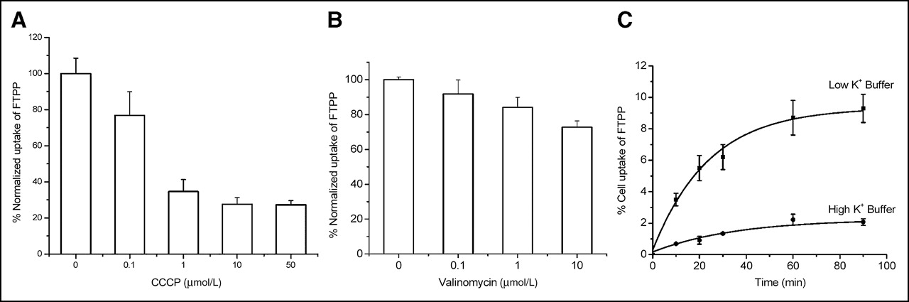
Effects of various concentrations of proton phore CCCP, K+ ionophore valinomycin, and high-K+ HEPES buffer on uptake of 5 μmol of FTPP per liter. Each value represents mean of 3 independent experiments. Values are expressed as mean percentage of normalized uptake ± SD of 3 independent experiments.
The effects of depolarizing the plasma membrane potential on only uptake of phosphonium cations by C6 cells were determined using a high-K+ HEPES, as shown in Figure 7C. For C6 rat glioma cells, depolarization of the plasma membrane decreased uptake of FTPP by 3.5-fold, compared with uptake in the low-K+ buffer at 90 min of exposure. Overall, these results clearly demonstrated that uptake of FTPP was electrogenic and driven by the plasma and mitochondrial membrane potentials.
DISCUSSION
One of the fruits of the Human Genome Project is that identification of new targets for diseases is being greatly accelerated (1), urging scientists to develop new techniques for rapid screening of molecules able to interact with these targets. Mass spectrometry is one such technique that has attracted significant interest because of its great potential for applications in high-throughput drug discovery and development (26). For instance, liquid chromatography combined with atmospheric pressure ionization mass spectrometric detection (LC-MS/MS) has actively been investigated for determination of the pharmacokinetic properties of drugs and their metabolites (26). Kerns et al. developed a novel method using LC-MS/MS to monitor the cellular uptake profile of a nonradioactive drug, paclitaxel (27). Typically, incubation of radiolabeled compounds with cells in culture is used to detect the amount of drug accumulated in cells. However, the radiolabeling of a series of compounds can be a time-consuming, expensive, and sometimes challenging task for organic radiosynthesis. The bioanalytic method reported by Kerns et al. avoids preparation of a radiolabeled version of the drug. The authors concluded that that method is rapid and sensitive and presents a unique advantage over traditional radioisotopic methods in that it can readily be used on a range of analogs without any additional synthetic effort (27).
Currently, LC-MS/MS is the most popular mass spectrometry technique for high-throughput assays. But the chromatography separation involved in this technique significantly extends the total analysis time. At least several minutes are required to finish a single run. Large amounts of solvent waste are also generated from the assay. Moreover, development and validation of the LC-MS/MS method are time consuming and require technically skilled scientists. MALDI-TOF-MS is another powerful analytic technique, with intrinsic advantages including a high speed of analysis (several seconds per sample) and a high sensitivity of detection (picomolar range), making it a potential tool for high-throughput screening of drugs. Despite the fact that quantitative determination using MALDI-TOF-MS techniques is complicated by several critical problems associated mainly with sample preparation, a variety of research reports still clearly demonstrate that MALDI-TOF-MS can be a powerful tool for quantitative analysis of low-molecular-weight compounds (4). For example, MALDI-TOF-MS has already been used successfully for quantitative analysis of an enzyme-catalyzed reaction (28), protease activity (29), and cyclosporin A in biologic samples (30). The method also was reported to have potential for detecting trace-level (in the parts-per-billion range) drug residues in complex environmental samples (31). Therefore, in this study, we used MALDI-TOF-MS to shorten the time line for developing molecular imaging agents. For proof of principle, the possibility of using MALDI-TOF-MS to screen and characterize uptake of small molecules, specifically phosphonium cations, was investigated.
First, the same amounts of nonradioactive TPP and 3H-TPP (1 nmol in 200 μL) were incubated with 0.5 × 10⁶ C6 cells. Uptake of 3H-TPP was easily determined for comparison with MALDI-TOF-MS analysis through routine scintillation counting of tritium-labeled compound. As for quantitative analysis of the TPP accumulation in the cells using MALDI-TOF-MS technique, calibration curves for the internal standard, MeTPP, and TPP and other phosphonium cations were first established (Fig. 4). The results showed a strong linear correlation and high accuracy (r2 > 0.98 for all curves) within the mass range applied for the analysis in this research (0.1–1.5 pmol for analytes and 1 pmol for the internal standard). After incubation with cells for different times, TPP from a 3,000-cell lysate and 1 pmol of MeTPP as an internal standard were sampled together for analysis. MALDI-TOF-MS spectra clearly revealed that TPP could easily be detected from the cell lysate even at 10 min after incubation (Fig. 2) and at levels of as low as 0.11 fmol TPP per cell (Fig. 5), as determined through the ion-intensity ratio and standard calibration curve. The time course of TPP influx determined by MALDI-TOF-MS and scintillation counting showed no significant difference statistically (P > 0.05 for all time points). Overall, these data validated MALDI-TOF-MS as an alternative approach for accurate measurement of the accumulation of phosphonium cations in cells.
The same procedure for MALDI-TOF-MS analysis was used to determine the accumulation of all other phosphonium cations in cells. Although the phosphonium analogs evaluated in this research displayed similar uptake kinetics, as presented in Figure 5, the absolute value of their uptake was different. CoTPP, a zwitterion under physiologic conditions, either does not accumulate or accumulates minimally in the cells during a 90-min incubation, presumably because of its zero net charge. TBuP is a phosphonium salt without any phenyl ring and shows low cellular accumulation (Figs. 3 and 6). Compared with TPP, this compound showed an approximately 18-fold decreased cellular accumulation (Fig. 6). Interestingly, cationic triarylalkylphosphonium salts (MeTPP, BrTPP, BuTPP, and PyTPP) displayed much lower uptake than did TPP or FTPP. These results highlight the importance of fine-tuning the structure of this class of compound to achieve the most efficient mitochondrial targeting and of carefully selecting which phosphonium may also serve as a carrier for drug delivery in nonimaging applications.
As shown in Figure 6, the percentage uptake of MeTPP, FTPP, and TPP was 1.06, 9.33, and 11.44, respectively, when they reached a plateau at ∼90 min of incubation. This information is of particular value for developing radiolabeled compounds as molecular probes for disease imaging. We have demonstrated that 3H-TPP accumulates specifically in tumors but minimally in inflammatory sites because of its mitochondria-targeting property (15). Therefore, TPP labeling with 18F may generate a novel molecular probe for mitochondrial dysfunction imaging in vivo using PET. The results shown in Figure 6 demonstrate that nonradioactive FTPP was still able to be taken up by cells at more than 80% of the level at which TPP was taken up. Moreover, the plasma membrane and mitochondrial membrane potential of C6 cells were modulated by using either high-K+ HEPES buffer or inhibitors such as CCCP and valinomycin. FTPP uptake profiles using these membrane potential-modulated cells verified that FTPP preserved the membrane potential-dependence property as TPP (24) in cell culture (Figs. 7A–7C) even though fluorine was incorporated into the parent molecule. In another study, using dynamic PET, 11C-MeTPP exhibited enhanced uptake and prolonged retention in canine brain glioma (12). Considering that accumulation in cell cultures is 8 times higher for FTPP than for MeTPP (Fig. 6) and that 18F has a longer half-life, further studies to develop 18F-labeled TPP as a mitochondria-targeting PET agent will be well justified after study of uptake of this probe by additional cell lines.
Rideout et al. previously reported that uptake of TPP by FaDu human hypopharyngeal carcinoma cells can be measured using MALDI-TOF-MS (32). We have further refined this strategy specifically for development of molecular imaging probes. Uptake of the compounds listed in Figure 1 was analyzed using a slightly revised protocol based on their report. The findings presented in Figure 5 agree well with the data reported by Rideout et al. Most important, we thoroughly studied the time course of uptake of a library of compounds using MALDI-TOF-MS, and analysis of the findings helped in the identification and proposal of new imaging probes for further development. The voltage-sensitive nature of the lead compound, FTPP, was also proven using MALDI-TOF-MS.
In routine cellular assays of radioactivity uptake, the amount of tracer to be used depends strongly on the specific activity of the radiolabeled agent and can range from picomolar to nanomolar. For instance, when 370 Bq (0.01 μCi) of 3H-TPP (1.11 TBq/mmol [30 Ci/mmol]) is incubated with cells in 100 μL of buffer, the concentration of 3H-TPP corresponds to 3.3 nmol/L. In this experiment, phosphonium cations and 3H-TPP at a 5 μmol/L concentration were used for incubation with cells. Although the concentration of cold phosphonium cations used for MALDI-TOF-MS cellular assays was more than a thousand times higher than that of radiotracers routinely used in cellular assays of radioactivity uptake, the procedure described in this paper can be fully optimized to scale down the initial incubation concentration of cold compounds. For instance, 0.5 × 10⁶ C6 cells were used for incubation with a 5 μmol/L concentration of phosphonium cations, and the cells were then lysed in 150 μL of water. Only a small fraction (1 μL, 0.67%) of the cell lysate was subjected to MALDI-TOF-MS analysis. Obviously, less water can be used for cell lysis or the cell lysate can be condensed through lyophilization and redissolving. Moreover, instead of only using 0.5 × 10⁶ cells, larger number of cells, for example, 5 × 10⁶ cells, can be applied for incubation. Through a combination of these 2 approaches, considerable improvements can be expected in the sensitivity of MALDI-TOF-MS for quantifying uptake of molecular probes by cells. In future studies, this hypothesis and other potential solutions will be explored in more detail so that much lower drug concentrations can be used for incubation with cells.
Imaging of abnormalities in small animals or patients at the molecular level with PET technology has shown significant potential and has attracted significant attention in biomedical research (1). Demand is increasing for more radiopharmaceuticals that can probe cellular or molecular targets. However, rapid and systematic development and characterization of radiolabeled agents present a formidable task to radiochemists. Liu et al. found radio-LC-MS to be a quick and accurate tool for determining the chemical composition of 99mTc radiopharmaceuticals at the tracer level (33). This technique can help with the quick identification of the right radiolabeled species during radiosynthesis and thus has the potential to speed the development of 99mTc-labeled agents. The research presented here demonstrates a strategy that may be used for high-throughput screening of PET and SPECT agents for much broader application to molecular imaging. This effort represents the first use, to the best of our knowledge, of mass spectrometry for preliminary evaluation of the suitability of a group of molecules for further radiolabeling development. The information documented here should help to significantly accelerate the design and characterization of molecular imaging probes.
CONCLUSION
MALDI-TOF-MS can be used for high-throughput screening and characterization of a library of phosphonium cations as mitochondrial targeting agents. Accumulations of phosphonium cations at subpicomolar levels are easily identified by the procedures described in this article. The high specificity, sensitivity, and speed of MALDI-TOF-MS render it a powerful tool for development of molecular probes and drugs. To our knowledge, this report is the first of the use of MALDI-TOF-MS for design and development of PET radiopharmaceuticals. The data on uptake of nonradioactive FTPP by cells warrant further synthesis and evaluation of 18F-FTPP as a molecular probe for imaging mitochondrial dysfunction.
Acknowledgments
The authors thank Dr. John Min for helpful discussions. This work was funded by the Doris Duke Charitable Foundation (SSG).
Footnotes
Received Oct. 11, 2004; revision accepted Dec. 15, 2004.
For correspondence or reprints contact: Sanjiv S. Gambhir, MD, PhD, Molecular Imaging Program at Stanford, Department of Radiology, and Bio-X Program, Stanford University, 318 Campus Dr., Clark Center, E-150, Stanford, CA 94305.
E-mail: sgambhir{at}stanford.edu


