


Article Figures & Data
Figures
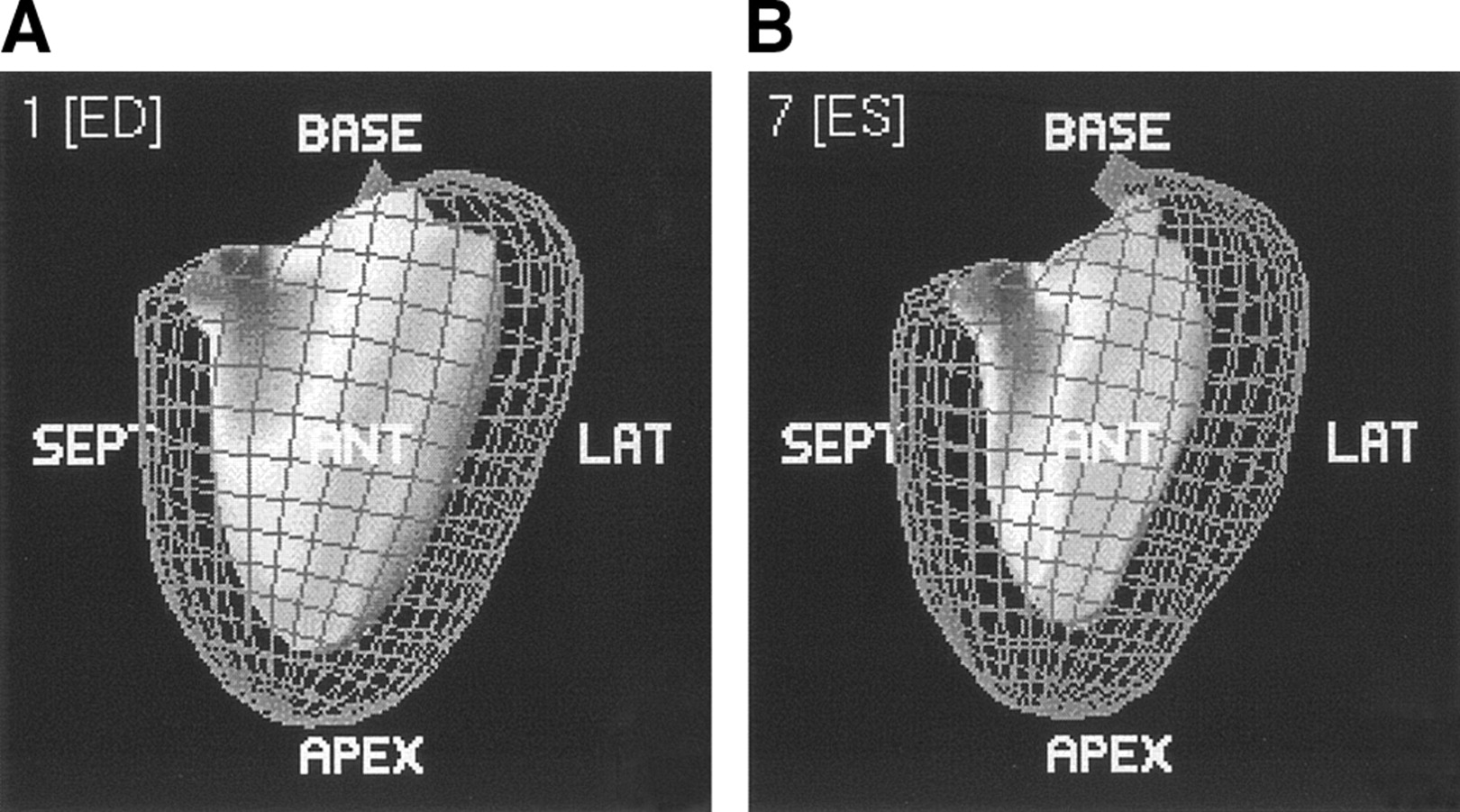
- FIGURE 1.
Images from QGS of end-diastolic (A) and end-systolic (B) phases showing epicardial and endocardial borders of the left ventricle. SEPT = septal; LAT = lateral.
- FIGURE 2.
Transverse 2-chamber view of end-diastolic (A) and end-systolic (B) phases of the left ventricle. Epicardial and endocardial contours are automatically drawn, but incorrect contours can be corrected manually.
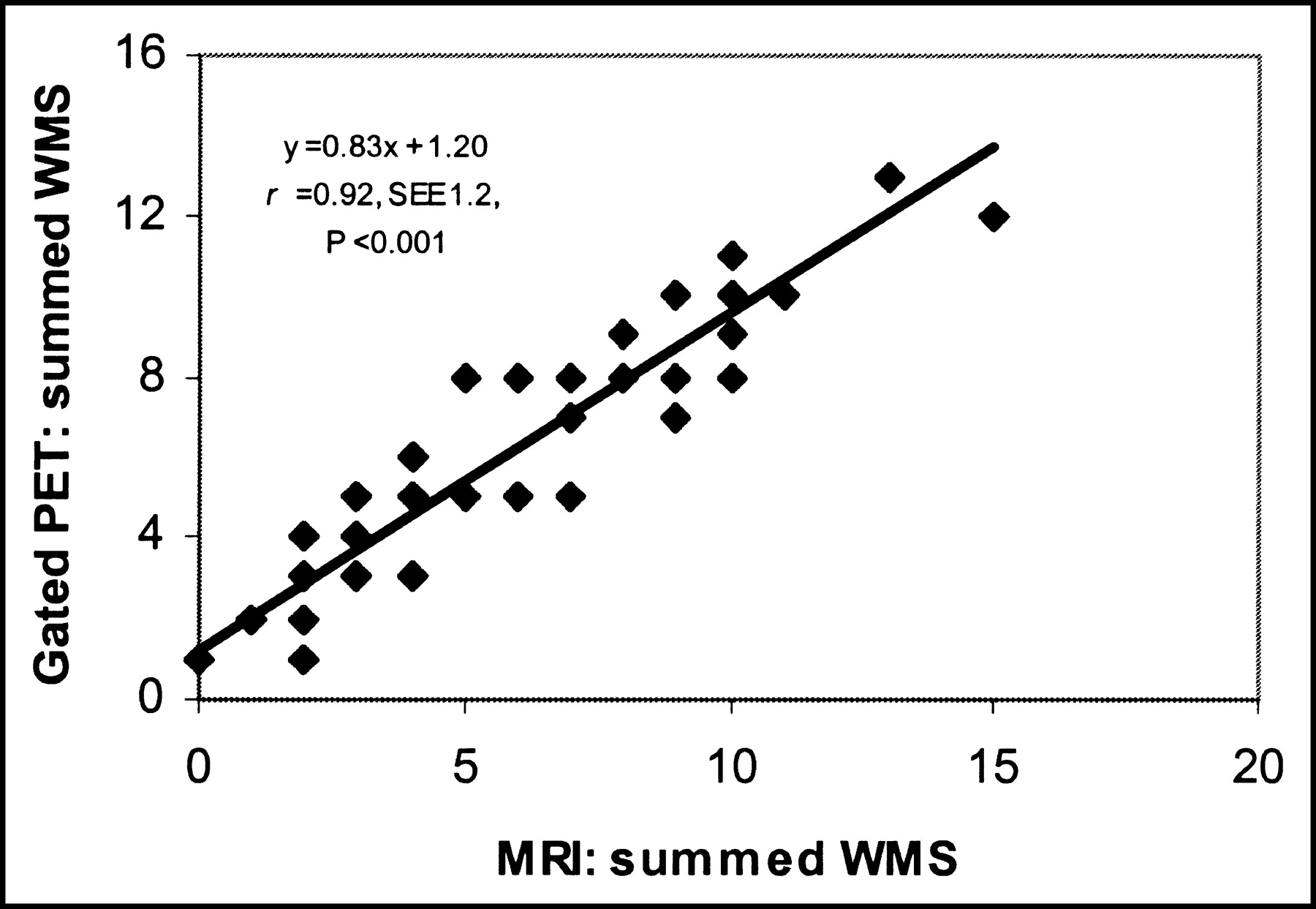
- FIGURE 3.
Relationship between summed wall motion scores (WMS) per patient on MRI and on gated 18F-FDG PET.
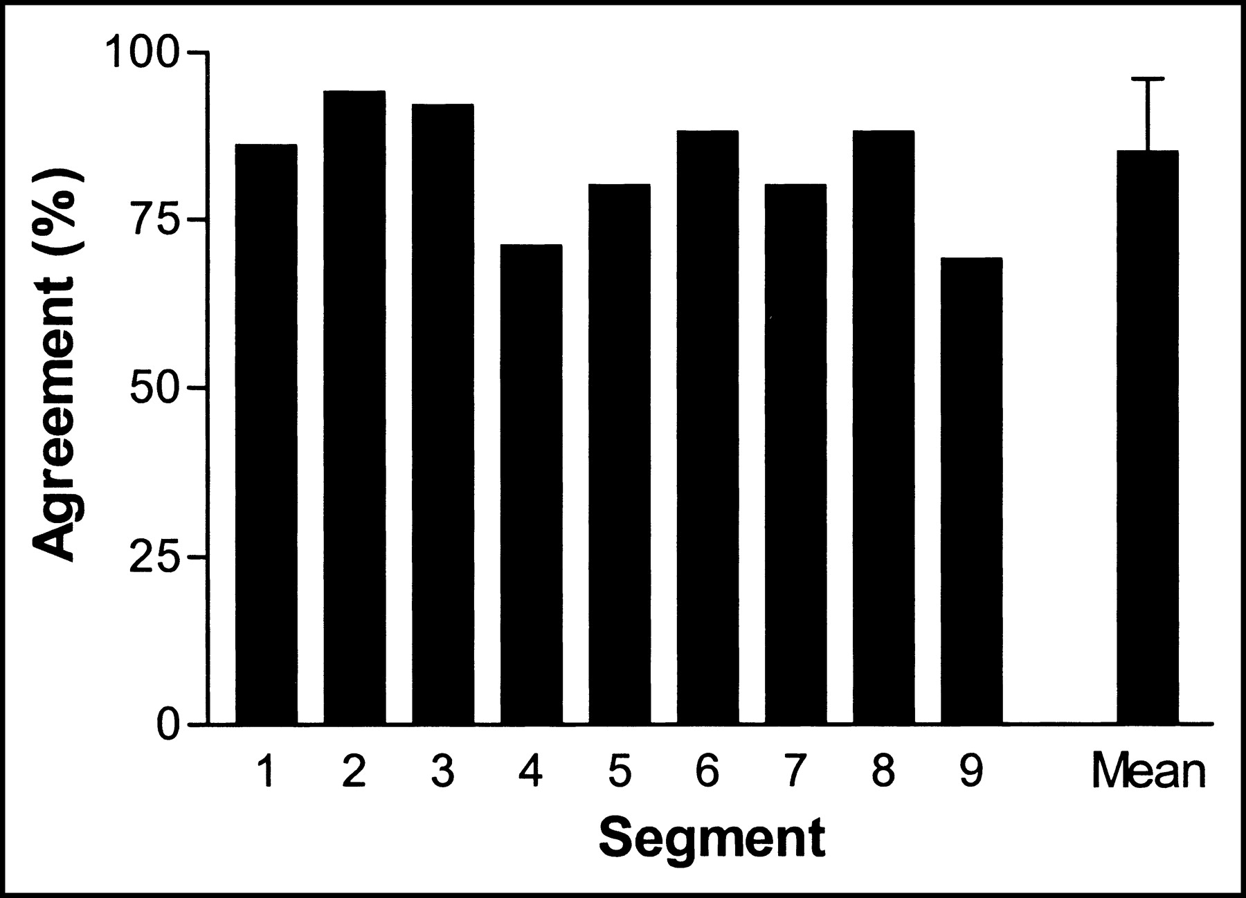
- FIGURE 4.
Agreement between regional wall motion scores per segment on MRI and on gated 18F-FDG PET. Basal segments: 1 = anterior; 2 = lateral; 3 = inferior; 4 = septal. Distal segments: 5 = anterior; 6 = lateral; 7 = inferior; 8 = septal. 9 = apical segment. Mean = mean agreement for all 9 segments ± SEE.
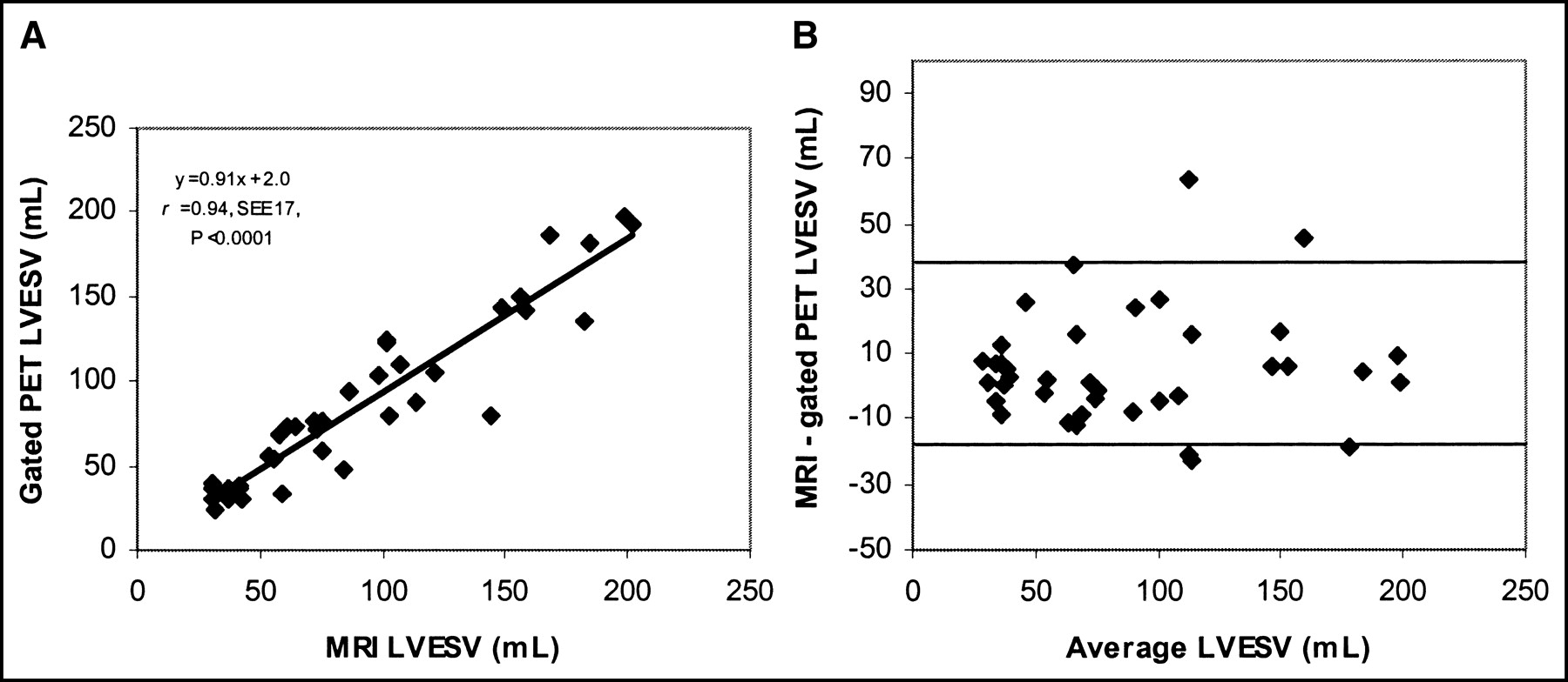
- FIGURE 5.
(A) Relationship between LVESV assessed by MRI and gated 18F-FDG PET. (B) Bland–Altman plot for LVESV without a systematic trend.
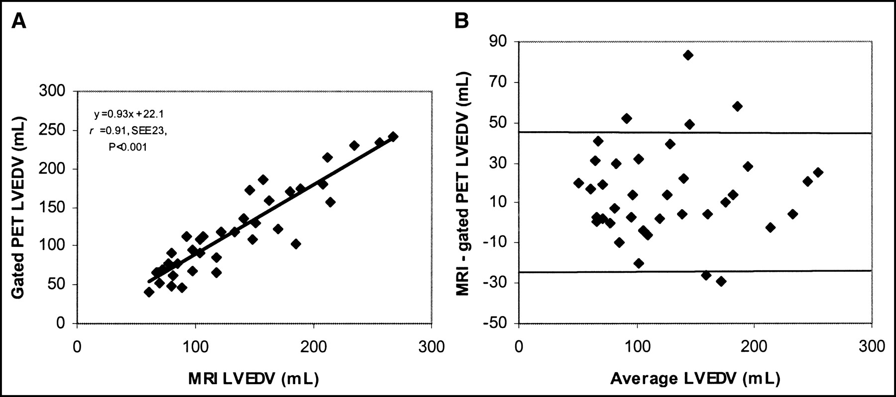
- FIGURE 6.
(A) Relationship between LVEDV assessed by MRI and gated 18F-FDG PET. (B) Bland–Altman plot for LVEDV without a systematic trend.
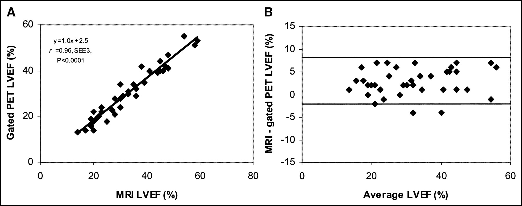
- FIGURE 7.
(A) Relationship between LVEF assessed by MRI and gated 18F-FDG PET. (B) Bland–Altman plot for LVEF without a systematic trend.


Tables
- TABLE 2
Agreement Between MRI and Gated 18F-FDG PET for Assessment of Regional Wall Motion Score (RWMS)
RWMS on MRI No. of segments with the following RWMS on gated PET: 0 1 2 Total 0 136 22 1 159 1 10 103 5 118 2 0 12 43 55 Total 146 137 49 332 Agreement was excellent (85%; κ-statistic, 0.79).
Measurement Value determined by: P MRI Gated PET LVESV Mean (mL) 91 ± 12 85 ± 51 0.57 Range (mL) 31–202 24–198 LVEDV Mean (mL) 131 ± 57 117 ± 56 <0.001 Range (mL) 61–267 41–242 LVEF Mean (%) 33 ± 12 30 ± 11 <0.001 Range 14–59 13–55


