Weissberg outlines the importance of underlying cell biology in understanding the potential for nuclear medicine imaging of atherosclerotic plaques.
Kumar and colleagues assess the predictive value of positive 18F-FDG PET findings after completion of chemotherapy in patients with gastrointestinal lymphomas and compare the ability of PET and CT to detect residual disease.
Kamel and colleagues report on the significance of incidentally detected gastrointestinal tract lesions and the effects of early identification on patient management and outcomes in a large screening population.

Heiss and colleagues evaluate the applications of a new scanner with improved spatial resolution that permits the determination of metabolic rates in small nuclei associated with various neurodegenerative disorders.
Ben-Haim and colleagues report on the ability of PET/CT to identify and localize focal vascular-wall 18F-FDG uptake and discuss the potential for early detection of patients at increased risk of future cardiovascular events.
Tiling and colleagues investigate whether tissue-specific parameters–Nin addition to lesion size–Nmay affect uptake of 99mTc-sestambi in scintimammography.
Tseng and colleagues assess the ability of PET to characterize biologic response of locally advanced breast cancer to chemotherapy using 15O-water–derived blood flow measurements and 18F-FDG–derived glucose metabolism rate parameters.

Yamane and colleagues look at changes in 18F-FDG uptake on PET as early as 1 day after initiation of chemotherapy in patients with malignant lymphoma and discuss the implications that these changes have for accurate diagnosis and staging.
Choi and colleagues identify the predictive elements that contribute to the success of 18F-FDG PET in providing noninvasively independent prognostic information in preoperative esophageal squamous cell carcinoma.
Bruehlmeier and colleagues discuss the use of late 18F-FMISO PET in providing a spatial description of hypoxia in brain tumors, independent of blood–brain barrier disruption and tumor perfusion.
Chiu and colleagues study patients with Alzheimer’s disease and evaluate results suggesting a relationship between education level and differences in regional cerebral flow as measured by 99mTc-HMPAO SPECT.
Love and colleagues compare coincidence detection–based 18F-FDG PET imaging with combined 111In-labeled leukocyte/99mTc-sulfur colloid marrow imaging in the diagnosis of infection in patients with failed prosthetic joints.
Krishnamurthy and colleagues detail the results of a long-term study on the constancy and variability of gallbladder ejection fraction in conditions such as chronic calculus cholecystitis and chronic acalculous cholecystitis.
Kasama and colleagues assess whether dobutamine stress 99mTc-tetrofosmin quantitative gated SPECT can be used to predict improvement of cardiac function and heart failure symptoms in patients receiving carvedilol therapy for idiopathic dilated cardiomyopathy.
Yoshinaga and colleagues use 11C-acetate PET to investigate differences in oxidative metabolic response in myocardium and discuss the implications of these findings for the interpretation of 18F-FDG PET cardiac images.
Wong and colleagues research the feasibility of using 18F-FDG PET to quantify reduction of hepatic tumor metabolism after 90Y-glass microsphere treatment for unresectable metastatic disease to the liver.

Davies and colleagues review the biology of atherosclerosis, conventional imaging techniques, and the potential of nuclear imaging to provide valuable information on the cellular, metabolic, and molecular composition of plaque.

Katoh and colleagues describe and illustrate the application of an algorithm that allows stable, rapid, and automated quantification of regional myocardial blood flow with 15O-water PET.
Miyagawa and colleagues compare 2 herpes simplex virus reporter gene expres-sion approaches for use in PET monitoring of gene therapy in rat and pig models.
Croteau and colleagues use small-animal PET to evaluate the effects of 2 anesthetic agents on myocardial perfusion and coronary reserve in rats under rest and stress conditions.
Spaeth and colleagues report on animal experiments designed to assess the potential of 18F-FET and 18F-FCH as PET tracers in differentiating radiation necrosis from tumor recurrence.
van Waarde and colleagues evaluate the in vitro and in vivo characteristics of 2 novel radioligands and discuss their potential for tumor detection and assessment of the σ-receptor occupancy of novel therapeutic drugs.
Murakami and colleagues report on a study designed to measure brain concentrations of an 11C-labeled immunosuppressive agent in monkeys and outline possible applications in PET imaging of stroke.
Marshall and colleagues investigate the accuracy of 18F-fluorodihydrorotenone as a deposited myocardial flow tracer and compare the results from investigations in the rabbit heart with those for 201Tl.
Hindorf and colleagues evaluate the influence of the mass, shape, and distances between organs on S values in radionuclide studies of mice.
Zanzonico and colleagues derive estimates of normal-tissue absorbed doses for 18F-FDHT PET imaging in patients with advanced prostate cancer.
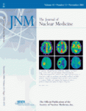
ON THE COVER
Visual comparison of 15O-H2O PET perfusion images and late 18F-fluoromisonidazole (FMISO) PET images of patients with glioblastoma showed a large range of tumor perfusion within areas of increased 18F-FMISO uptake (i.e., hypoxia was present in both hypoperfused and hyperperfused tumor areas). Generally, increased 18F-FMISO uptake was found in the tumor margin but not in the tumor center. Tumor centers of all glioblastomas showed decreased radioactivity in both 15O-H2O and 18F-FMISO PET images. The perfusion–hypoxia patterns suggested that hypoxia in these tumors may develop irrespective of the magnitude of perfusion.


