Index by author
Cover image

ON THE COVER
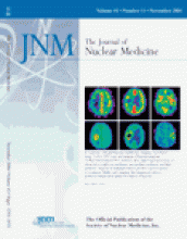
Visual comparison of 15O-H2O PET perfusion images and late 18F-fluoromisonidazole (FMISO) PET images of patients with glioblastoma showed a large range of tumor perfusion within areas of increased 18F-FMISO uptake (i.e., hypoxia was present in both hypoperfused and hyperperfused tumor areas). Generally, increased 18F-FMISO uptake was found in the tumor margin but not in the tumor center. Tumor centers of all glioblastomas showed decreased radioactivity in both 15O-H2O and 18F-FMISO PET images. The perfusion-hypoxia patterns suggested that hypoxia in these tumors may develop irrespective of the magnitude of perfusion.


