


Abstract
Coexpression of a reporter gene and a therapeutic gene may allow for noninvasive monitoring of cardiac gene therapy. We sought to evaluate the usefulness of an adenoviral vector expressing mutant herpesviral thymidine kinase reporter gene (HSV1-sr39tk) and vascular endothelial growth factor (VEGF) 121 in independent expression cassettes (Ad4tk). Methods: Accumulation of 14C-2′-fluoro-5-methyl-1-β-d-arabinofuranosyluracil (FIAU) and 9-(4-18F-fluoro-3-hydroxymethylbutyl)guanine (FHBG) as reporter probes, and secretion of VEGF into medium, were determined for Ad4tk-infected H9c2 rat cardiac cells in vitro. Results: In vitro tracer uptake increased with increasing vector concentration and over time. It was comparable to cells infected with adenovirus expressing only wild-type HSV1-tk (reporter probe: 14C-FIAU) or mutant HSV1-sr39tk (reporter probe: 18F-FHBG). No significant uptake was observed in cells infected with adenovirus expressing VEGF alone. With increasing vector concentration, Ad4tk-infected cells increasingly released VEGF into medium. VEGF production correlated significantly with cellular reporter probe uptake (r = 0.93; P = 0.0003). Conclusion: The usefulness of a vector coexpressing HSV1-tk and VEGF for noninvasive imaging of expression of a therapeutic transgene has been demonstrated in vitro. This approach may allow for future in vivo monitoring of cardiac angiogenesis gene therapy.
The feasibility of noninvasive imaging of cardiac transgene expression using PET and radiolabeled reporter probes for detection of adenovirally transfected herpesviral thymidine kinase reporter gene (HSV1-tk) has recently been demonstrated in small animals (1) and large animals (2). Reporter gene imaging is thought to be promising for in vivo monitoring of the location, magnitude, and persistence of cardiac transgene expression.
Although the basic methodology has been validated, further studies are needed for translation to applications in cardiac gene therapy. A next step is to coexpress a reporter gene with a therapeutic gene and to demonstrate that therapeutic gene expression can be detected via imaging of reporter gene expression.
We chose the vascular endothelial growth factor (VEGF) gene as the therapeutic gene because it has been applied successfully in trials of clinical gene therapy for treatment of ischemic heart disease (3,4) and because it is considered to be nearing large-scale human application (5). The aim of this study was to evaluate the usefulness of an adenoviral vector coexpressing VEGF and HSV1-tk for noninvasive imaging of cardiac therapeutic gene transfer.
MATERIALS AND METHODS
Vectors
Ad4tk, a replication-deficient adenovirus expressing mutant HSV1-sr39tk and VEGF 121 independently, each under control of cytomegalovirus promoter and followed by SV40 polyadenylation signal, was constructed at Weill Medical College. Both cassettes were in left-to-right orientation; the HSV1-sr39tk expression cassette was 5′ to the VEGF expression cassette, replacing the E1 region.
Adenovirus expressing only wild-type HSV1-tk (6), mutant HSV1-sr39tk (kindly provided by Sanjiv S. Gambhir, UCLA) (1), or VEGF (7) were used as controls.
In Vitro Studies
H9c2 rat embryonic cardiac cells were infected as described previously (6). RNA isolation, preparation of cellular extract, and uptake studies were performed 24 h after infection with an increasing multiplicity of infection (MOI). The time course of gene expression was measured at an MOI of 25.
Northern Blot.
RNA was extracted as described previously (8) and was hybridized first with HSV-tk cDNA (1.1 kbp), then with VEGF cDNA (0.45 kbp), and then with 18S-RNA-coding DNA as a loading control. Images were obtained using an FLA-2000 PhosphorImager (Fuji) and quantified with AIDA software (Raytest).
Western Blot.
Infected cells were treated with radioimmunoprecipitation assay buffer and applied to sodium dodecylsulfate–polyacrylamide gel electrophoresis. Western blot was performed according to the standard protocol after incubation of HSV1-tk–specific polyclonal antibody (provided by William Summers, Yale University) and goat antirabbit IgG-peroxidase–coupled secondary antibody (Oncogene Science, Inc.).
Enzyme-Linked Immunosorbent Assay (ELISA).
VEGF secretion into the medium of infected cells was determined using a human VEGF ELISA kit (Oncogene Science, Inc.). For analysis, serum concentration was reduced from 10% to 1% immediately after infection.
Reporter Probe Uptake.
Uptake of reporter probes was measured as previously described (6) and was normalized to the number of viable cells as determined by trypan blue staining. 14C-2′-Fluoro-5-methyl-1-β-d-arabinofuranosyluracil (FIAU; specific activity 2 GBq/μmol) was obtained commercially (Hartmann Analytic). 9-(4-18F-Fluoro-3-hydroxymethylbutyl)guanine (FHBG) was synthesized according to a protocol developed at Forschungszentrum Rossendorf. Tosylated and methoxytritylated precursor was radiolabeled using a K18F-F/Kryptofix 2.2.2 complex (Merck), followed by splitting off of protection groups under acidic conditions and purification by high-performance liquid chromatography, yielding 18F-FHBG at an average specific activity of 19 GBq/μmol. When indicated, 18F-FHBG was added to incubation medium together with 14C-FIAU. After 1 h of incubation, 18F-FHBG accumulation was measured using a γ-counter before addition of tissue solubilizer (Soluene-350; Packard). Scintillation fluid was added 20 h after cell incubation, to allow for decay of 18F. Subsequently, β-counting was performed as previously described (6).
RESULTS
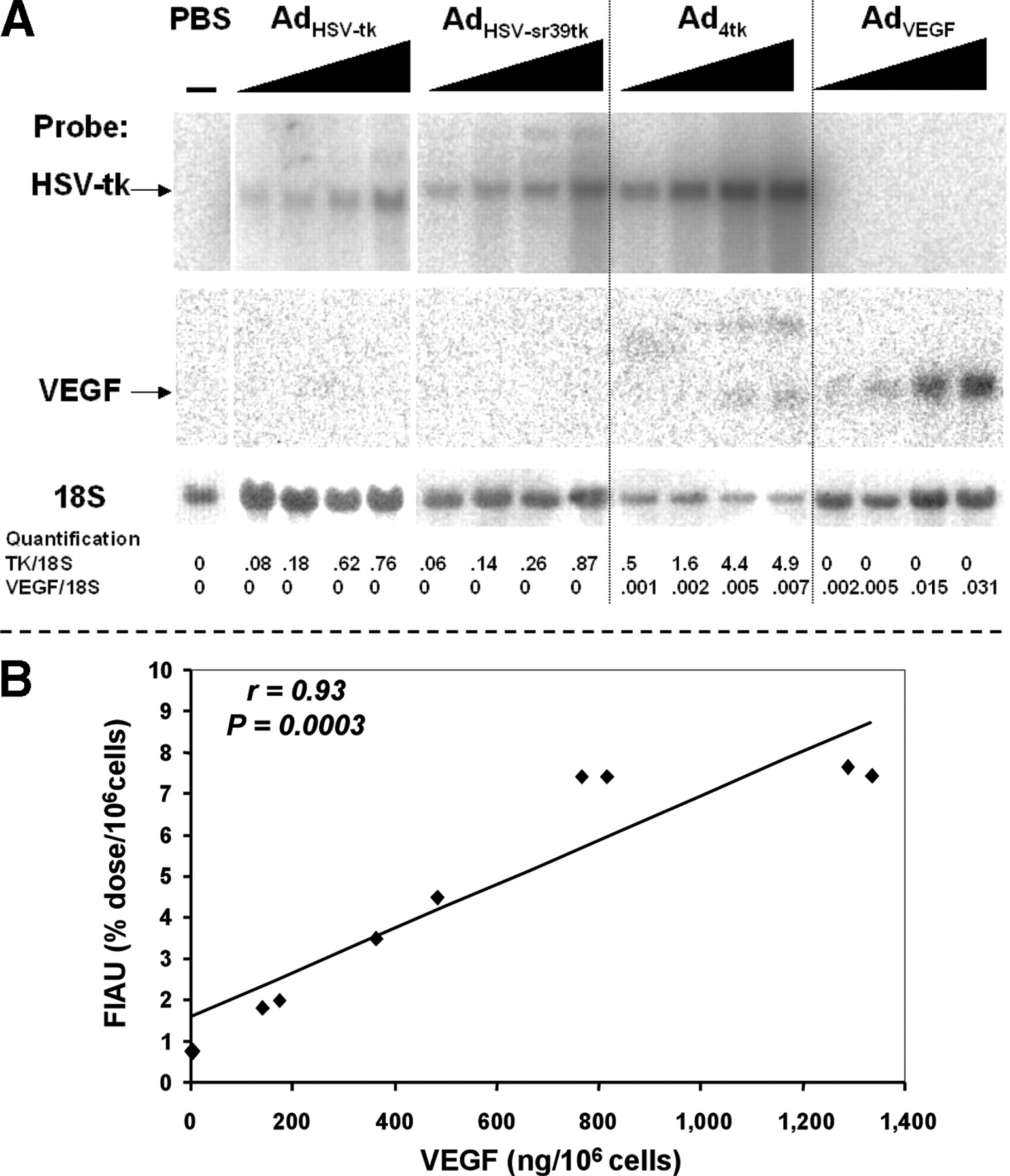
Northern blot analysis revealed increasing HSV1-tk and VEGF RNA expression with an increasing MOI of Ad4tk (Fig. 1A). In parallel experiments on protein expression level, Western blots for HSV1-tk similarly revealed an increasing signal with increasing MOI (data not shown).

Correlation of HSV1-tk and VEGF expression. (A) Results of analysis of transgene mRNA by Northern blot of H9c2 cells infected with an increasing MOI (12.5, 25, 50, 100) of the respective adenoviral vector or not infected (phosphate-buffered saline). Detection is with HSV1-tk (top), VEGF probe (middle), or 18S-fragment for normalization (bottom). (B) Results of transgene product analysis, showing a good correlation between 14C-FIAU uptake and VEGF secretion into the medium of cells infected with Ad4tk.
Accumulation of 14C-FIAU and 18F-FHBG generally increased with increasing vector concentration in the presence of HSV1-tk (Table 1) and exhibited only the background level in negative controls. Accumulation of 14C-FIAU was comparable in cells infected with Ad4tk and in adenovirus-expressing wild-type HSV1-tk only. For 18F-FHBG, uptake in Ad4tk-infected cells was the highest at all MOIs.
Reporter Probe Accumulation in H9c2 Cells
VEGF secretion into medium also increased with an increasing MOI for Ad4tk (Table 2) and was higher than for VEGF-expressing control vector. Infection with HSV1-tk–expressing adenovirus yielded no detectable VEGF secretion.
Secretion of VEGF
14C-FIAU accumulation and VEGF secretion into medium increased over time after Ad4tk infection at a constant MOI (Table 3).
Time Course of VEGF Secretion and FIAU Accumulation of Ad4tk-Infected Cells
When the data from increasing MOI and time course after Ad4tk infection were combined, an excellent correlation between VEGF secretion and 14C-FIAU accumulation was obtained (r = 0.93; P = 0.0003) (Fig. 1B).
DISCUSSION
In summary, the in vitro data suggest that expression of HSV1-tk reporter gene closely reflects therapeutic VEGF gene expression when both genes are coexpressed in separate expression cassettes using a single adenoviral vector. The feasibility of reporter gene coexpression for noninvasive monitoring of the location, magnitude, and persistence of cardiac therapeutic gene expression in vivo is suggested.
VEGF gene therapy is currently applied in clinical trials to treat ischemic heart disease, but success in human applications is monitored only indirectly via improvement of symptoms or clinical results such as those of exercise testing or perfusion scintigraphy (3,4). A method that allows for quantitative in vivo visualization of the success of gene transfer and the time course of subsequent transgene expression would be of considerable value. PET performed after coexpression with a reporter gene may not only enhance monitoring of clinical therapeutic gene delivery but also allow linkage of clinical observations and functional test results to transgene expression, thus refining understanding of therapeutic mechanisms. As a next step, a larger animal series will be required to confirm that in vitro correlation between transgenes can be transferred to the in vivo setting.
In several studies on tumor cells, 2 imaging reporter genes have been coexpressed and correlated. Application of equivalent titers of separate adenoviral vectors expressing HSV1-tk or dopamine D2 receptor yielded a good correlation for the uptakes of specific reporter probes (9), but the success of this approach is not guaranteed for infection of all cells with both vectors. Other studies used bicistronic vectors coexpressing both reporter genes linked by internal ribosomal entry site (10). This results in excellent correlation, but the absolute level of expression of the second gene is generally reduced. Finally, the approach of the present study—a single vector with 2 expression cassettes—has also been evaluated previously and showed a good correlation between expression of 2 reporter genes (11). To our knowledge, this is the first report on coexpression of a therapeutic gene and a reporter gene for nuclear imaging in cardiac cells.
We evaluated uptake of the pyrimidine derivative FIAU and the acycloguanosine derivative FHBG. Both have been demonstrated to be useful reporter probes for imaging cardiac gene products (1,2). Previous observations are confirmed and extended by demonstration of a relationship between reporter probe uptake and expression of a cotransfected therapeutic gene. More detailed comparison of the 2 probes (and alternative approaches) is beyond the scope of this communication but may be performed in the future to further refine the methodology for imaging cardiac reporter genes.
CONCLUSION
The usefulness of a vector coexpressing VEGF and HSV1-tk reporter gene has been demonstrated in this in vitro study. Further application to in vivo monitoring of gene therapy in models of ischemic heart disease is warranted.
Acknowledgments
This study was supported by the Deutsche Forschungsgemeinschaft (Be 2217/4-1).
Footnotes
Received Mar. 9, 2004; revision accepted May 20, 2004.
For correspondence or reprints contact: Frank M. Bengel, MD, Nuklearmedizinische Klinik und Poliklinik, Technische Universität München, Klinikum rechts der Isar, Ismaninger Strasse 22, 81675 München, Germany.
E-mail: frank.bengel{at}lrz.tum.de
REFERENCES
In this issue


Jump to section
Related Articles
Cited By...
- Reporter Gene PET for Monitoring Survival of Transplanted Endothelial Progenitor Cells in the Rat Heart After Pretreatment with VEGF and Atorvastatin
- Multimodality Cardiovascular Molecular Imaging, Part II
- Noninvasive Characterization of Myocardial Molecular Interventions by Integrated Positron Emission Tomography and Computed Tomography
- PET of Cardiac Transgene Expression: Comparison of 2 Approaches Based on Herpesviral Thymidine Kinase Reporter Gene