


Article Figures & Data
Figures
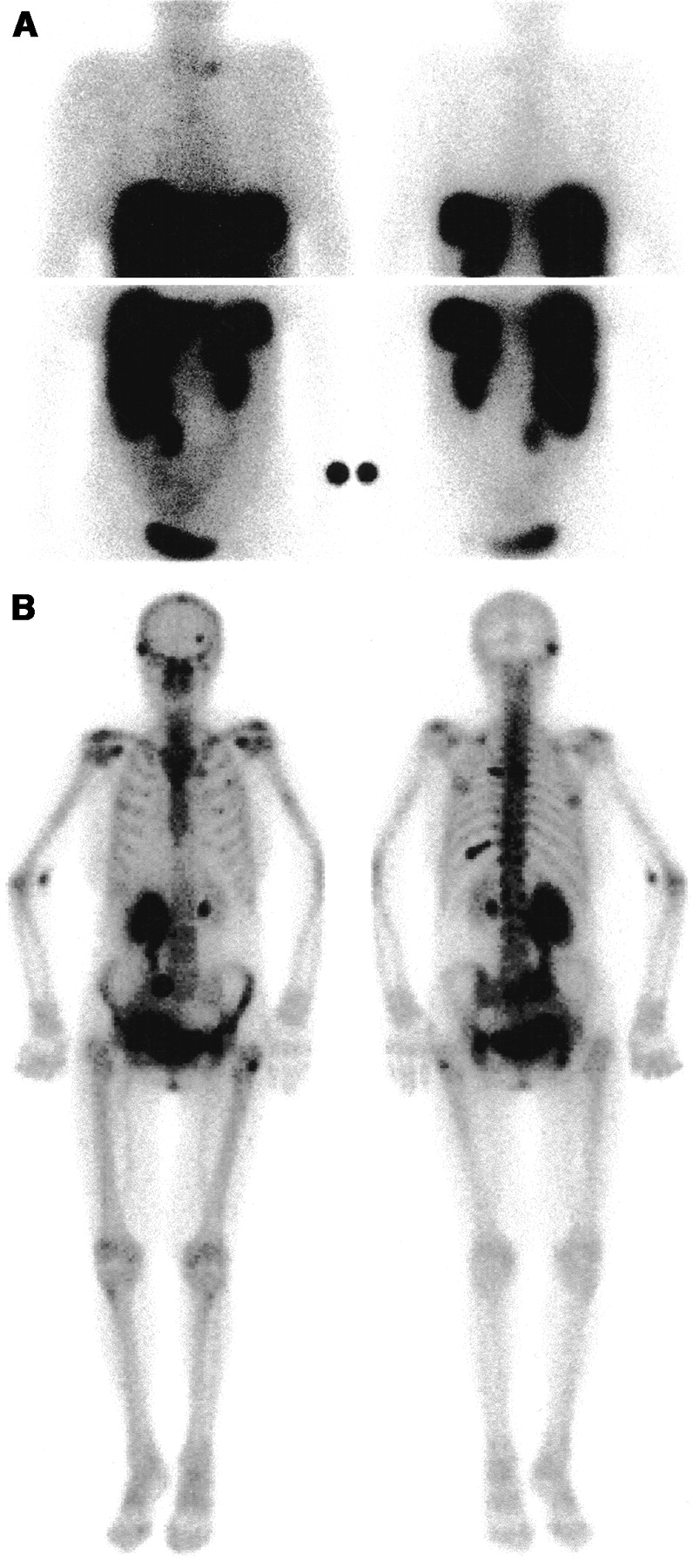
- FIGURE 1.
Visualization of bone metastases by bone scintigraphy (B) but not by octreotide scintigraphy (A) in 70-y-old female midgut carcinoid patient (patient 9). Upper panel (A) and left panel (B) represent anterior images. Bone lesions are present in vertebral body thoracic 5, dorsal part of left costa 5 and costa 10. Lesions located in skull and left femur are not accurately visualized with octreotide scintigraphy. Note hydronephrosis of right kidney.
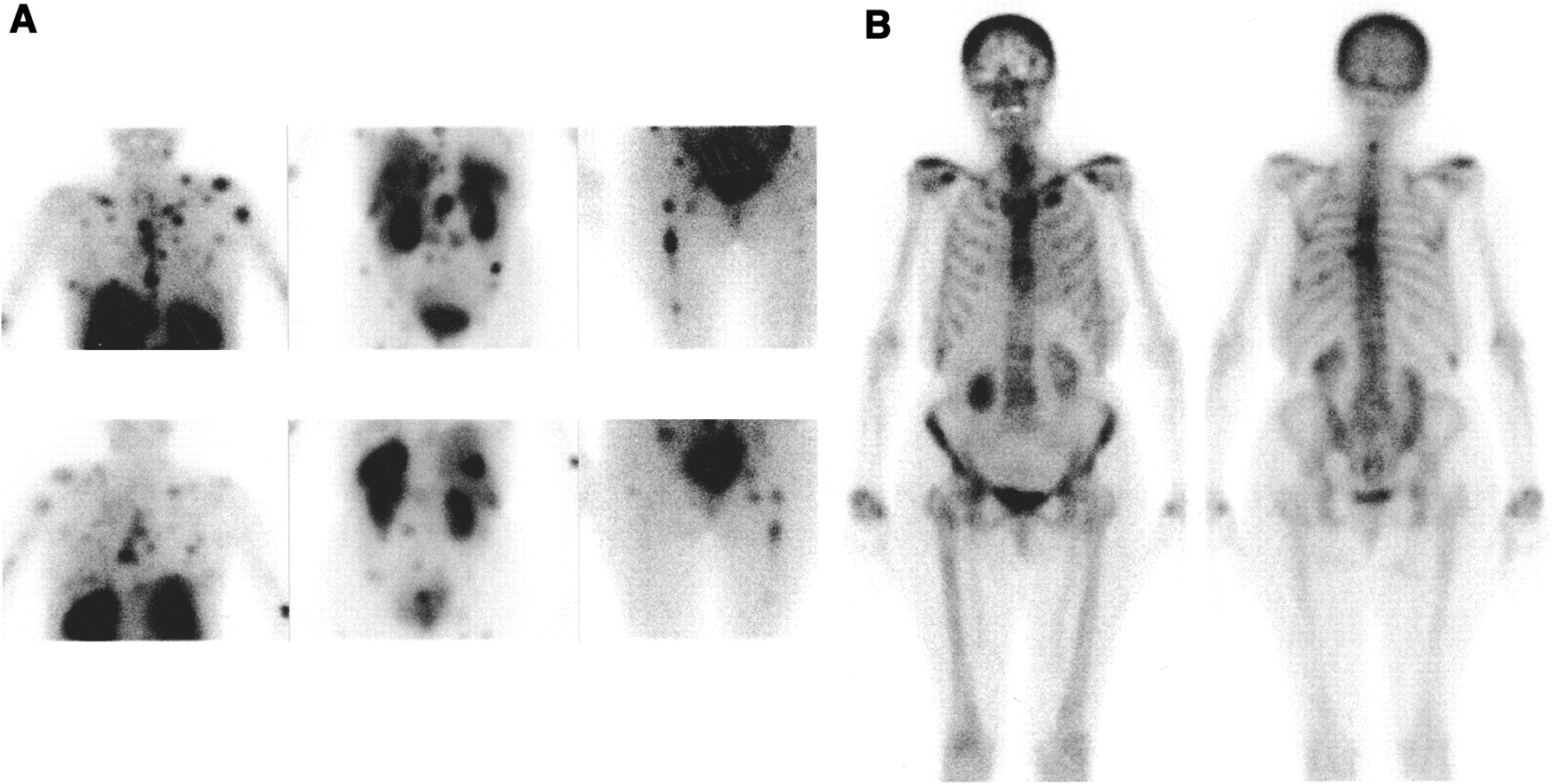
- FIGURE 2.
Complementary visualization of bone metastases by octreotide scintigraphy (A) and bone scintigraphy (B) in 63-y-old female midgut carcinoid patient (patient 6). Octreotide scintigraphy visualizes bone metastases in left humerus, pelvis, and bilateral femur. Cervical vertebral hot spot is visualized by bone scintigraphy but not by octreotide scintigraphy. Hot spots located in thoracic skeleton and lumbar spine are visible with both investigations.
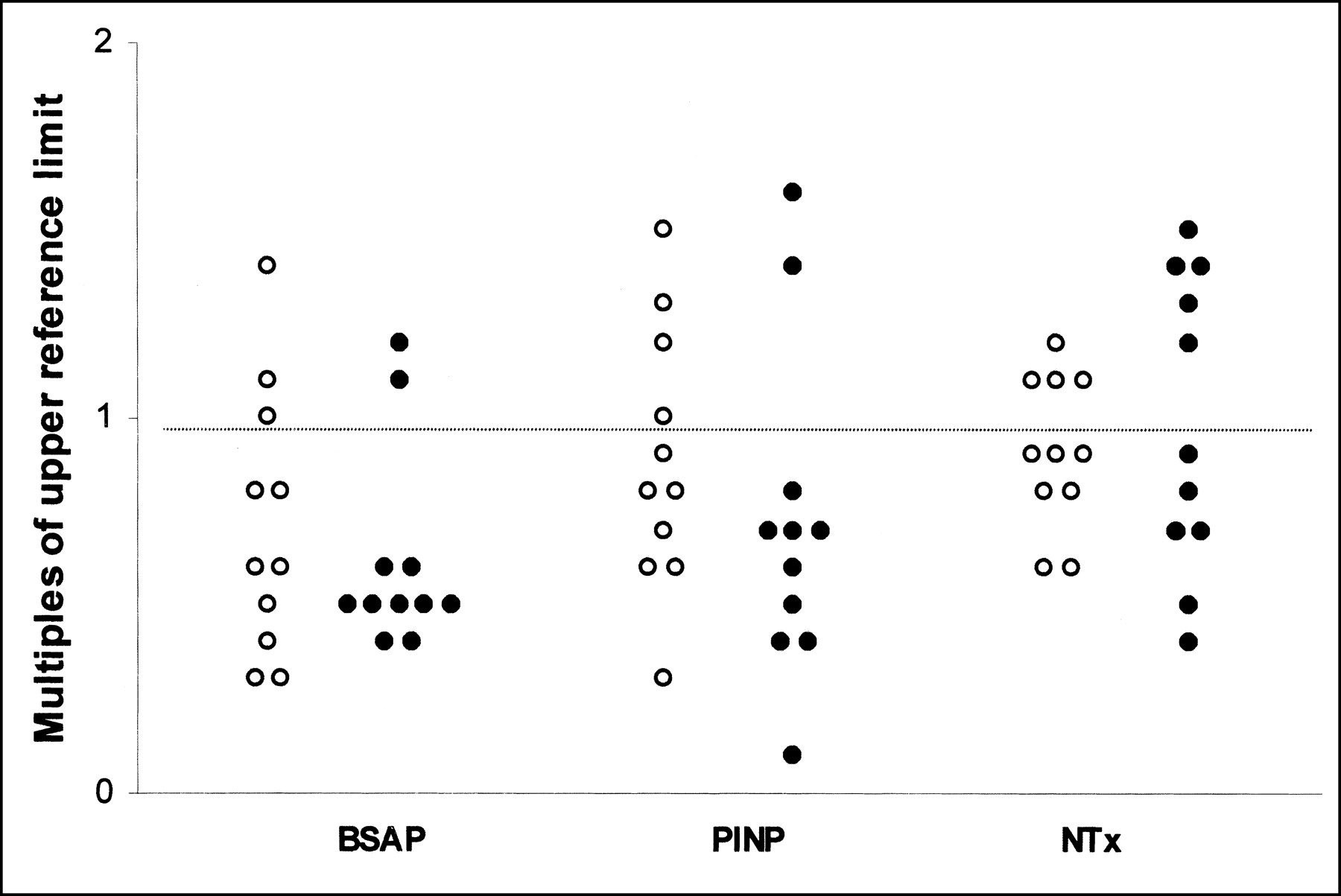
- FIGURE 3.
Multiples of upper reference limits of BSAP, PINP, and NTx. (○), Patients without bone metastases; (•), patients with bone metastases. Horizontal dotted line represents upper reference limit.
Tables
- TABLE 2
Clinical and Biochemical Characteristics of Carcinoid Patients With and Without Bone Metastases
Characteristic Patients with bone metastases Control patients No. of patients 11 11 Males 3 3 Females 8 8 Age (y) (range) 65 (36–75) 69 (46–75) Duration of disease (mo) (range) 17 (0–239) 25 (0–126) Midgut primary site (no. of patients) 11 11 Carcinoid syndrome present (no. of patients) 11 9 Site of metastases (no. of patients) Liver 10 7 Intraabdominal 8 6 Mediastinal 3 3 Pulmonary 3 1 Skeleton 11 0 Other 4 4 Treatment (no. of patients) Somatostatin analog 7 5 Interferon-α 0 4 Somatostatin analog and interferon-α 3 1 None 1 1 Laboratory (median) (range) 5-HIAA (mmol/mol creatinine) 23.1 (4.0–143.9) 29.9 (5.9–286.5) Platelet serotonin (nmol/109 platelets) 23.0 (4.6–60.1) 27.6 (17.1–50.4) PINP (μg/L) 47.8 (9.7–110.0) 58.2 (21.0–104.0) BSAP (U/L) 12.2 (8.4–26.7) 12.8 (7.6–33.3) INTP (BCE/L) 20.8 (8.7–35.1) 22.1 (13.4–29.6) INTP = amino-terminal telopeptide of type I collagen; BCE = bone collagen equivalents.
- TABLE 3
Clinical Characteristics and Outcome of Imaging Techniques in Patients with Bone Metastases of Carcinoid Tumors
Patient no. Sex Age (y) Time since diagnosis (mo) Site of bone metastases Plain radiography MRI Bone scintigraphy Octreotide scintigraphy 1 M 55 239 Lumbar spine, chest, skull, extremities N — P P 2 M 59 19 Total spine, chest N P P P 3 M 69 17 Total spine, chest, pelvis, extremities P P P P 4 F 36 0 Pelvis P — P N 5 F 58 21 Skull, extremities — — — P 6 F 63 0 Total spine, chest, pelvis, extremities N P P P 7 F 65 65 Total spine, chest, pelvis, skull, extremities N P P — 8 F 69 6 Thoracic spine, chest, pelvis P — P P 9 F 70 18 Thoracic spine, chest, pelvis, skull, extremities — P P N 10 F 72 9 Pelvis N — N P 11 F 75 6 Thoracic spine P P P N Positive (performed) technique 4 (9) 6 (6) 9 (10) 7 (10) Sensitivity (%) 44 100 90 70 95% CI of sensitivity (%) 12–76 61–100 72–100 35–93 N = negative; — = not done; P = positive.
- TABLE 4
Visualization of Bone Metastases: Bone Scintigraphy Compared with Octreotide Scintigraphy
Patient no.* Bone metastases visible with Total BS and OS BS OS 1 1 4 1 6 2 6 2 8 3 7 7 4 1 1 6 6 1 8 15 8 2 1 3 9 3 3 10 1 1 11 1 1 Total 22 10 13 45 % of total (= 45) 49 22 29 100 95% CI (%) 34–64 10–34 16–42 ↵* Patients 5 and 7 did not undergo both procedures.
BS = bone scintigraphy; OS = octreotide scintigraphy.
In this issue


Jump to section
Related Articles
Cited By...
- Bone metastases and skeletal-related events from neuroendocrine tumors
- Tumor Response Assessment to Treatment with [177Lu-DOTA0,Tyr3]Octreotate in Patients with Gastroenteropancreatic and Bronchial Neuroendocrine Tumors: Differential Response of Bone Versus Soft-Tissue Lesions
- Response and Long-Term Control of Bone Metastases After Peptide Receptor Radionuclide Therapy with 177Lu-Octreotate
- Bone Metastases in Patients with Neuroendocrine Tumor: 68Ga-DOTA-Tyr3-Octreotide PET in Comparison to CT and Bone Scintigraphy
- Metastatic Carcinoid Tumors: A Clinical Review