
Main menu

  • Home
  • Content
    • Current
    • Ahead of print
    • Past Issues
    • JNM Supplement
    • SNMMI Annual Meeting Abstracts
    • Continuing Education
    • JNM Podcasts
  • Subscriptions
    • Subscribers
    • Institutional and Non-member
    • Rates
    • Journal Claims
    • Corporate & Special Sales
  • Authors
    • Submit to JNM
    • Information for Authors
    • Assignment of Copyright
    • AQARA requirements
  • Info
    • Reviewers
    • Permissions
    • Advertisers
  • About
    • About Us
    • Editorial Board
    • Contact Information
  • More
    • Alerts
    • Feedback
    • Help
    • SNMMI Journals
  • SNMMI
    • JNM
    • JNMT
    • SNMMI Journals
    • SNMMI

User menu

  • Subscribe
  • My alerts
  • Log in
  • My Cart

Search

  • Advanced search
Journal of Nuclear Medicine
  • SNMMI
    • JNM
    • JNMT
    • SNMMI Journals
    • SNMMI
  • Subscribe
  • My alerts
  • Log in
  • My Cart
Journal of Nuclear Medicine

Advanced Search

  • Home
  • Content
    • Current
    • Ahead of print
    • Past Issues
    • JNM Supplement
    • SNMMI Annual Meeting Abstracts
    • Continuing Education
    • JNM Podcasts
  • Subscriptions
    • Subscribers
    • Institutional and Non-member
    • Rates
    • Journal Claims
    • Corporate & Special Sales
  • Authors
    • Submit to JNM
    • Information for Authors
    • Assignment of Copyright
    • AQARA requirements
  • Info
    • Reviewers
    • Permissions
    • Advertisers
  • About
    • About Us
    • Editorial Board
    • Contact Information
  • More
    • Alerts
    • Feedback
    • Help
    • SNMMI Journals
  • View or Listen to JNM Podcast
  • Visit JNM on Facebook
  • Join JNM on LinkedIn
  • Follow JNM on Twitter
  • Subscribe to our RSS feeds
OtherBasic Science Investigations

Quantitative 89Zr Immuno-PET for In Vivo Scouting of 90Y-Labeled Monoclonal Antibodies in Xenograft-Bearing Nude Mice

Iris Verel, Gerard W.M. Visser, Ronald Boellaard, Otto C. Boerman, Julliette van Eerd, Gordon B. Snow, Adriaan A. Lammertsma and Guus A.M.S van Dongen
Journal of Nuclear Medicine October 2003, 44 (10) 1663-1670;
Iris Verel
  • Find this author on Google Scholar
  • Find this author on PubMed
  • Search for this author on this site
Gerard W.M. Visser
  • Find this author on Google Scholar
  • Find this author on PubMed
  • Search for this author on this site
Ronald Boellaard
  • Find this author on Google Scholar
  • Find this author on PubMed
  • Search for this author on this site
Otto C. Boerman
  • Find this author on Google Scholar
  • Find this author on PubMed
  • Search for this author on this site
Julliette van Eerd
  • Find this author on Google Scholar
  • Find this author on PubMed
  • Search for this author on this site
Gordon B. Snow
  • Find this author on Google Scholar
  • Find this author on PubMed
  • Search for this author on this site
Adriaan A. Lammertsma
  • Find this author on Google Scholar
  • Find this author on PubMed
  • Search for this author on this site
Guus A.M.S van Dongen
  • Find this author on Google Scholar
  • Find this author on PubMed
  • Search for this author on this site
  • Article
  • Figures & Data
  • Info & Metrics
  • PDF
Loading

Article Figures & Data

Figures

  • FIGURE 1.
    • Download figure
    • Open in new tab
    • Download powerpoint
    FIGURE 1.

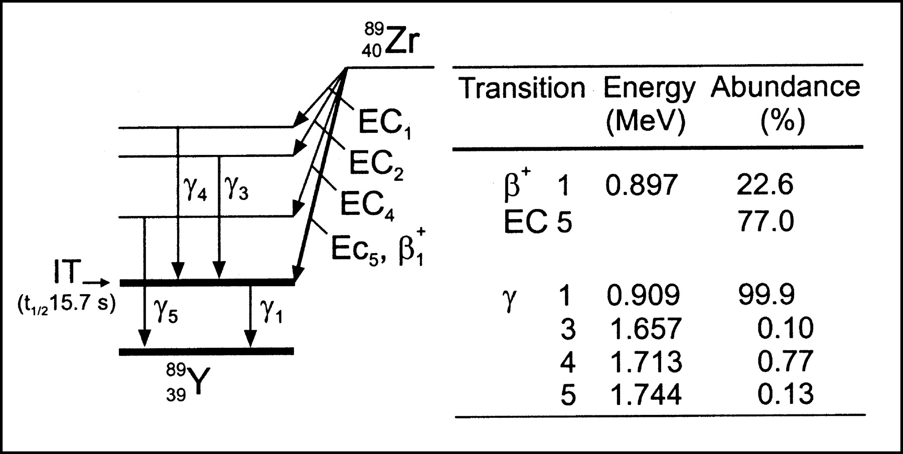
    Simplified 89Zr decay scheme (modified from ICRP publication (9)). Only transitions in excess of 0.1% abundance are shown. EC = electron capture; IT = isomeric transition; t1/2 = half-life.

  • FIGURE 2.
    • Download figure
    • Open in new tab
    • Download powerpoint
    FIGURE 2.

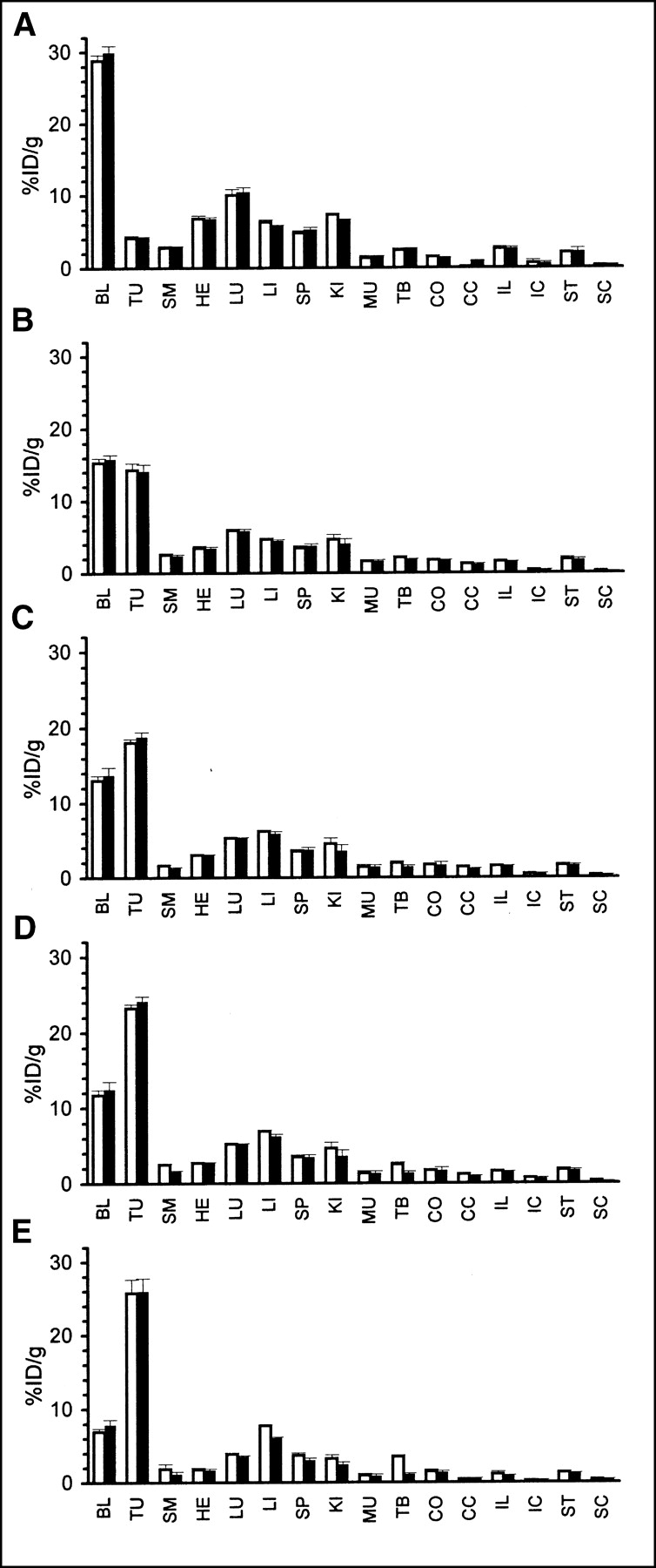
    Biodistribution of coinjected cmAb U36-N-sucDf-89Zr (0.37 MBq, white bars) and cmAb U36-p-BSCN-Bz-DOTA-88Y (0.13 MBq, black bars) in HNX-OE xenograft-bearing mice at 3 h (A), 24 h (B), 48 h (C), 72 h (D), and 144 h (E) after injection. At the indicated time points, 4 mice were bled, sacrificed, and dissected, and radioactivity levels (%ID/g ± SE) of blood, tumor, organs, and gastrointestinal contents were assessed. BL = blood; TU = tumor; SM = sternum; HE = heart; LU = lung; LI = liver; SP = spleen; KI = kidney; MU = muscle; TB = thighbone; CO = colon; CC = colon content; IL = ileum; IC = ileum content; ST = stomach; SC = stomach content.

  • FIGURE 3.
    • Download figure
    • Open in new tab
    • Download powerpoint
    FIGURE 3.

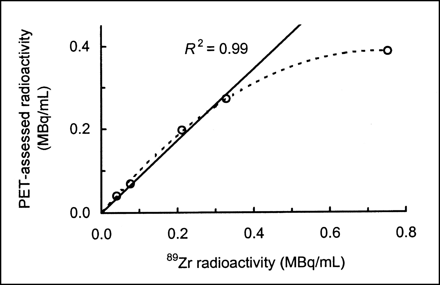
    89Zr-counting-rate linearity determination with HRRT PET camera. Plot is of PET-assessed radioactivity in cylinder phantom versus radioactivity based on dose calibrator measurements. Note presence of linearity, except for highest radioactivity measurement.
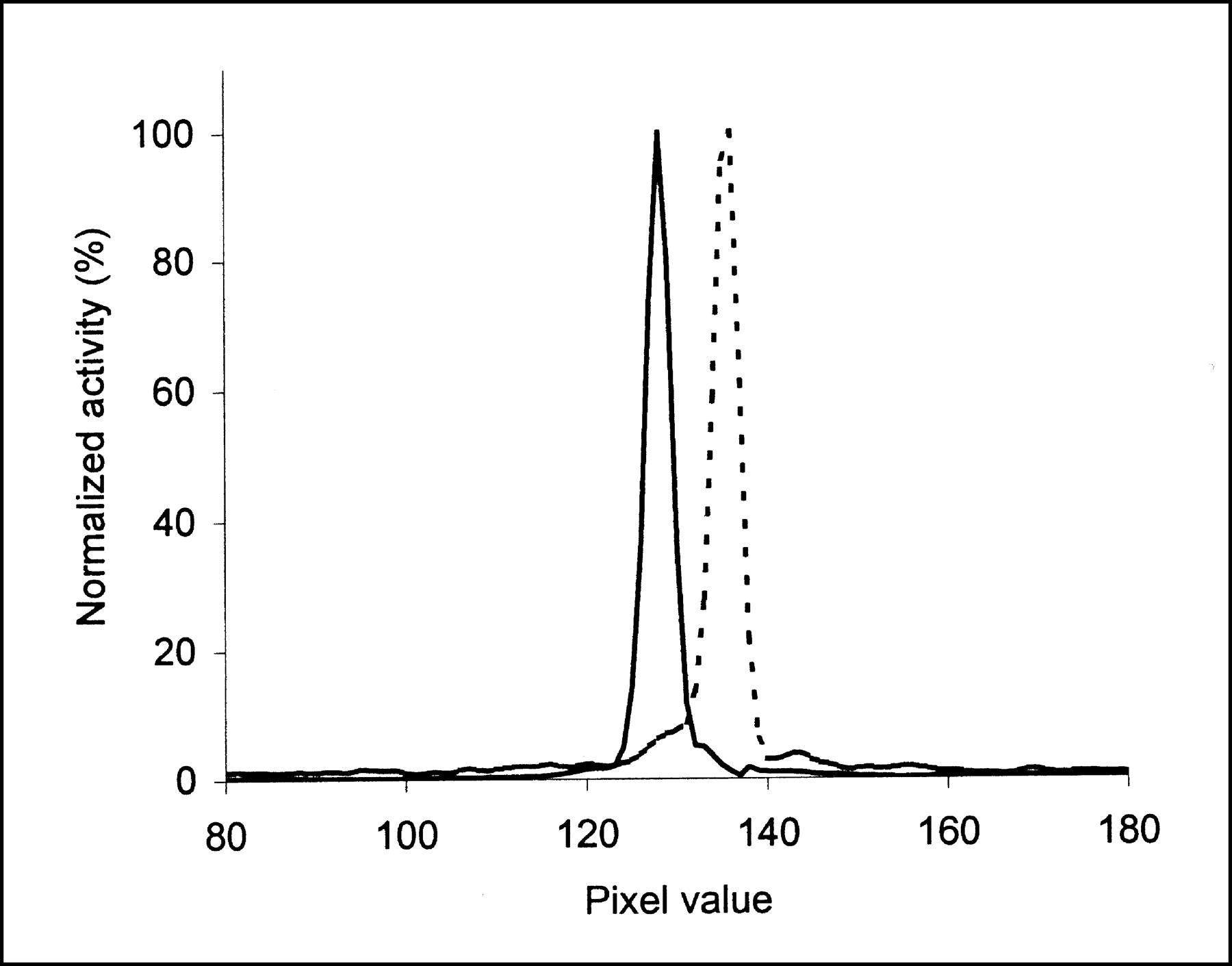
  • FIGURE 4.
    • Download figure
    • Open in new tab
    • Download powerpoint
    FIGURE 4.

    Spatial resolution determination for 89Zr (dotted line) with HRRT PET camera. Line profile was drawn through image of line source containing 89Zr. For comparison, line profile for 18F (solid line) is also shown.

  • FIGURE 5.
    • Download figure
    • Open in new tab
    • Download powerpoint
    FIGURE 5.

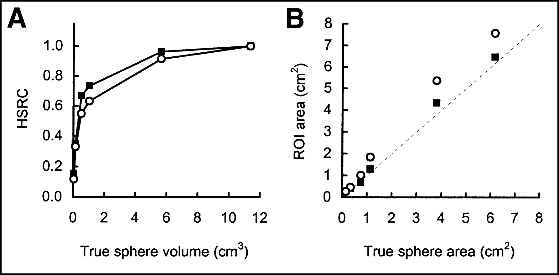
    HSRCs (A) and sphere size estimations (B) for 89Zr (○) with HRRT PET camera. For comparison, 18F (▪) data are also shown. For this purpose, 50% isocontour ROIs were drawn around spheres of Jaszczak phantom.
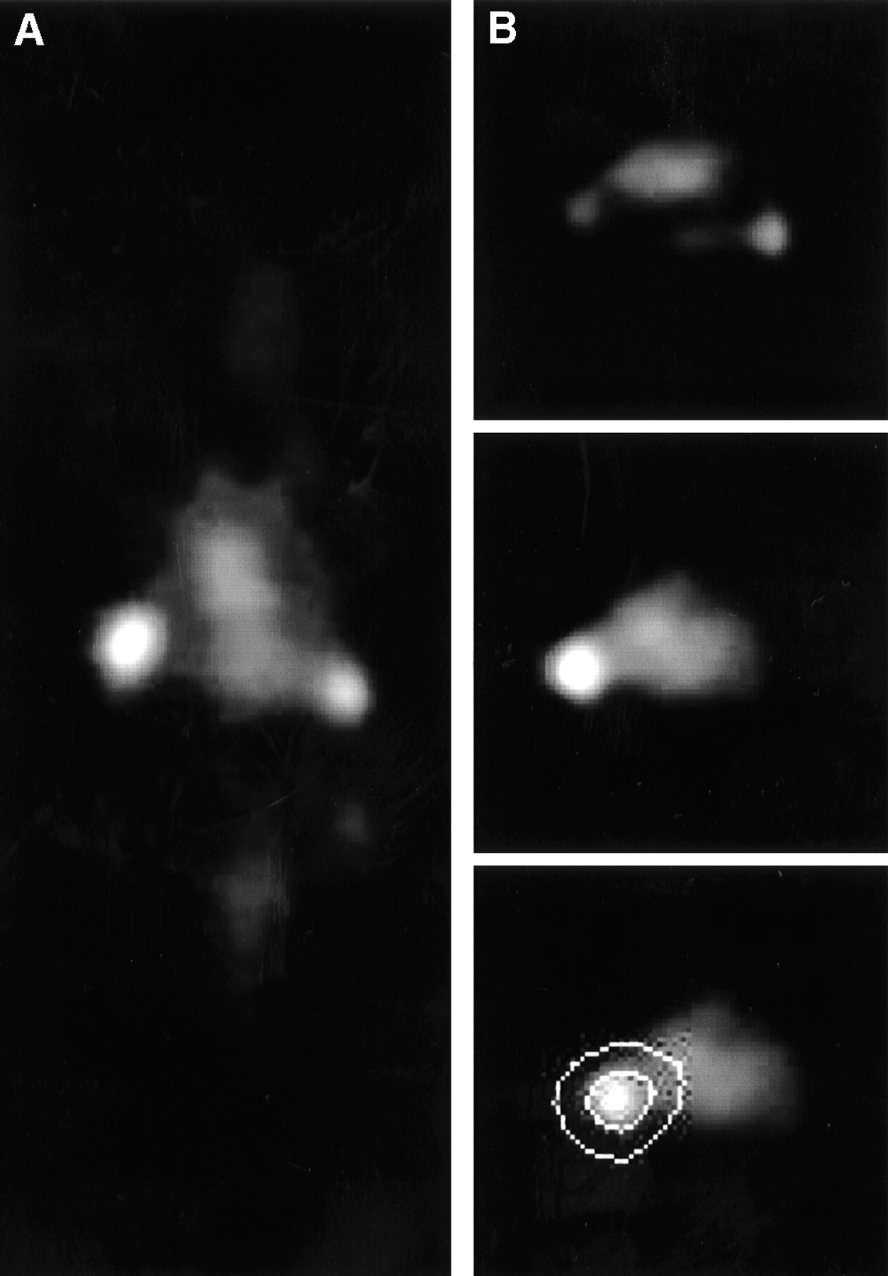
  • FIGURE 6.
    • Download figure
    • Open in new tab
    • Download powerpoint
    FIGURE 6.

    HRRT PET images of HNX-OE xenograft-bearing mouse injected with cmAb U36-N-sucDf-89Zr (3.7 MBq) at 72 h after injection. Coronal image plane (A) in which both tumors (left, 124 mg; right, 26 mg) were visible was chosen. Transaxial image planes in which right tumor was optimally visible (B, top) or left tumor was optimally visible (B, middle and bottom) were chosen. Bottom panel of B illustrates approach to arrive at ring-shaped area used for surroundings determination.
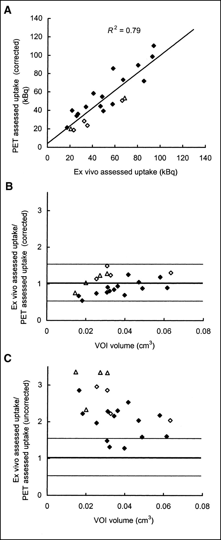
  • FIGURE 7.
    • Download figure
    • Open in new tab
    • Download powerpoint
    FIGURE 7.

    Correlation between PET-assessed tumor uptake and ex vivo-assessed tumor uptake. HNX-OE xenograft-bearing mice were injected with cmAb U36-N-sucDf-89Zr (3.7 MBq) and scanned at 1 d (⋄, n = 2), 2 d (▵, n = 2), or 3 d (♦, n = 8) with HRRT PET camera. Immediately after being scanned, mice were dissected and radioactivity levels in tumors were determined with γ-counter. After reconstruction of images, VOIs were drawn over tumors and radioactivity amounts were calculated. (A) After correction for partial-volume effects, PET-assessed (image-derived) tumor radioactivity values were plotted as function of ex vivo-assessed (γ-counter-derived) tumor radioactivity values. Ratio of ex vivo-assessed and PET-assessed tumor radioactivity values, corrected (B) or not corrected (C) for partial-volume effects, was plotted as function of VOI.

PreviousNext
Back to top

In this issue

Journal of Nuclear Medicine
Vol. 44, Issue 10
October 1, 2003
  • Table of Contents
  • Index by author
Print
Download PDF
Article Alerts
Sign In to Email Alerts with your Email Address
Email Article

Thank you for your interest in spreading the word on Journal of Nuclear Medicine.

NOTE: We only request your email address so that the person you are recommending the page to knows that you wanted them to see it, and that it is not junk mail. We do not capture any email address.

Enter multiple addresses on separate lines or separate them with commas.
Quantitative 89Zr Immuno-PET for In Vivo Scouting of 90Y-Labeled Monoclonal Antibodies in Xenograft-Bearing Nude Mice
(Your Name) has sent you a message from Journal of Nuclear Medicine
(Your Name) thought you would like to see the Journal of Nuclear Medicine web site.
Citation Tools
Quantitative 89Zr Immuno-PET for In Vivo Scouting of 90Y-Labeled Monoclonal Antibodies in Xenograft-Bearing Nude Mice
Iris Verel, Gerard W.M. Visser, Ronald Boellaard, Otto C. Boerman, Julliette van Eerd, Gordon B. Snow, Adriaan A. Lammertsma, Guus A.M.S van Dongen
Journal of Nuclear Medicine Oct 2003, 44 (10) 1663-1670;

Citation Manager Formats

  • BibTeX
  • Bookends
  • EasyBib
  • EndNote (tagged)
  • EndNote 8 (xml)
  • Medlars
  • Mendeley
  • Papers
  • RefWorks Tagged
  • Ref Manager
  • RIS
  • Zotero
Share
Quantitative 89Zr Immuno-PET for In Vivo Scouting of 90Y-Labeled Monoclonal Antibodies in Xenograft-Bearing Nude Mice
Iris Verel, Gerard W.M. Visser, Ronald Boellaard, Otto C. Boerman, Julliette van Eerd, Gordon B. Snow, Adriaan A. Lammertsma, Guus A.M.S van Dongen
Journal of Nuclear Medicine Oct 2003, 44 (10) 1663-1670;
Twitter logo Facebook logo LinkedIn logo Mendeley logo
  • Tweet Widget
  • Facebook Like
  • Google Plus One
Bookmark this article

Jump to section

  • Article
    • Abstract
    • MATERIALS AND METHODS
    • RESULTS
    • DISCUSSION
    • CONCLUSION
    • Acknowledgments
    • Footnotes
    • REFERENCES
  • Figures & Data
  • Info & Metrics
  • PDF

Related Articles

  • No related articles found.
  • PubMed
  • Google Scholar

Cited By...

  • PD-L1 PET/CT Imaging with Radiolabeled Durvalumab in Patients with Advanced-Stage Non-Small Cell Lung Cancer
  • Study of 89Zr-Pembrolizumab PET/CT in Patients With Advanced-Stage Non-Small Cell Lung Cancer
  • Bevacizumab Targeting Diffuse Intrinsic Pontine Glioma: Results of 89Zr-Bevacizumab PET Imaging in Brain Tumor Models
  • Immuno-PET Imaging of CD30-Positive Lymphoma Using 89Zr-Desferrioxamine-Labeled CD30-Specific AC-10 Antibody
  • Immuno-PET of the Hepatocyte Growth Factor Receptor Met Using the 1-Armed Antibody Onartuzumab
  • 89Zr-DFO-J591 for ImmunoPET of Prostate-Specific Membrane Antigen Expression In Vivo
  • Cerenkov Luminescence Imaging of Medical Isotopes
  • Immuno-PET Quantitation of de2-7 Epidermal Growth Factor Receptor Expression in Glioma Using 124I-IMP-R4-Labeled Antibody ch806
  • Development and Characterization of Clinical-Grade 89Zr-Trastuzumab for HER2/neu ImmunoPET Imaging
  • Disparity Between In Vivo EGFR Expression and 89Zr-Labeled Cetuximab Uptake Assessed with PET
  • Immuno-PET: A Navigator in Monoclonal Antibody Development and Applications
  • In vitro and In vivo Characterization of 64Cu-Labeled AbegrinTM, a Humanized Monoclonal Antibody against Integrin {alpha}v{beta}3
  • Potential of immuno-positron emission tomography for tumor imaging and immunotherapy planning.
  • Performance of immuno-positron emission tomography with zirconium-89-labeled chimeric monoclonal antibody u36 in the detection of lymph node metastases in head and neck cancer patients.
  • 89Zr as a PET Surrogate Radioisotope for Scouting Biodistribution of the Therapeutic Radiometals 90Y and 177Lu in Tumor-Bearing Nude Mice After Coupling to the Internalizing Antibody Cetuximab
  • Quantitative Immuno-Positron Emission Tomography Imaging of HER2-Positive Tumor Xenografts with an Iodine-124 Labeled Anti-HER2 Diabody
  • The Promise of Immuno-PET in Radioimmunotherapy
  • Google Scholar

More in this TOC Section

  • Synthesis and Biologic Evaluation of Novel 18F-Labeled Probes Targeting Prostate-Specific Membrane Antigen for PET of Prostate Cancer
  • Tumor-Specific Binding of Radiolabeled PEGylated GIRLRG Peptide: A Novel Agent for Targeting Cancers
  • PET/MRI of Hypoxic Atherosclerosis Using 64Cu-ATSM in a Rabbit Model
Show more Basic Science Investigations

Similar Articles

SNMMI

© 2025 SNMMI

Powered by HighWire