


Article Figures & Data
Figures
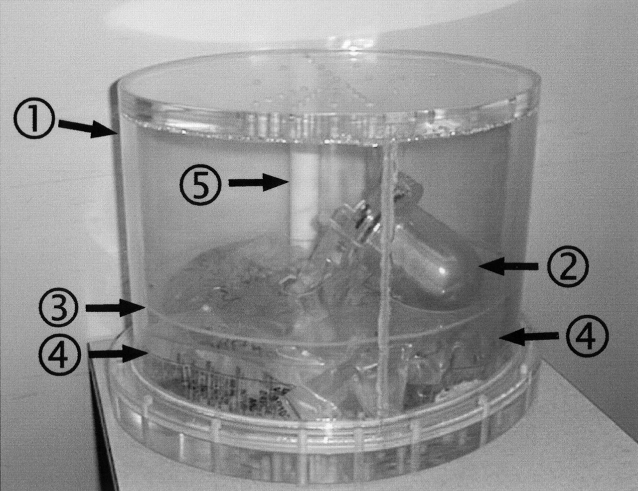
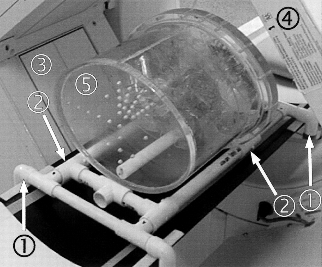
- FIGURE 1.
Photograph of phantom shows its components: 1, acrylic tub; 2, cardiac insert; 3, acrylic diaphragm; 4, saline bags; 5, Teflon (DuPont, Wilmington, DE) spine.
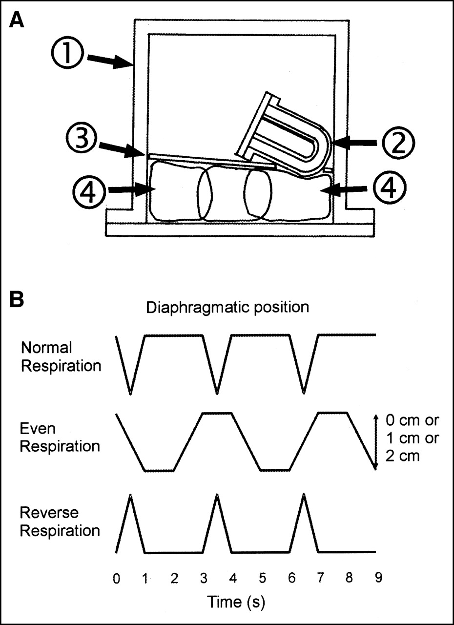
- FIGURE 2.
(A) Schematic diagram of midcoronal section through phantom shows relationship between saline bags and cardiac insert. Components of phantom are numbered same as in Figure 1. (B) Respiratory patterns used for phantom experiments. x-axis: time (s); y-axis: position of diaphragm. Cranial is up.
- FIGURE 3.
Photograph of gantry assembly on gamma-camera bed: 1, fixed frame; 2, sliding frame; 3, L-shaped detectors; 4, transmission source; 5, phantom (oriented supine, cranial to left). Push-pull handle has been removed for photography.
- FIGURE 4.
Four illustrative slices of transmission map for vertical short axis with no motion (top left) and with 2 cm of motion (bottom left) and for vertical long axis with no motion (top right) and with 2 cm of motion (bottom right). Heart was at 30° caudal angulation; cold liver and spleen inserts were present. Respiratory pattern was normal respiration.
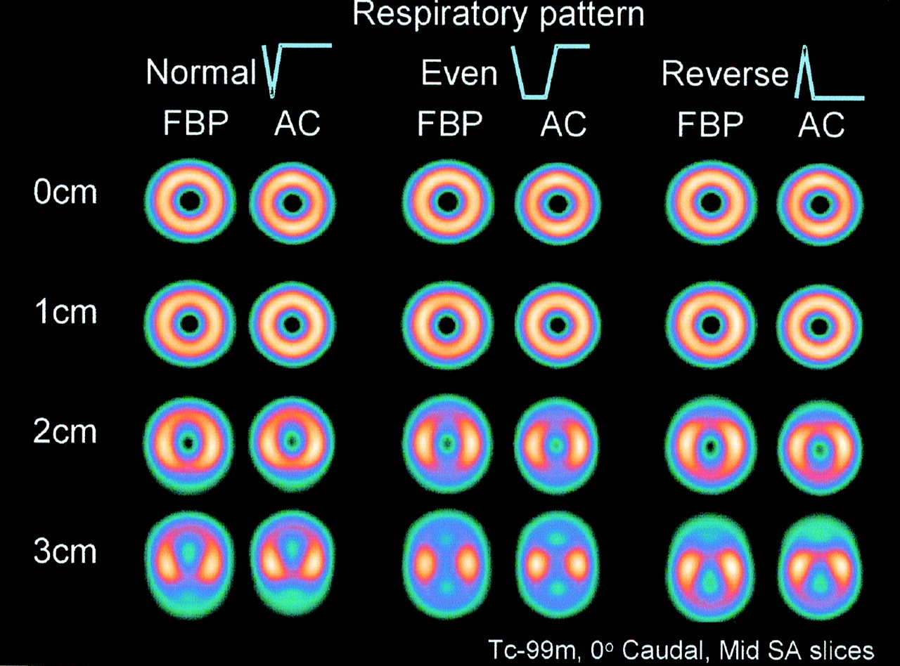
- FIGURE 5.
Demonstration of respiratory motion artifact and its dependence on respiratory pattern. For these experiments, myocardial inferior wall self-attenuation was avoided by positioning heart truly horizontally (perpendicular to face of detector), and no liver or spleen inserts were present. Myocardial insert contained 40 MBq 99mTc. Midshort-axis (SA) slices are illustrated. Normal, Even, Reverse = respiratory pattern (see Fig. 2B); AC = measured transmission AC with iterative reconstruction and scatter correction; 0, 1, 2, and 3 cm = respiratory amplitude of phantom.
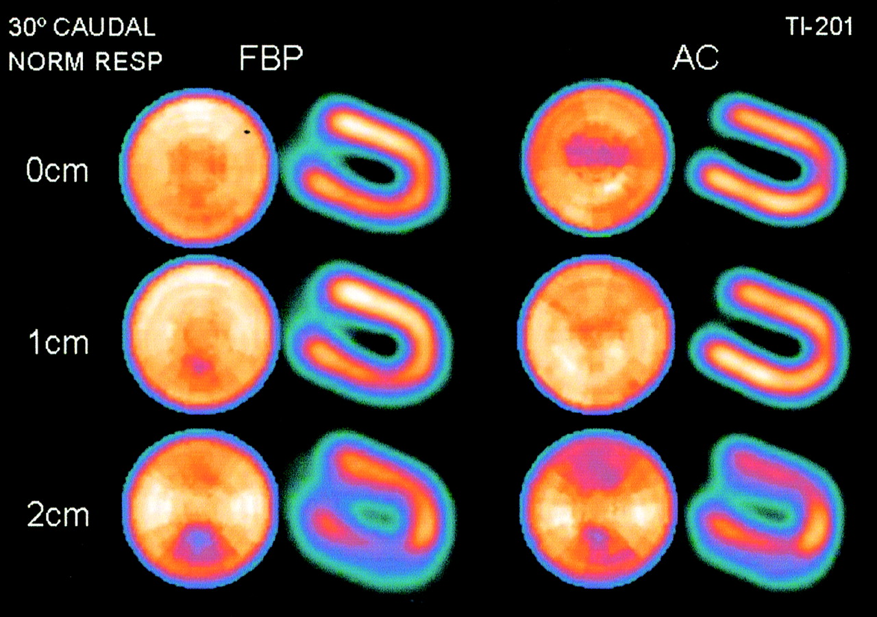
- FIGURE 6.
Interaction of attenuation artifact and respiratory motion artifact. Heart was at 30° caudal angulation; cold liver and spleen inserts were present. Myocardial insert contained 4.5 MBq 201Tl. Respiratory pattern was normal respiration. Distance-weighted polar plots and midvertical long-axis slices are illustrated. Normal = respiratory pattern (see Fig. 2B); AC = measured transmission AC with iterative reconstruction and scatter correction; 0, 1, and 2 cm = respiratory amplitude of phantom.
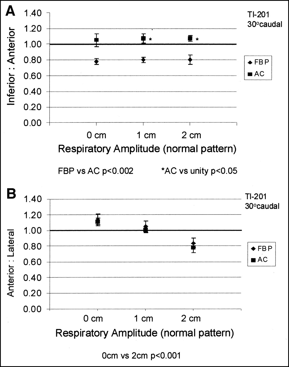
- FIGURE 7.
Representative graphs of inferior-to-anterior myocardial activity ratio (A) and anterior-to-lateral activity ratio (B) as function of respiratory amplitude. Heart was at 30° caudal angulation; cold liver and spleen inserts were present. Myocardial insert contained 4.5 MBq 201Tl. Respiratory pattern was normal respiration. FBP = FBP processing; AC = measured transmission AC with iterative reconstruction and scatter correction processing.


Tables
- TABLE 1
Effect of Ventricle Caudal Angulation and Respiratory Amplitude on Inferior-to-Anterior and Anterior-to-Lateral Wall Ratios
201Tl wall ratio Amplitude (cm) Angle 15° 30° 45° FBP AC FBP AC FBP AC Inferior-to-anterior* 0 0.97 ± 0.04† 1.11 ± 0.07†‡ 0.78 ± 0.04 1.05 ± 0.08 0.72 ± 0.10 0.98 ± 0.13 1 0.94 ± 0.04† 1.11 ± 0.04†‡ 0.80 ± 0.03 1.07 ± 0.06‡ 0.75 ± 0.11 0.92 ± 0.12 2 0.96 ± 0.07† 1.22 ± 0.05†‡ 0.80 ± 0.06 1.07 ± 0.04‡ 0.69 ± 0.13 0.95 ± 0.16 Anterior-to-lateral§ 0 1.00 ± 0.07¶ 1.10 ± 0.07¶ 1.14 ± 0.07¶ 1.11 ± 0.05¶ 1.05 ± 0.07¶ 1.05 ± 0.06¶ 1 0.91 ± 0.08 0.88 ± 0.04 1.05 ± 0.07 1.01 ± 0.04 0.95 ± 0.07 0.92 ± 0.08 2 0.75 ± 0.05 0.74 ± 0.03 0.84 ± 0.06 0.78 ± 0.06 0.88 ± 0.06 0.80 ± 0.08 ↵* All inferior-to-anterior FBP vs. AC pairs, P < 0.002.
↵† All inferior-to-anterior 15° vs. 45° pairs, P < 0.05.
↵‡ Inferior-to-anterior AC vs. unity, P < 0.05.
↵§ All anterior-to-lateral FBP vs. AC pairs, P = not significant.
↵¶ All anterior-to-lateral 0-cm vs. 2-cm pairs, P < 0.001.
Data are expressed as mean ± 1 SD. Cold liver and spleen inserts were present. Respiratory pattern was normal respiration.
In this issue


Jump to section
Related Articles
Cited By...
- Correction of Heart Motion Due to Respiration in Clinical Myocardial Perfusion SPECT Scans Using Respiratory Gating
- A Method to Remove Artifacts in Attenuation-Corrected Myocardial Perfusion SPECT Introduced by Misalignment Between Emission Scan and CT-Derived Attenuation Maps
- Human-Observer Receiver-Operating-Characteristic Evaluation of Attenuation, Scatter, and Resolution Compensation Strategies for 99mTc Myocardial Perfusion Imaging