


Article Figures & Data
Figures
- FIGURE 1.
Scheme of synthesis of FECh and 18F-FECh. Fluoroethanol method produces nonradioactive FECh tosylate as final product. TBA method produces radioactive 18F-FECh tosylate as final product.
- FIGURE 2.
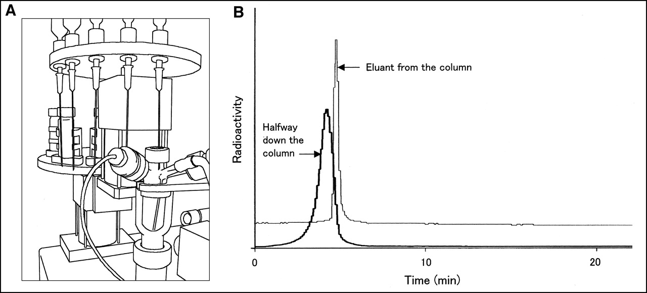
(A) Automated apparatus for 18F-FECh synthesis. Reaction vessel is shown close up. Purification module is out of view. (B) Preparative HPLC of 18F-FECh in automated apparatus for 18F-FECh synthesis. Column used was ODS-silica gel column, 250 × 10 mm; solvent, 50 mmol/L phosphoric acid + 1 mmol/L 2-naphthalenesulfonic acid; flow rate, 5 mL/min. Radioactivity “halfway down the column” is reading of detector (detector 1) placed on side of column to watch approach of 18F-FECh. Radioactivity of effluent from column was monitored by another detector (detector 2).
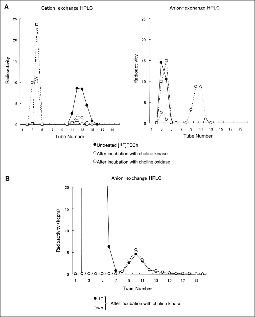
- FIGURE 3.
(A) HPLC after incubation of 18F-FECh with choline kinase and with choline oxidase. No-carrier-added 18F-FECh chloride was incubated with yeast choline kinase and 15 μmol ATP or with bacterial choline oxidase. From incubated specimen, methanol-water–soluble component was separated and fractionated on cation- and anion-exchange HPLC. 18F radioactivity was measured in each fraction. (B) HPLC after incubation of 18F-FECh with choline kinase and γ-32P-ATP. No-carrier-added 18F-FECh chloride (approximately 0.005 μmol) was incubated with yeast choline kinase and 0.01 μmol γ-32P-ATP. From incubated specimen, methanol-water–soluble component was separated and fractionated on anion-exchange HPLC. 18F and 32P radioactivities were measured in each fraction. kcpm = kilocounts per minute.
- FIGURE 4.
HPLC of methanol-water layer after incubation of 18F-FECh with Ehrlich ascites tumor cells. No-carrier-added 18F-FECh chloride was incubated with Ehrlich ascites tumor cells (approximately 106 cells/mL) in glucose-fortified Hanks’ solution for 30 min. Methanol-water–soluble component was separated from incubated cells and divided into halves. One half was analyzed on HPLC using cation-exchange resin and other half was analyzed on HPLC using anion-exchange resin. 18F radioactivity was measured in every fraction.
- FIGURE 5.
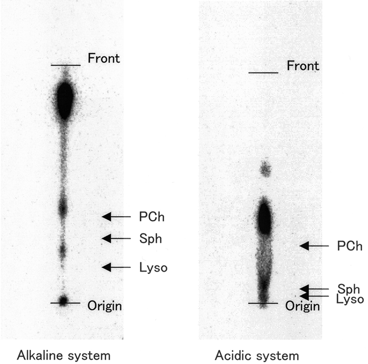
TLC of chloroform layer after incubation of 18F-FECh with Ehrlich ascites tumor cells (autoradiography). No-carrier-added 18F-FECh chloride was incubated with Ehrlich ascites tumor cells (approximately 106 cells/mL) in glucose-fortified Hanks’ solution for 30 min. Chloroform-soluble fraction of cell extract was concentrated and developed on TLC silica gel plates. Solvent was as follows: for alkaline system, chloroform, methanol, and 28% ammonia (65:35:5); for acidic system, benzene, pyridine, and formic acid (50:40:10). Autoradiography of TLC plates was performed using imaging plate detector system. Authentic choline-containing phospholipids were developed in same way as above and are indicated by arrows. PCh = phosphatidylcholine; Sph = sphingomyelin; Lyso = lysophosphatidylcholine.
- FIGURE 6.
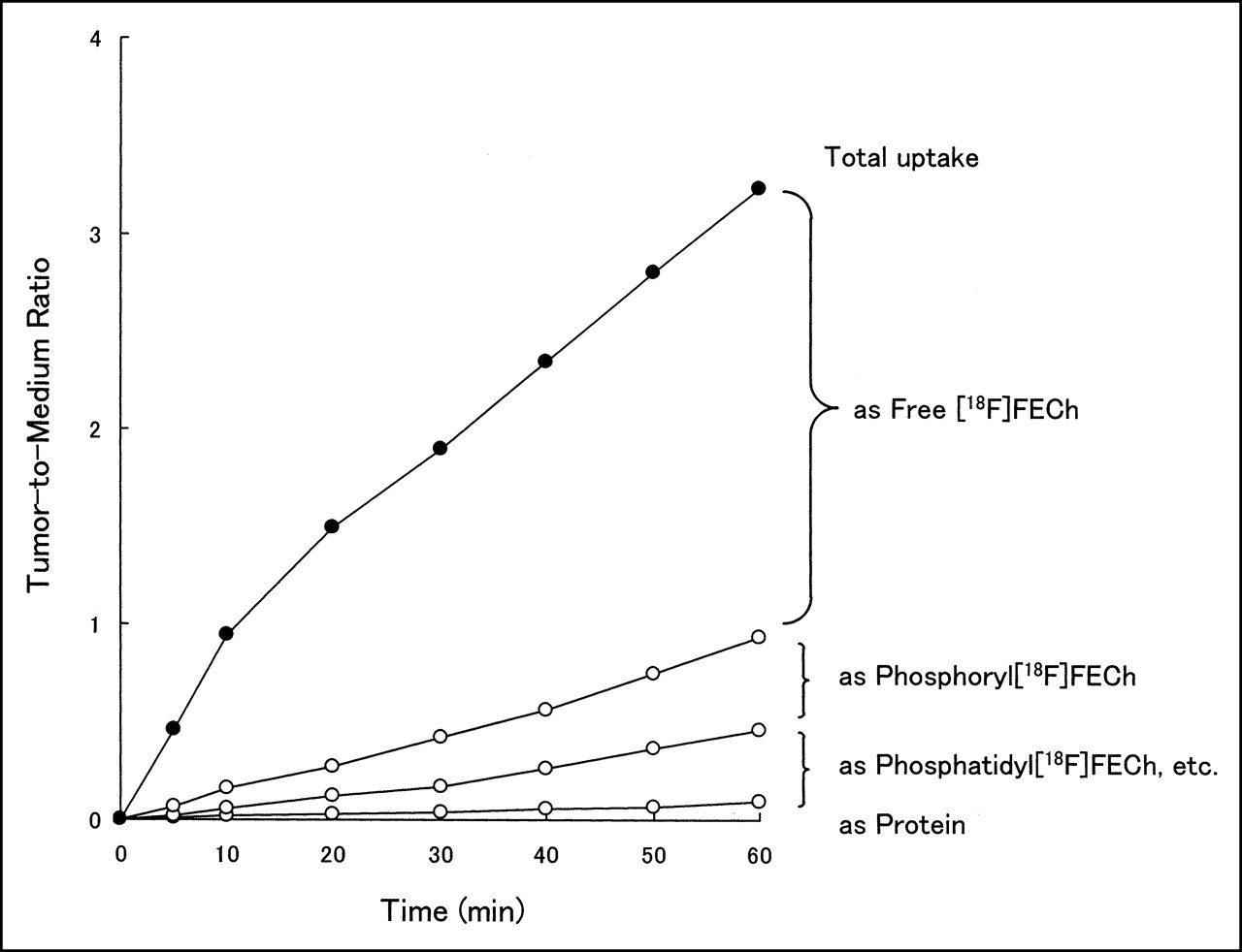
Time course of uptake and metabolism of 18F-FECh in Ehrlich ascites tumor cells. No-carrier-added 18F-FECh chloride was incubated with Ehrlich ascites tumor cells (approximately 106 cells/mL) in glucose-fortified Hanks’ solution for various periods up to 60 min. Total 18F uptake was measured after washing cells by centrifugation, and cell-to-medium ratio was calculated after hematocrit determination. After treatment of cells with methanol and chloroform, radioactivities in free 18F-FECh, phosphoryl-18F-FECh, 18F-labeled phospholipids (phosphatidyl-18F-FECh and so forth), and proteins were measured.
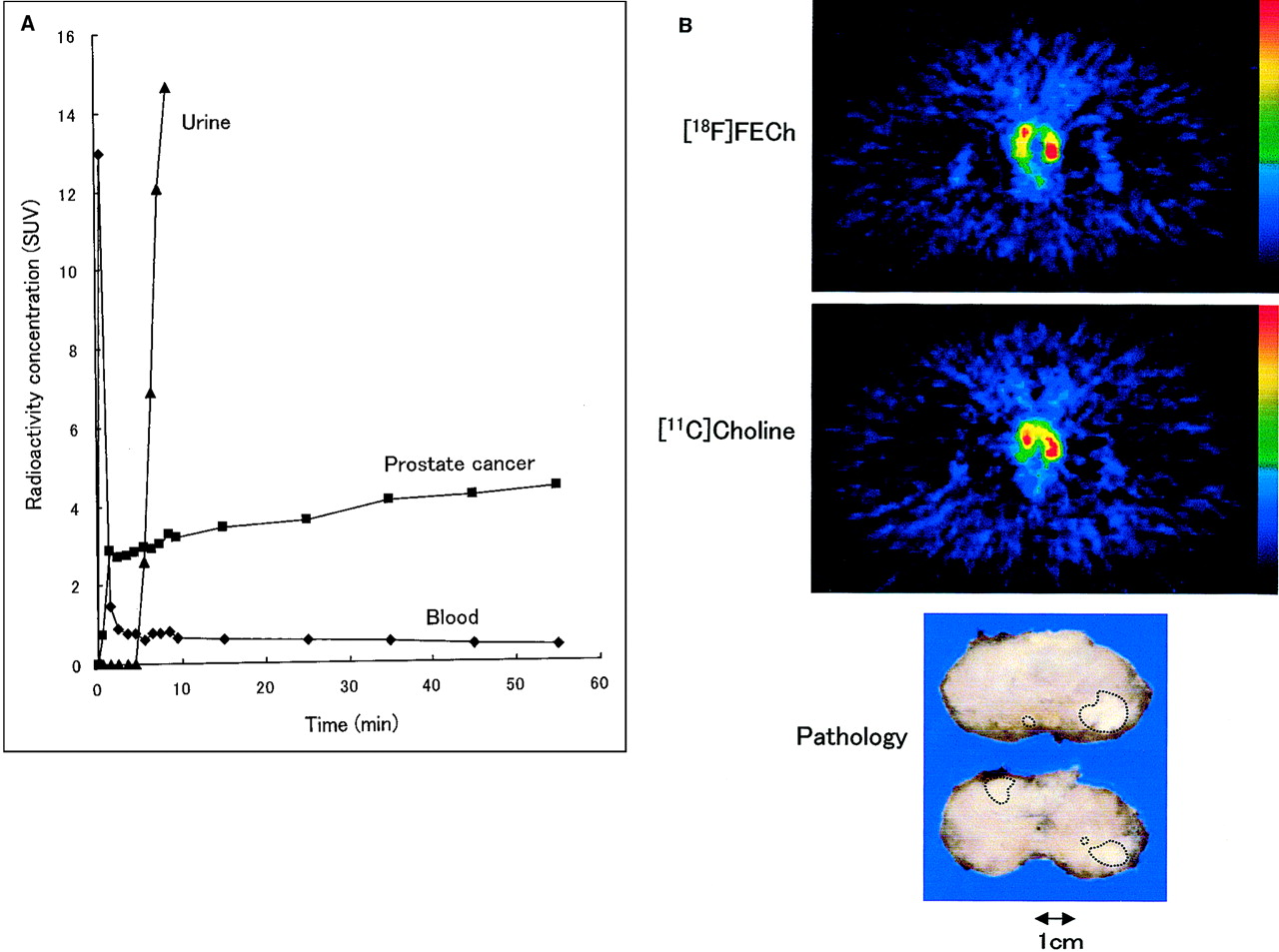
- FIGURE 7.
(A) Time course of radioactivity concentration in blood, prostate cancer, and urine after intravenous injection of 18F-FECh to untreated prostate cancer patient. SUV was calculated from PET data. (B) PET images of prostate cancer with 18F-FECh and 11C-choline and pathologic specimen of resected prostate (patient 5). 18F-FECh and 11C-choline images were taken at 60 and 20 min after injection, respectively. Red represents SUV of ≥4.0. Under diagnosis of stage B2, total prostatectomy and bilateral pelvic lymph node dissection were performed. Pathologic examination indicated adenocarcinoma of prostate in both lobes (Gleason score, 7) and no metastasis in pelvic lymph nodes (pathologic stage, pT2b, pN0). Largest tumor mass, 1.2 cm in diameter, was located in peripheral zone of left lobe.


Tables
Patient no.* Age (y) PSA (ng/mL) Positive biopsy Histology Clinical stage SUV in most radioactive area 18F-FECh 11C-Choline 30 min 60 min 5 min 20 min 1 67 4.6 1/6 Mod. diff. B1 2.84 3.25 3.43 4.75 2 71 5.5 3/6 Well-diff. B1 1.97 1.86 1.66 1.86 3 75 6.6 3/6 Well-diff. B1 2.78 2.83 2.85 2.75 4 63 7.7 2/6 Well-diff. B1 2.50 2.90 2.48 2.48 5 62 8.4 4/6 Mod. diff. B2 4.08 4.25 4.37 4.07 6 70 19.5 3/7 Well-diff. B1 5.39 5.14 4.79 5.43 7 78 19.6 3/7 Mod. diff. D2 4.25 4.36 3.10 4.90 8 77 36.0 7/7 Poor diff. D1 3.12 3.27 3.93 4.67 9 75 41.8 3/6 Poor diff. B1 2.71 3.30 3.28 3.28 10 68 45.7 5/6 Poor diff. D2 3.63 3.97 4.71 5.21 11 72 45.9 3/6 Mod. diff. B1 3.43 2.55 4.56 4.51 12 72 51.3 6/6 Poor diff. D2 5.51 6.46 7.22 7.58 13 64 80.0 6/6 Mod. diff. C 3.42 3.35 3.88 4.08 14 78 104.0 5/6 Poor diff. D2 4.41 4.66 3.17 3.57 15 68 126.3 6/6 Mod. diff. D2 4.88 4.70 4.94 5.77 16 82 242.6 4/4 Mod. diff. D2 6.47 7.50 6.04 7.11 ↵* Patient 5 underwent total prostatectomy and all other patients received hormonal therapy after PET study.
Mod. = moderately; diff. = differentiated; poor = poorly.
In this issue


Jump to section
Related Articles
Cited By...
- Molecular Imaging of Prostate Cancer: Tapping into the Opportunities
- Combined PET/MRI Improves Diagnostic Accuracy in Patients with Prostate Cancer: A Prospective Diagnostic Trial
- Reduced 64Cu Uptake and Tumor Growth Inhibition by Knockdown of Human Copper Transporter 1 in Xenograft Mouse Model of Prostate Cancer
- Detection of Recurrent Prostate Cancer After Radical Prostatectomy: Comparison of 11C-Choline PET/CT with Pelvic Multiparametric MR Imaging with Endorectal Coil
- Choline phosphorylation and regulation of transcription of choline kinase {alpha} in hypoxia
- Novel Tracers and Their Development for the Imaging of Metastatic Prostate Cancer
- Gene Expression Patterns and Tumor Uptake of 18F-FDG, 18F-FLT, and 18F-FEC in PET/MRI of an Orthotopic Mouse Xenotransplantation Model of Pancreatic Cancer
- Tumor Cell Metabolism Imaging
- Choline Kinase Down-regulation Increases the Effect of 5-Fluorouracil in Breast Cancer Cells
- Initial Experience with the Radiotracer Anti-1-Amino-3-18F-Fluorocyclobutane-1-Carboxylic Acid with PET/CT in Prostate Carcinoma
- Imaging Prostate Cancer with 11C-Choline PET/CT
- Value of 11C-Choline PET and Contrast-Enhanced CT for Staging of Bladder Cancer: Correlation with Histopathologic Findings
- Localization of Primary Prostate Cancer with Dual-Phase 18F-Fluorocholine PET
- Phosphorylation of the Yeast Choline Kinase by Protein Kinase C: IDENTIFICATION OF Ser25 AND Ser30 AS MAJOR SITES OF PHOSPHORYLATION
- microPET and Autoradiographic Imaging of GRP Receptor Expression with 64Cu-DOTA-[Lys3]Bombesin in Human Prostate Adenocarcinoma Xenografts
- PET Scan Detects Prostate Cancer in a Patient with Hodgkins Lymphoma
- 11C-Acetate PET Imaging of Prostate Cancer: Detection of Recurrent Disease at PSA Relapse
- PET Imaging of Prostate Cancer with 11C-Acetate
- Phosphorylation of Saccharomyces cerevisiae Choline Kinase on Ser30 and Ser85 by Protein Kinase A Regulates Phosphatidylcholine Synthesis by the CDP-choline Pathway
- PET for Prostate Cancer Imaging: Still a Quandary or the Ultimate Solution?