


Article Figures & Data
Figures
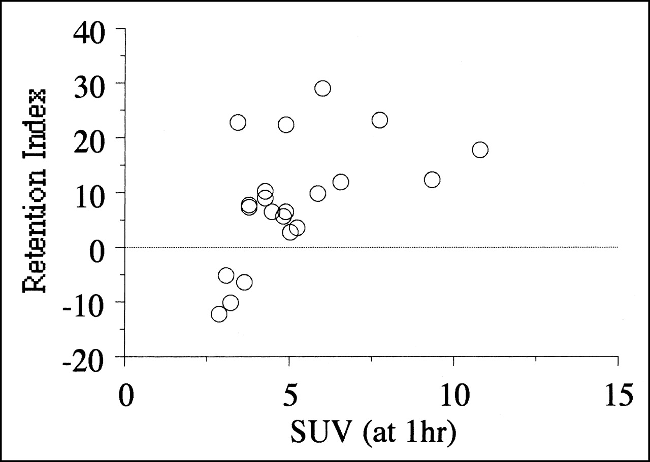
- FIGURE 1.
Results of SUV at 1 h and RI of dual-phase 18F-FDG PET. Positive correlation existed between SUV at 1 h and RI (P < 0.05). Four patients with negative RI showed low SUVs at 1 h.
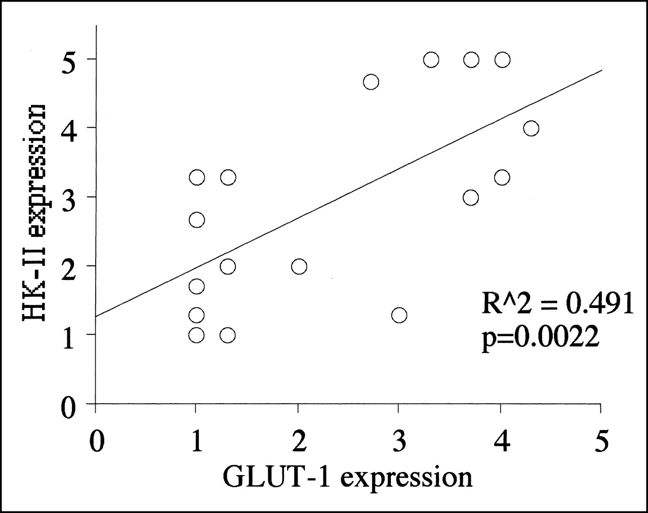
- FIGURE 2.
Results of immunohistochemical staining using anti-GLUT-1 and anti-HK-II. They correlated closely (P < 0.005).
- FIGURE 3.
Results of comparative analysis between quantitative values of dual-phase 18F-FDG PET and immunohistochemical staining. SUV at 1 h showed a weak positive correlation with GLUT-1 glucose transporter expression (P = 0.055), whereas no significant relationship was found between SUV at 1 h and HK-II hexokinase expression. However, RI had no significant relationship with GLUT-1 glucose transporter expression, whereas RI and HK-II hexokinase expression had significant positive relationship (P < 0.05).
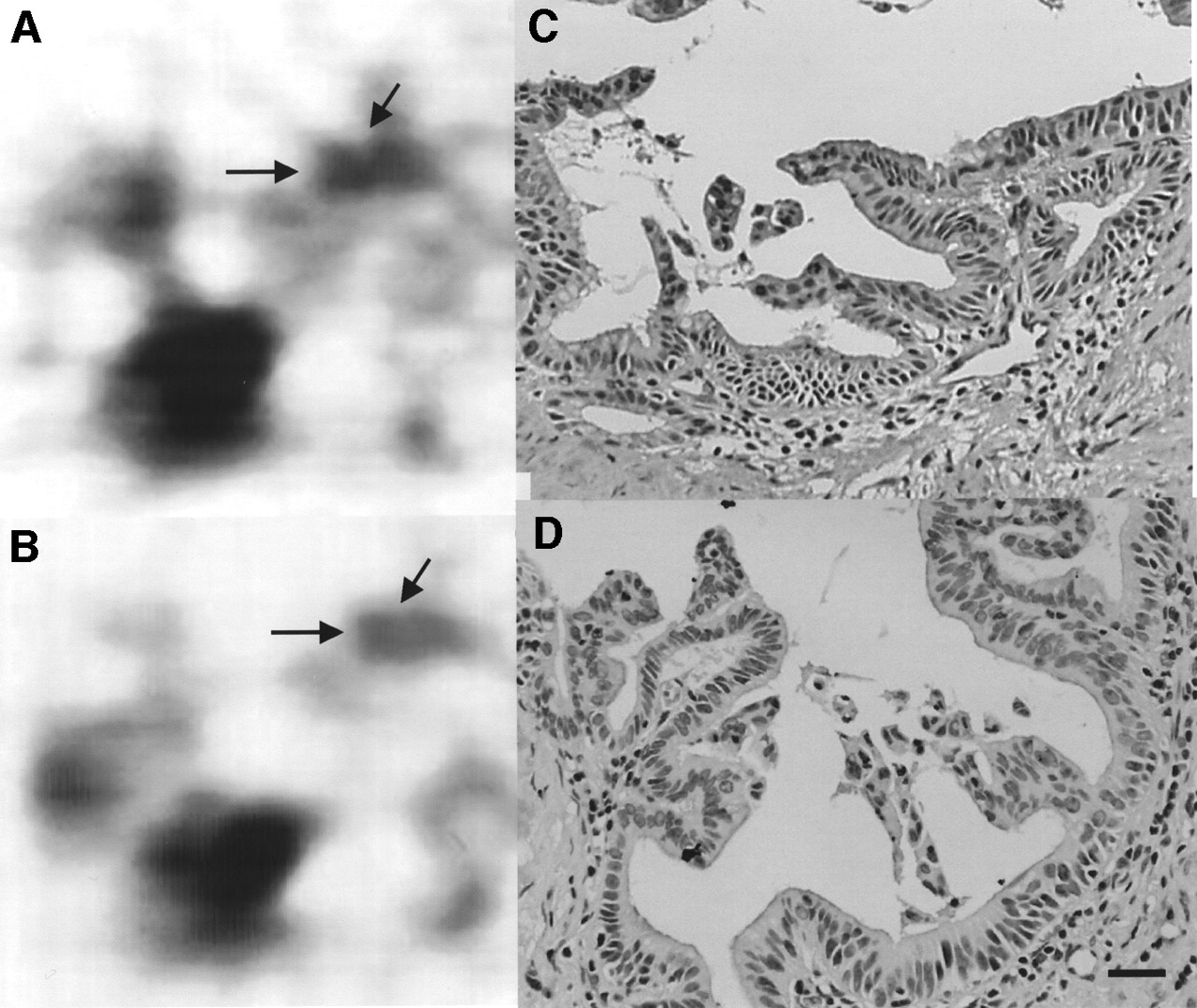
- FIGURE 4.
Invasive ductal adenocarcinoma (arrows) in pancreatic head of patient 1. 18F-FDG PET images obtained at 1 h, when tumor SUV was 2.82 (A), and at 2 h, when tumor SUV was 2.48 (B), with RI of −12.1%. Immunohistochemical staining of GLUT-1 (C) and of HK-II (D) shows no significant strong expression of GLUT-1 or HK-II. Percentages of strong expressed cells of both proteins were scored as <20%. (×400; bar = 20 μm)
- FIGURE 5.
Invasive ductal adenocarcinoma (arrows) in pancreatic head of patient 17. 18F-FDG PET images obtained at 1 h, when tumor SUV was 6.0 (A), and at 2 h, when tumor SUV was 7.75 (B), with RI of 29.2%. Immunohistochemical staining of GLUT-1 (C) and of HK-II (D) shows overexpression of GLUT-1 and HK-II. Expression of GLUT-1 is observed mainly in membrane, whereas that of HK-II is observed mainly in cytoplasm. Percentages of strong expressed cells of GLUT-1 were scored as 50%–80%, and those of HK-II were scored as >80%. (×400; bar = 20 μm)


Tables
Patient no. Sex Age (y) Histologic diagnosis Differentiation Operation SUV (at 1 h) SUV (at 2 h) RI 1 F 71 Invasive ductal carcinoma Well Resectable 2.82 2.48 −12.06 2 M 68 Invasive ductal carcinoma Poorly Unresectable* 3.16 2.84 −10.13 3 F 58 Invasive ductal carcinoma Poorly Unresectable* 3.58 3.35 −6.42 4 M 52 Invasive ductal carcinoma Moderately Resectable 3.06 2.90 −5.23 5 F 66 Invasive ductal carcinoma Moderately Resectable 4.99 5.14 3.01 6 M 58 Invasive ductal carcinoma Poorly Unresectable* 5.24 5.43 3.63 7 M 70 Invasive ductal carcinoma Poorly Resectable 4.78 5.06 5.86 8 M 62 Invasive ductal carcinoma Moderately Resectable 4.42 4.72 6.79 9 M 62 Invasive ductal carcinoma Moderately/poorly Unresectable* 3.76 4.04 7.45 10 M 49 Invasive ductal carcinoma Poorly Unresectable* 3.73 4.02 7.77 11 M 57 Invasive ductal carcinoma Poorly Unresectable* 5.85 6.44 10.09 12 F 82 Invasive ductal carcinoma Moderately Unresectable* 4.26 4.70 10.33 13 M 53 Invasive ductal carcinoma Poorly Resectable 9.34 10.51 12.53 14 F 68 Invasive ductal carcinoma Moderately/poorly Unresectable* 10.76 12.67 17.75 15 M 58 Invasive ductal carcinoma Moderately Resectable 4.86 5.96 22.63 16 F 62 Invasive ductal carcinoma Moderately Unresectable* 7.68 9.47 23.31 17 M 54 Invasive ductal carcinoma Well Unresectable* 6.00 7.75 29.17 18 M 59 Mucinous cystadenocarcinoma Well Resectable 4.24 4.63 9.20 19 F 64 Mucinous cystadenocarcinoma Well Unresectable* 6.54 7.34 12.23 20 F 40 Mucinous cystadenocarcinoma Well Resectable 3.41 4.19 22.87 21 M 63 Intraductal papillary carcinoma Well Resectable 4.83 5.16 6.83 ↵* Needle biopsy under laparotomy.
Patient no. GLUT-1 (G-index) HK-II (HK-index) Cellularity (C-index) Average Counting trial Average Counting trial Average Counting trial First Second Third First Second Third First Second 1 1.0 1 1 1 1.0 1 1 1 3.0 3 3 2 1.0 1 1 1 1.3 1 2 1 1.5 1 2 3 3.0 3 3 3 1.3 2 1 1 5.0 5 5 4 1.0 1 1 1 3.3 3 3 4 2.5 3 2 5 1.3 1 2 1 1.0 1 1 1 2.5 2 3 6 2.7 2 3 3 4.7 4 5 5 5.0 5 5 7 4.0 4 4 4 3.3 3 4 3 1.5 1 2 8 5.0 5 5 5 4.0 3 4 5 2.5 3 2 9 1.0 1 1 1 1.3 2 1 1 2.0 2 2 10 3.3 4 3 3 5.0 5 5 5 5.0 5 5 11 3.7 4 3 4 3.0 4 2 3 5.0 5 5 12 1.0 1 1 1 2.7 4 2 2 3.0 3 3 13 5.0 5 5 5 5.0 5 5 5 4.5 5 4 14 2.0 3 2 1 2.0 2 2 2 5.0 5 5 15 4.3 4 4 5 4.0 3 4 5 3.0 3 3 16 1.3 1 2 1 3.3 3 4 3 4.0 4 4 17 3.7 3 4 4 5.0 5 5 5 2.5 3 2 18 1.0 1 1 1 1.7 3 1 1 4.0 5 3 19 1.3 1 2 1 2.0 2 2 2 2.0 2 2 20 2.7 2 3 3 4.7 4 5 5 4.0 4 4 21 4.0 4 4 4 5.0 5 5 5 2.0 2 2
In this issue


Jump to section
Related Articles
Cited By...
- Prognostic Significance of 18F-FDG PET/CT and Tumor Metabolic Changes in Patients With Pancreatic Ductal Adenocarcinoma
- Identification of Keratin 19-Positive Cancer Stem Cells Associating Human Hepatocellular Carcinoma Using 18F-Fluorodeoxyglucose Positron Emission Tomography
- Oncogene Pathway Activation in Mammary Tumors Dictates FDG-PET Uptake
- Relationship Between 18F-FDG Accumulation and Lactate Dehydrogenase A Expression in Lung Adenocarcinomas
- Prognostic Implication of Dual-Phase PET in Adenocarcinoma of the Lung
- Evaluation of the Role of Hexokinase Type II in Cellular Proliferation and Apoptosis Using Human Hepatocellular Carcinoma Cell Lines
- 18F-FDG PET Findings and GLUT-1 Expression in IPMNs of the Pancreas
- Colorectal Tumor Cells Treated with 5-FU, Oxaliplatin, Irinotecan, and Cetuximab Exhibit Changes in 18F-FDG Incorporation Corresponding to Hexokinase Activity and Glucose Transport
- Clinical applications and advances of positron emission tomography with fluorine-18-fluorodeoxyglucose (18F-FDG) in the diagnosis of liver neoplasms
- Different Glucose Uptake and Glycolytic Mechanisms Between Hepatocellular Carcinoma and Intrahepatic Mass-Forming Cholangiocarcinoma with Increased 18F-FDG Uptake
- Biologic Correlates of Intratumoral Heterogeneity in 18F-FDG Distribution with Regional Expression of Glucose Transporters and Hexokinase-II in Experimental Tumor
- Retrospective Digital Image Fusion of Multidetector CT and 18F-FDG PET: Clinical Value in Pancreatic Lesions--A Prospective Study with 104 Patients
- 18F-FDG Uptake in Squamous Cell Carcinoma of the Cervix Is Correlated with Glucose Transporter 1 Expression
- Delayed 18F-FDG PET for Detection of Paraaortic Lymph Node Metastases in Cervical Cancer Patients
- Value of Dual-Phase 2-Fluoro-2-Deoxy-D-Glucose Positron Emission Tomography in Cervical Cancer
- 18F-Fluoro-2-deoxyglucose positron emission tomography in the evaluation of gastrointestinal malignancies