


Article Figures & Data
Figures
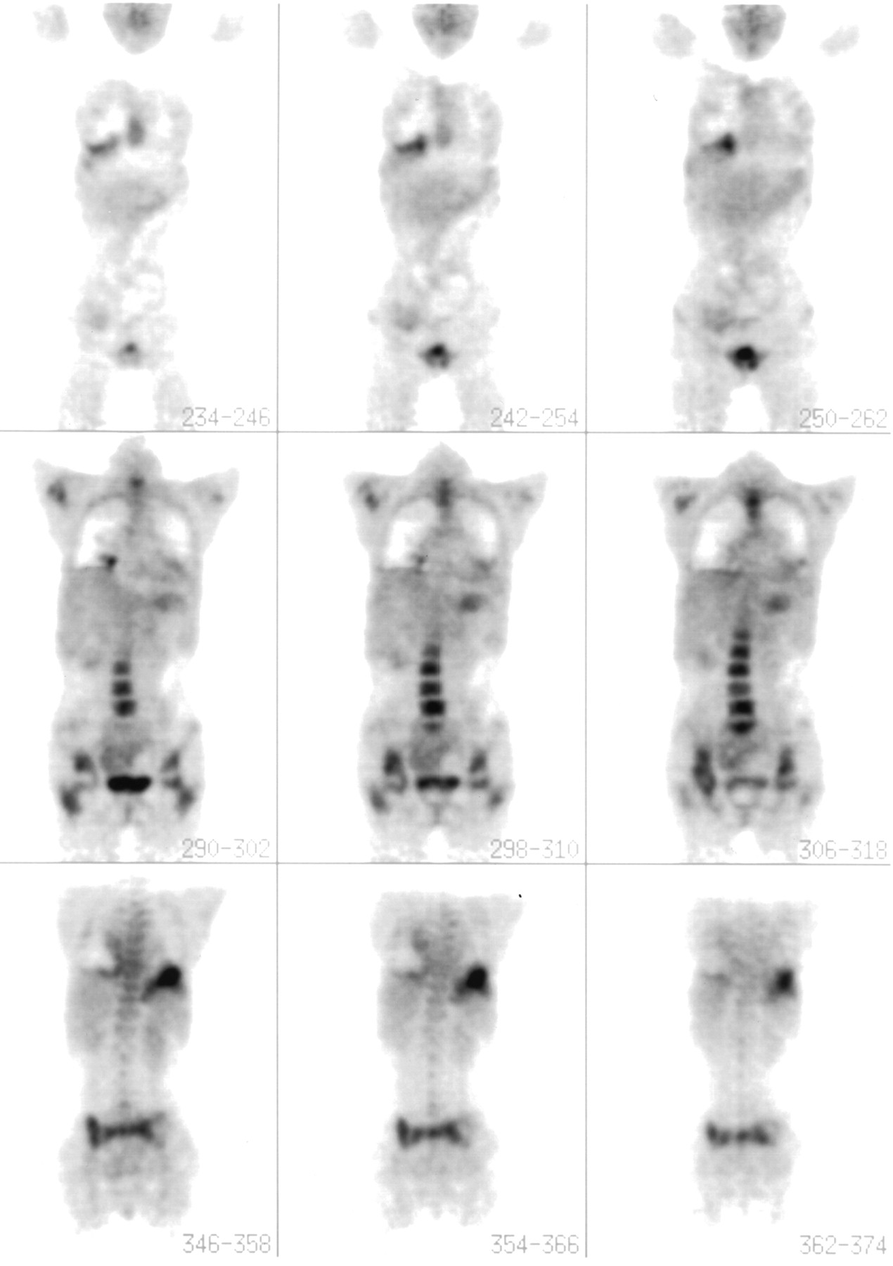
- FIGURE 1.
Patient with IgA λ-chain myeloma, stage IIIA. Radiographs revealed multiple lytic lesions in areas that showed positive findings on 18F-FDG PET imaging (e.g., spine, pelvis, and hips). Plasmacytoma in left lung was confirmed on biopsy. Plasmacytomas were noted in left apex and hilar regions on CT scanning. Splenic myeloma was also suspected but not directly confirmed.
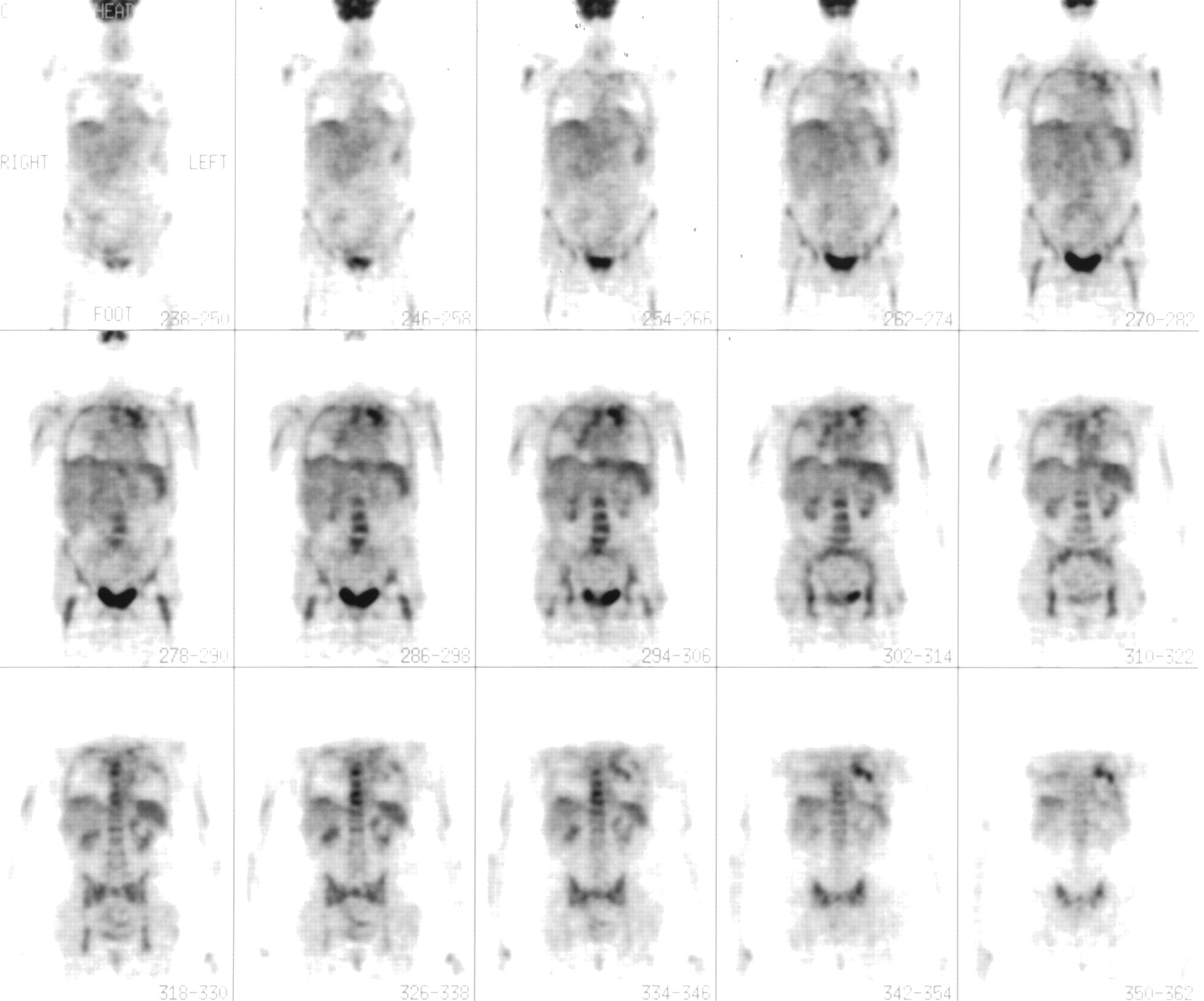
- FIGURE 2.
Patient with Bence Jones protein λ-chain myeloma. Extensive disease is seen in spine, and plasmacytomas are seen in right lower lung and left upper abdomen.
- FIGURE 3.
Patient with relapse after stem cell transplantation. Dysphagia developed and was associated with positive finding in distal esophagus. Direct biopsy showed plasma cells with local amyloid deposition. This responded well to local irradiation.


Tables
Disease status Result of baseline study Result of follow-up study MGUS 14/14 (100%) negative; followed 3–43+ mo In 1/14 (7%), myeloma developed at 8 mo; in 1 patient, focal plasmacytoma with amyloid developed* Active myeloma 16/16 (100%) positive; 4/16 (25%) had negative radiography results but multiple focal lesions on scan In 1 patient, disease was upstaged from stage I to III: Multiple new disease sites were identified 4/16 (25%) had extramedullary disease Poor outcome with extramedullary disease Remission In 6/10 (60%), new lesions developed over 6–42+ mo Local irradiation used for focal relapse; nonsecretory relapse detected and treated Relapse 21/26 (81%) had new sites of disease Anatomic distribution of lesions documented 6/26 (23%) had extramedullary disease Very poor survival with extramedullary disease: median, 7 mo Suspected disease status Clinical question Potential role of 18F-FDG PET MGUS* How likely is progression? If 18F-FDG PET results are negative, stable MGUS is likely Active myeloma* Is disease smoldering or active? If 18F-FDG PET results are positive, active disease is likely, even if radiography results are negative What is prognosis? Extramedullary disease indicates poor prognosis Remission Is there true complete remission? Positive scan findings after transplantation indicate persistent disease or relapse and poorer outcome Is there new disease? New focal disease (especially nonsecretory) can be documented and treated Relapse Is there unsuspected new disease? Can detect clinically important disease requiring unanticipated therapy (e.g., ureteral obstruction) What is baseline restaging at relapse? Can establish baseline for ongoing monitoring in clinical trials ↵* Working definitions for MGUS vs. active myeloma are currently under review by International Prognostic Index Study Group. It is possible that 18F-FDG PET scanning can contribute to diagnostic or discriminatory criteria.
In this issue


Jump to section
Related Articles
Cited By...
- Multiple myeloma with central nervous system relapse
- Multiple Myeloma, Version 3.2017, NCCN Clinical Practice Guidelines in Oncology
- Multiple Myeloma, Version 2.2016
- In Silico Modeling-based Identification of Glucose Transporter 4 (GLUT4)-selective Inhibitors for Cancer Therapy
- In vivo molecular imaging of chemokine receptor CXCR4 expression in patients with advanced multiple myeloma
- Impact of Initial FDG-PET/CT and Serum-Free Light Chain on Transformation of Conventionally Defined Solitary Plasmacytoma to Multiple Myeloma
- 11C-Acetate PET/CT for Metabolic Characterization of Multiple Myeloma: A Comparative Study with 18F-FDG PET/CT
- The role of positron emission tomography-computed tomography and magnetic resonance imaging in diagnosis and follow up of multiple myeloma
- Imaging in myeloma
- Multiple myeloma presenting as plasmacytoma of the jaws showing prominent bone formation during chemotherapy
- Imaging of Multiple Myeloma and Related Plasma Cell Dyscrasias
- Prognostic relevance of 18-F FDG PET/CT in newly diagnosed multiple myeloma patients treated with up-front autologous transplantation
- Multiple Myeloma
- Consensus recommendations for standard investigative workup: report of the International Myeloma Workshop Consensus Panel 3
- Prognostic Significance of Focal Lesions in Whole-Body Magnetic Resonance Imaging in Patients With Asymptomatic Multiple Myeloma
- Multiple Myeloma
- Targeting Glucose Consumption and Autophagy in Myeloma with the Novel Nucleoside Analogue 8-Aminoadenosine
- A new pet for myeloma
- NCCN Task Force: Clinical Utility of PET in a Variety of Tumor Types
- FDG-positron-emission tomography for staging and therapeutic assessment in patients with plasmacytoma
- Hyperattenuating bone marrow abnormalities in myeloma patients using whole-body non-enhanced low-dose MDCT: correlation with haematological parameters
- 18F-FDG PET/CT, 99mTc-MIBI, and MRI in Evaluation of Patients with Multiple Myeloma
- Multiple Myeloma Presenting With [18F]Fluorodeoxyglucose Avid Liver Lesions Diagnosed on Positron Emission Tomography Scan
- A prospective comparison of 18F-fluorodeoxyglucose positron emission tomography-computed tomography, magnetic resonance imaging and whole-body planar radiographs in the assessment of bone disease in newly diagnosed multiple myeloma
- Artifactual Spinal Metastases Imaged by PET/CT: A Case Report
- Imaging of Malignant Bone Involvement by Morphologic, Scintigraphic, and Hybrid Modalities
- Plasma Cell Problems: CASE 3. Plasmacytoma Mimicking a Paraganglioma of the Skull Base: Diagnostic Value of Somatostatin Receptor Scintigraphy
- Side Effects and Good Effects from New Chemotherapeutic Agents: CASE 3. Bortezomib in Primary Refractory Plasmacytoma
- Magnitude of Response With Myeloma Frontline Therapy Does Not Predict Outcome: Importance of Time to Progression in Southwest Oncology Group Chemotherapy Trials
- Unusual Presentations of Hematologic Malignancies: CASE 1. Solitary Bone Plasmacytoma: Role of Magnetic Resonance Imaging and Positron Emission Tomography
- Treatment of multiple myeloma