


Article Figures & Data
Figures
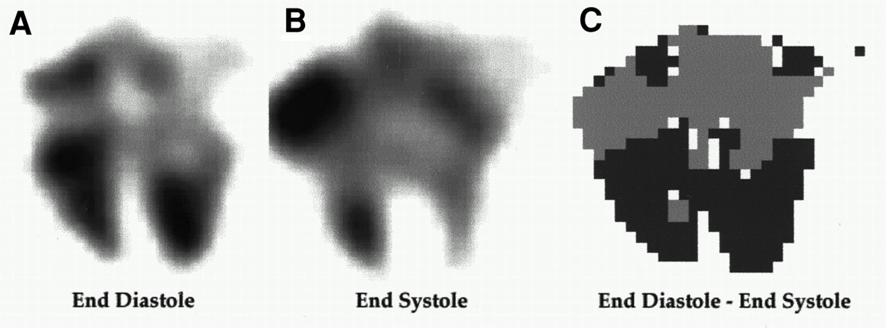
- FIGURE 1.
Horizontal long-axis slice at end-diastole (A) and end-systole (B). Subtracting end-systolic image from end-diastolic image (C) results in positive values around ventricles (dark gray) and negative values around auricles (light gray).
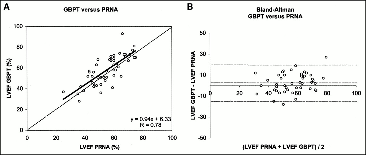
- FIGURE 2.
Detecting septum is done on best septal view at end-diastole. By summing all voxels along x-axis, profile is generated. Maximum (A) is selected, and another profile along this location is created. Local minimum (B) is selected as septum, and local maximum on left side of septum (C) is selected as center of left ventricle.
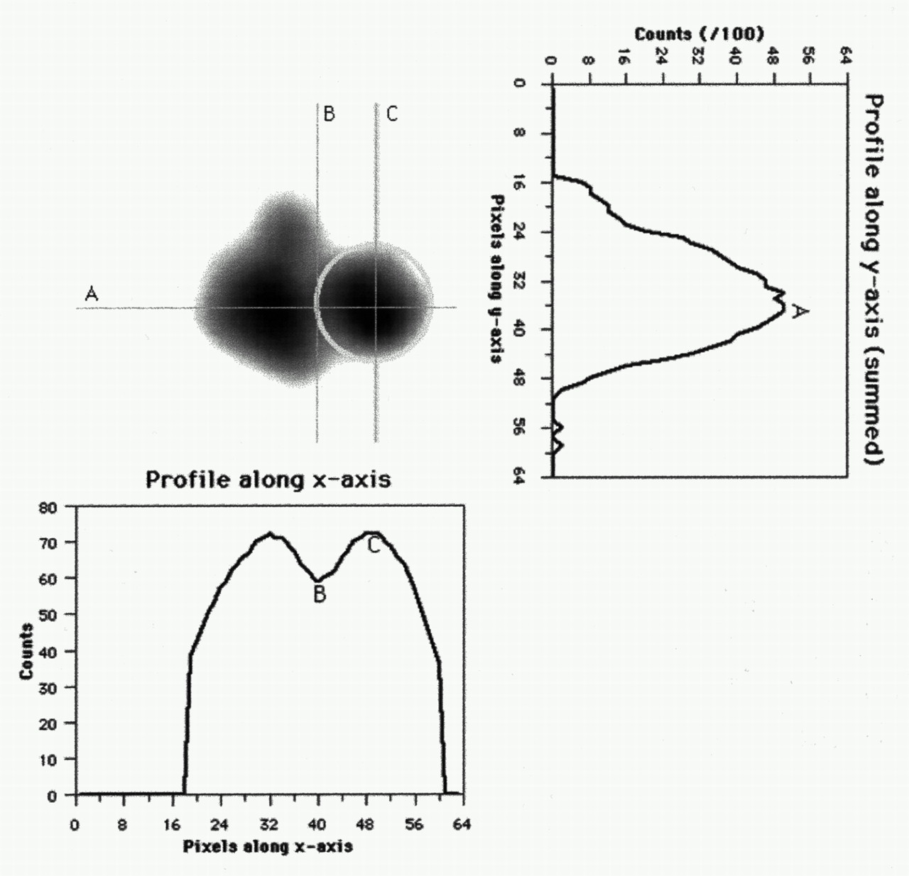
- FIGURE 3.
Summary of algorithm. Initially, end-diastolic image (A) is segmented (B) and segments are identified (C) as left and right ventricle. Optimal threshold is found when corresponding ventricular wall (D) best fits first derivative of end-diastolic count distribution (E).
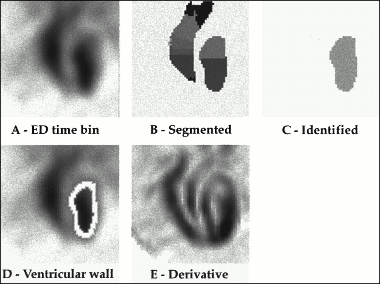
- FIGURE 4.
Linear regression analysis (A) and Bland-Altman plot (B) between LVEF determined by GBPT and LVEF determined by PRNA.
- FIGURE 5.
Intraobserver reproducibility. Linear regression (A) and Bland-Altman analysis (B) between original LVEFs measured (LVEF 1) and ejection fraction measured 3 mo later (LVEF 2).
- FIGURE 6.
Agreement between LVEFs measured by two independent observers.


Tables
Cutoff frequency Cycles per centimeter 0.25 0.30 0.35 0.40 0.45 r 0.78 0.88 0.89 0.88 0.86 Slope 1.51 1.30 1.09 1.10 1.11 Intercept −19.58 −11.21 −0.72 −0.67 −1.26 Systematic error (%) +6.95 +4.55 +3.80 +4.45 +4.50 Random error (%) 15.99 9.32 6.76 7.27 7.92 P 0.07 0.04 0.02 0.01 0.02
In this issue


Jump to section
Related Articles
Cited By...
- Diagnosis of Diffuse and Localized Arrhythmogenic Right Ventricular Dysplasia by Gated Blood-Pool SPECT
- Accuracy of 4 Different Algorithms for the Analysis of Tomographic Radionuclide Ventriculography Using a Physical, Dynamic 4-Chamber Cardiac Phantom
- Validation of Gated Blood-Pool SPECT Cardiac Measurements Tested Using a Biventricular Dynamic Physical Phantom
- Left Ventricular Ejection Fraction and Volumes from Gated Blood-Pool SPECT: Comparison with Planar Gated Blood-Pool Imaging and Assessment of Repeatability in Patients with Heart Failure
- Quantitative Evaluation of Myocardial Blood Flow and Ejection Fraction with a Single Dose of 13NH3 and Gated PET