


Article Figures & Data
Figures
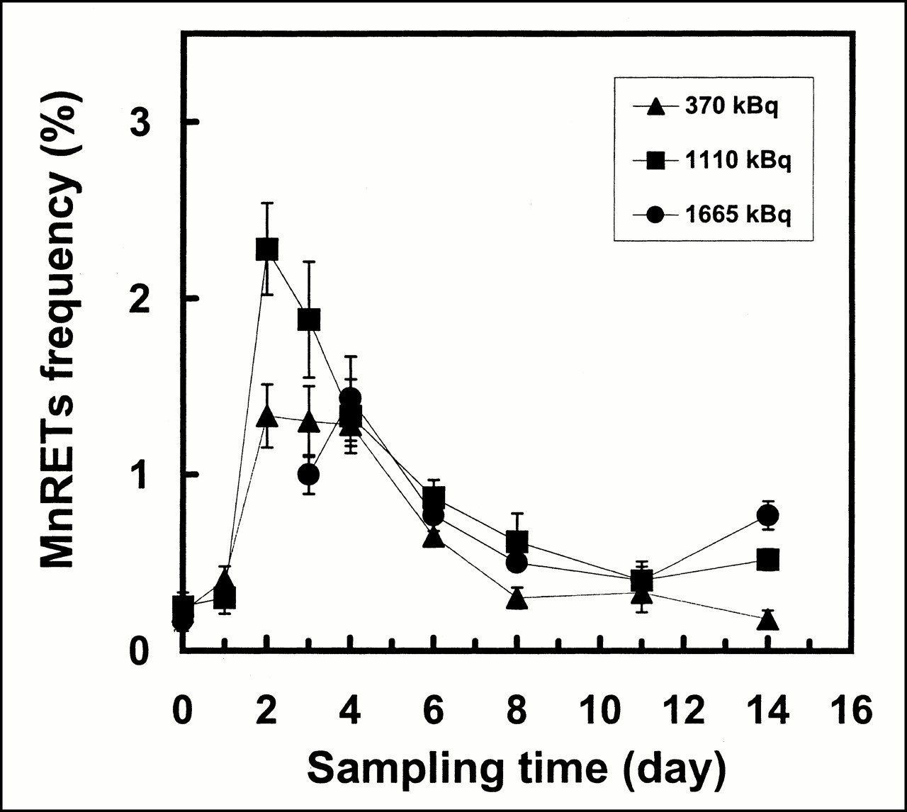
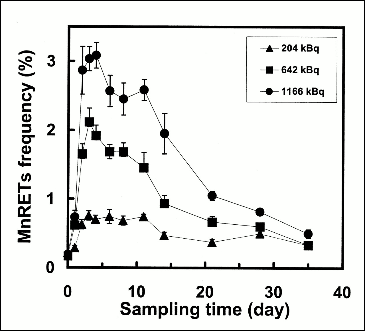
- FIGURE 1.
Frequency of induction of MnRETs in peripheral blood of mice as function of time after intravenous injection of 32P-orthophosphate. Results are shown for three different injected activities. Error bars represent SEs of mean for two independent experiments.
- FIGURE 2.
Frequency of induction of MnRETs in peripheral blood of mice as function of time after intravenous injection of 90Y-citrate. Response curves are shown for three different injected activities. Error bars represent SEs of mean for two independent experiments.
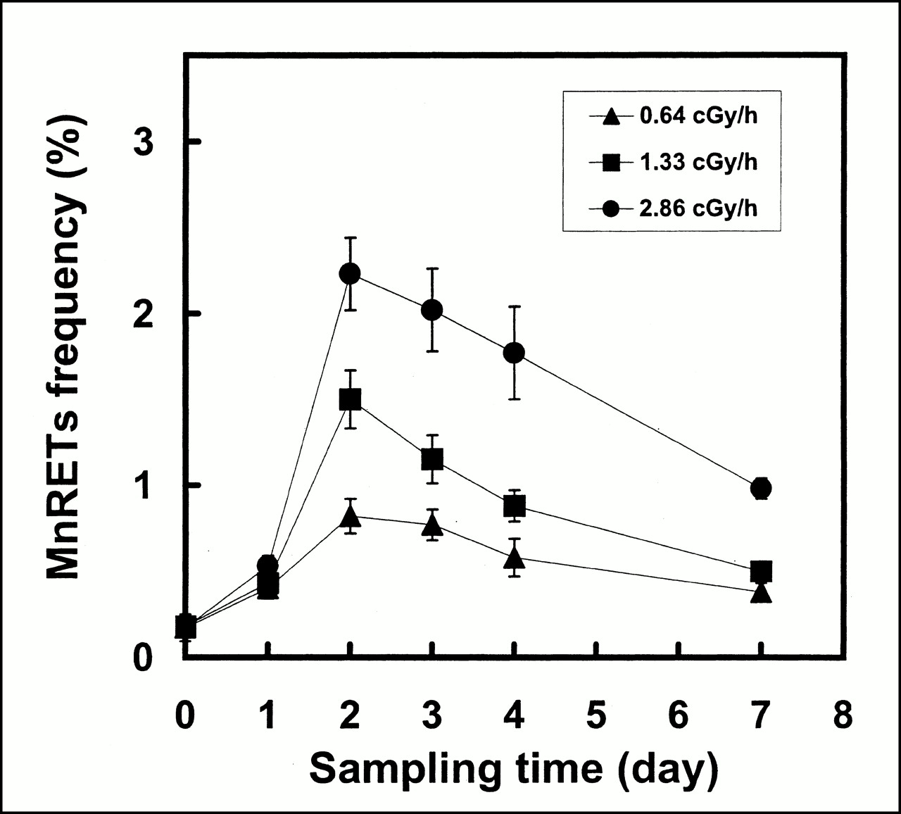
- FIGURE 3.
Frequency of induction of MnRETs in peripheral blood of mice as function of time after initiating irradiation with external γ-rays from 137Cs irradiator. Dose rate was decreased exponentially, with Td of 255 h, which corresponds to Te from femurs of mice that were injected with 32P-orthophosphate. Data points represent mean ± SE of mean of two independent experiments.
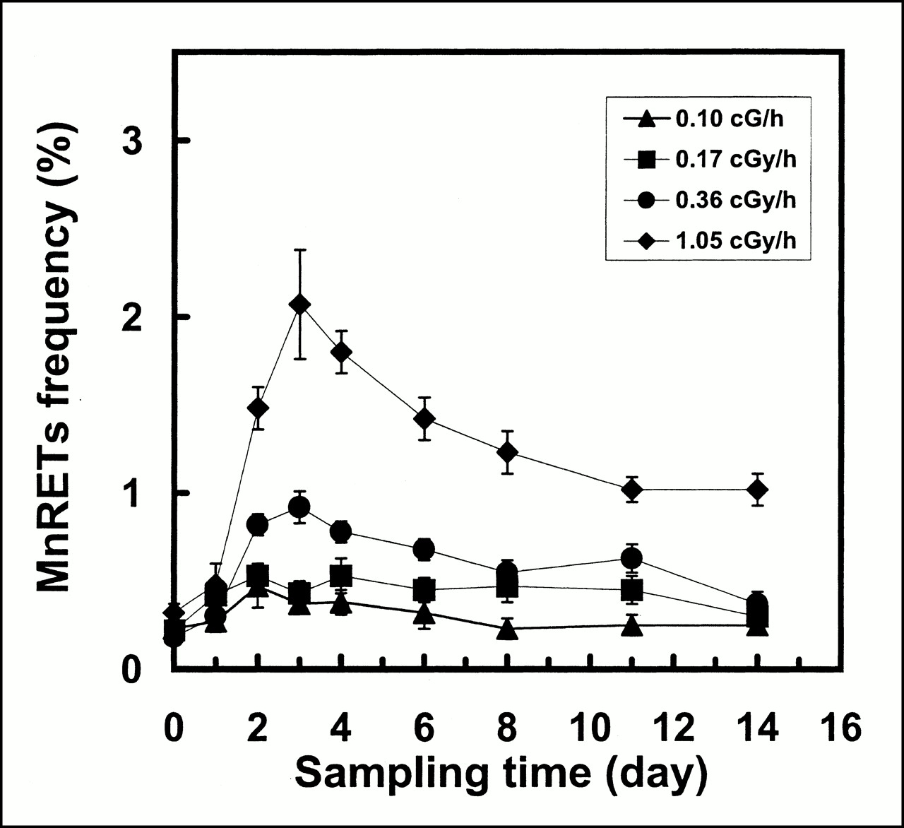
- FIGURE 4.
Frequency of induction of MnRETs in peripheral blood of mice as function of time after initiating irradiation with external γ-rays from 137Cs irradiator. Dose rate was decreased exponentially, with Td of 64 h, which corresponds to Te of radioactivity from the femurs of mice that were injected with 90Y-citrate. Data points represent mean ± SE of mean of two independent experiments.
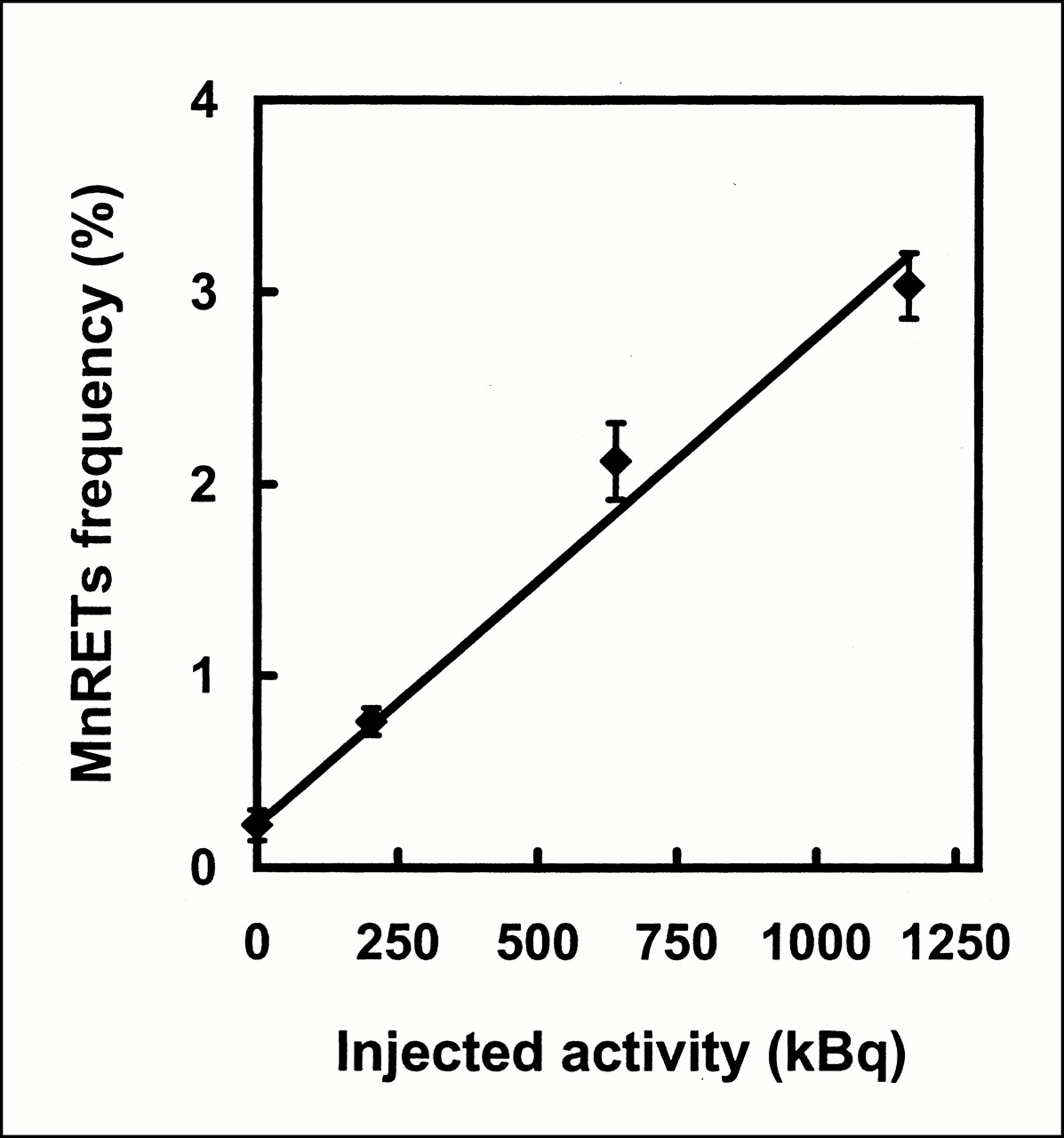
- FIGURE 5.
Frequency of induction of MnRETs in peripheral blood of mice as function of AInj of 32P-orthophosphate on third day after administration. Error bars represent SEs of mean for two independent experiments.
- FIGURE 6.
Frequency of induction of MnRETs in peripheral blood of mice as function of AInj of 90Y-citrate on second day after administration. Error bars represent SEs of mean for two independent experiments.
- FIGURE 7.
Frequency of induction of MnRETs in peripheral blood of mice as function of initial dose rate from 137Cs–γ-rays on third day after initiating irradiation. External γ-rays were delivered with exponentially decreasing dose rate, with Td of 255 h. Error bars represent SEs of mean for two independent experiments.
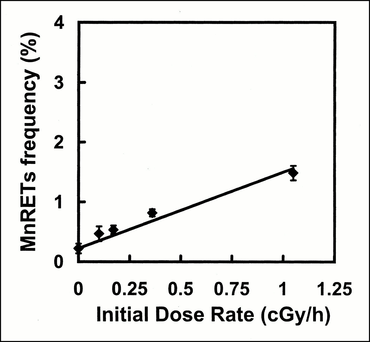
- FIGURE 8.
Frequency of induction of MnRETs in peripheral blood of mice as function of initial dose rate from 137Cs–γ-rays on second day after initiating irradiation. External γ-rays were delivered with exponentially decreasing dose rate, with Td of 64 h. Error bars represent SEs of mean for two independent experiments.


